Fluorography is a diagnostic procedure that is mandatory during preventive medical examinations. This method became widely used to detect chest diseases in the 1950s. The high incidence of tuberculosis in our country has made fluorography a necessary examination that must be completed before employment, admission to the hospital, and during professional medical examinations. Pregnant women are exempt from it. What to do if an examination is necessary or a woman finds out about pregnancy after having a fluorography?

Fluorography: principle of action, indications for research
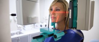
Fluorography is x-ray photography of an image formed on a fluorescent screen, which is obtained when x-rays pass through a human being. When rays pass through, organs and tissues transmit them differently, which is displayed as light and dark areas in the photo. In this way, doctors can determine the presence of the disease and its location.
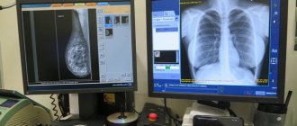
The most common is film X-ray photography, but in recent years it has begun to be replaced by digital. In digital photography, a CCD matrix is used instead of X-ray film. Another method of digital radiography is based on the principle of layer-by-layer scanning of the chest.

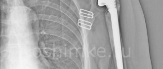
The main indication for fluorography is the detection of tuberculosis, cancer and other lung diseases. In addition, it allows you to detect infectious foci in the chest organs, changes in the heart, bones and muscles. Thanks to the penetrating ability of X-rays, it is possible to detect scars, cords, adhesions, sclerosis, and radiance of lung tissue. The image shows cysts, emphysematous phenomena, and infiltrates.
Doctors recommend being examined once a year for prevention and be sure to go to the X-ray room if you have had contact with a patient with tuberculosis. When living in unfavorable regions, it is allowed to take radio photography twice a year.
Fluorography at the pregnancy planning stage
Is it worth taking x-rays at the planning stage of a child? Future parents fear that radiation will affect the formation of germ cells. Their concerns are partly justified because ionizing radiation negatively affects growing and developing cells. Women should not visit the X-ray room in the first days of the cycle, when the oocyte is just forming in the follicle. Men should wait a few days after fluorography so that the sperm have time to renew themselves. The minimum period that must be waited before conceiving is 1 month.
However, you should not give up radio photography completely. It allows you to identify complex and severe illnesses that are best treated before pregnancy. Tuberculosis, pneumonia, lung tumors - these diseases can cause much more harm to the mother and unborn child than fluorography. If a married couple is worried about the effect of radiation on the germ cells, afraid that it will cause a chromosomal mutation and the child will be born sick, the diagnosis is carried out several months before the expected conception.

How can fluorography affect pregnancy if done early?
Why is X-ray photography harmful in early pregnancy? In the first weeks of gestation, the active formation of organ rudiments in the embryo occurs. From a zygote - a single-celled embryo - a blastocyst consisting of several cells is produced by division. The cells of the embryo begin to specialize and continue to divide, thus forming primary tissues from which the rudiments of organs are formed. By mid-term, the fetus has already formed most of its organs.
Young dividing cells are most susceptible to the negative effects of ionizing radiation. This is why many doctors are categorically against X-ray photography of pregnant women. They believe that such exposure can lead to an abnormal pregnancy. It is recommended to refrain from chest examination until 25 weeks. In the second half of pregnancy, radiation will not harm the fetus.
How justified are these warnings? Some doctors question such fears. They are confident that it is possible to have a pregnant woman undergo fluorography once; it will not harm the child, because in medical practice there is not a single case recorded when radiophotography would lead to disruption of the baby’s intrauterine development. In addition, during the procedure, the woman’s stomach is covered with a lead apron, through which X-rays do not pass.
Is it possible to refuse the examination and is it necessary to do so?
Fluorography of the husband during pregnancy is, in principle, not necessary, but it is extremely unwise to refuse it. Firstly, he is responsible for the health of his wife and child, so he must be confident in his health. Secondly, the procedure is painless, practically harmless and takes little time. Any future father, even a very busy one, will be able to set aside time for the sake of his family.
Possible negative consequences for the mother in labor. Some medical institutions refuse to admit a woman without the appropriate certificate. They can also refer her to the prenatal infectious diseases department to reduce the possible risk for other women in labor and newborns.
Of course, a woman will not remain on the street to give birth, but troubles may arise. That is why it is easier to undergo the procedure and get the opportunity to see your child in the first minutes of his life.
What to do if the study was carried out when the woman did not know that she was pregnant?
Not all women know about pregnancy from the first weeks. With an irregular cycle, when a delay is common, and small bleeding that the expectant mother may mistake for menstruation, the woman may not even suspect that she is pregnant. At this time, she can undergo a medical examination and undergo a routine examination, including radio photography.
If fluorography was done before the expectant mother found out about pregnancy, she should immediately consult a doctor. After listening to an explanation of the situation, the doctor will decide whether to prescribe additional diagnostics, ultrasound or genetic analysis. Women over 35 years of age, as well as those with relatives with chromosomal abnormalities, are at risk. The risk of pathologies in a child may increase under the influence of radiation.
What you definitely shouldn’t do is worry too much. Stress has a much worse effect on the course of gestation than a single dose of ionizing radiation.
Reviews from women about the procedure

Elena, 24 years old: “I had fluorography done during pregnancy, when I still didn’t know about my situation. Then my mother-in-law scared me that the child would be born with disabilities and I consulted a doctor. They said that nothing bad would happen, and so it was. Just unnecessary worries.”
Irina, 30 years old: “Since I work in the maternity ward, I often undergo fluorography. Before going on maternity leave, I also underwent the study on time - I can’t jeopardize my health and the health of our patients. I know that research does no harm, so I’m not afraid of it.”
Olga, 27 years old: “The story of my pregnancy was not entirely cloudless. My ex-husband suffered from tuberculosis, which I learned about later. The doctors suspected the disease and, fortunately, they did not find tuberculosis in me. That’s how we separated from my husband; he comes from a dysfunctional family, and the child was born healthy. Now I’m again expecting an increase and I know that fluorography is harmless - if necessary, it can be done without fear.”
Is fluorography prescribed for pregnant women, in what cases and how is it performed?
Pregnant women are not routinely sent for fluorography, even if it is necessary during a medical examination for the workplace. However, this diagnosis cannot be avoided if there are suspicions of diseases of the chest organs.
X-ray photography is done only for vital indications, when the harm from the lack of correct diagnosis and treatment is much higher than from X-rays. Indications:
- pulmonary tuberculosis in a pregnant woman or her loved one with whom she has been in contact;
- pneumonia;
- neoplasms in the lungs;
- enlarged heart;
- entry of a foreign body into the lower respiratory tract.
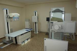
When choosing between irradiation methods, you should choose digital fluorography, which is performed using a scanner method. During the procedure, the chest is scanned layer by layer in the horizontal plane with a fan-shaped beam of X-rays. Unfortunately, not all hospitals have new equipment; most have old film machines.
The table shows a comparison of two X-ray photography methods:
| Diagnostic method | Radiation dose | Percentage of permissible annual radiation dose |
| Film | 0.5–0.8 mSv | 50–80% |
| Digital | 0.05 mSv | 5% |
The doctor giving the referral for the examination and the nurse performing the procedures must know that the patient is pregnant. During the irradiation process, the woman's abdomen and pelvis will be covered with a lead apron.
Is it possible to replace radiophotography with other diagnostic methods? Depending on the disease, the doctor may prescribe other examinations:
- Ultrasound. Ultrasound will not help detect tuberculosis or pneumonia, but it is effective in detecting changes in the heart muscle.
- Blood chemistry. Allows you to identify infectious and inflammatory diseases and suspect the presence of tumors.
- PCR swab. For diseases of the lungs and bronchi, sputum is examined. This analysis allows you to detect a specific pathogen.

What is more dangerous – x-ray or fluorography?
Another diagnostic method that uses x-rays is a chest x-ray. How does it differ from fluorography?
Features of radiography:
- Allows you to identify those ailments that are not visible on a fluorogram. X-rays are necessary for suspected pathologies of the cardiovascular system, injuries and bone diseases.
- Detects smaller formations. The minimum lesion visible on fluorogram has a diameter of 5 mm. X-ray is capable of recognizing neoplasms and lesions with a diameter of 2 mm. This is very important for the early diagnosis of tuberculosis and lung cancer.
- The costs of producing an image are higher. To obtain a fluorogram, the film can be developed directly in rolls; less of it is required, which makes the diagnostic method cheaper. X-rays are developed using special baths, so x-rays take longer to take and are more expensive.
- The dose of ionizing radiation is higher. X-rays are prescribed only if serious pathologies are suspected, because the radiation dose is 2 times higher. This is necessary to ensure greater penetration of X-rays through organ tissue.
So should pregnant women refuse fluorography or not? If a preventive examination can be postponed, then the diagnosis prescribed by the doctor cannot be refused. Radiography allows early detection of diseases that, without treatment, can lead to serious consequences.
Second medical opinion on the image
Remember that, if necessary, the obtained X-ray images can be re-analyzed by the right specialist! You can order a repeat interpretation of an X-ray, CT or MRI using the National Teleradiology Network. A team of highly specialized radiologists will help clarify your diagnosis and provide a detailed description of the submitted x-rays and other studies within 24 hours.
Pavel Popov
Candidate of Medical Sciences, Member of the European Society of Radiologists