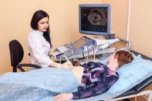
For the last 20 years, all pregnant women have been required to undergo an ultrasound examination. And if previously the capabilities of ultrasound were limited - the doctor saw only a dark lump on the monitor, now both the gynecologist and parents, with the help of 4D ultrasound, can see the baby in volume and color.
COST of 4D ultrasound with viewing on a large screen from 2000 rubles. DISC WITH RECORDING AS A GIFT
CLICK TO MAKE AN APPOINTMENT, TEST OR ULTRASOUND
This procedure is unique. It not only allows expectant mothers to look at their baby before birth, but also to find out if everything is fine with him. In memory of these events, a video is recorded so that parents can see after many years what their son or daughter was like before he was born.
What is 4D ultrasound: features of the technique
The content of the article
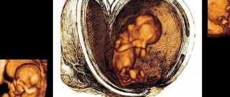
Four-dimensional ultrasound is the latest ultrasound examination, with the help of which a complete examination of the fetus is carried out using a volumetric scanning method. The examination allows the specialist to study images from different angles, in different spatial dimensions in real time.
4-D ultrasound makes it possible to:
- Carry out a qualitative check of fetal development, and in case of pathology, detect it in the early stages.
- Examine the future baby in detail, find out its gender, analyze all organs separately, and even determine which parent it will resemble.
- This technique allows you to restore the psycho-emotional contact between future parents and the baby.
One of the advantages of 4-D ultrasound is that when the fetus is moving, it is more convenient for the doctor to examine its skeleton and proportions.
When a four-dimensional ultrasound is performed, how many times is the procedure needed?
Ultrasound examination is prescribed to pregnant women 3 times throughout the entire period. If there are no additional indications, ultrasound screenings are performed three times:
- at 11-13 weeks;
- at 18-21 weeks;
- at 30-34 weeks.
However, according to obstetric practice, ultrasound examinations are carried out more often, because if there are any concerns about the health of the mother and child, one has to find out how the baby “lives” in the mother’s body.
Evaluation of the embryo using a 4D scanner is usually carried out from the 20th to 24th week; at later stages, the image may be less clear due to the large weight of the fetus. It is also possible to use this method in certain cases and in the early stages.
Ultrasound diagnostics makes it possible to identify severe fetal pathologies in the early stages and, in this case, terminate the pregnancy in a timely manner. Thanks to ultrasound screening, the number of newborns with Down syndrome and other severe genetic pathologies has decreased by one and a half times.
When can you undergo a 4D ultrasound during pregnancy?
The content of the article
According to the classic pregnancy protocol, ultrasound is performed at least 2-3 times during pregnancy - at 10-12 and 20-24 weeks of pregnancy. The specialist prescribes additional ultrasound diagnostics at 37-38 weeks of pregnancy to determine the possibility of a natural birth.
Doctors recommend turning to the 4D ultrasound method when conducting studies for periods longer than 20 weeks. This is due to the fact that after 20 weeks the fetus is already fully formed, which ensures adequate visualization of the baby.
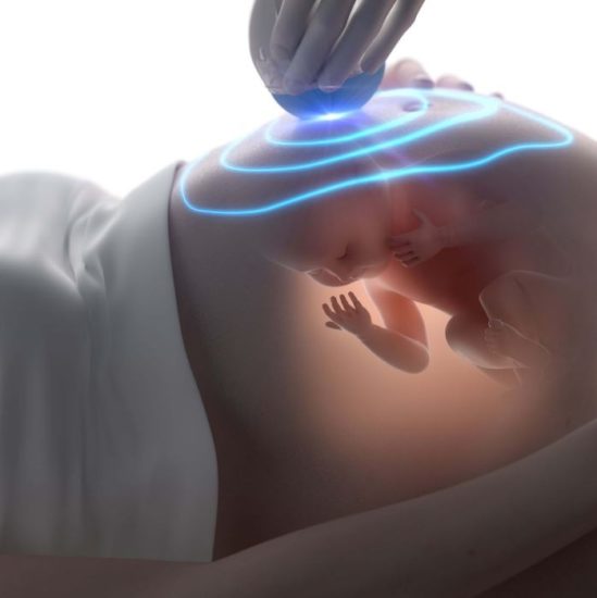
Operating principle of 4D ultrasound
The research technique in a four-dimensional format involves the use of ultrasounds based on echolocation. Ultrasound waves emanating from the organs and tissues of the fetus are captured by a highly efficient sensor. Thanks to this, specialists receive a high-quality image, which makes it possible to examine all internal organs, recognize any pathology, and also conduct Doppler mapping of blood vessels.
The difference between 4-D ultrasound and 3-D ultrasound is that in the first case, the image of the embryo is recorded as a video film on disk, and in the second, the fetus is additionally displayed in separate images. This method is considered completely safe for both the pregnant woman and the unborn child.
The duration of the procedure is about 40-50 minutes, in some cases the examination may last several minutes longer.
What can be seen with 4D ultrasound
The method allows you to expand the image and see the child from all sides.
It turns out to be a kind of “movie” about the development of a baby, which can be watched many times. The video image shows how the baby moves and his emotions change. This indirectly shows whether he is comfortable, because on a child’s face, pain is reflected in the same way as on the face of an adult. Previously, a simple ultrasound was used for this purpose, during which a two-dimensional “flat” image appeared on the screen, which did not provide complete information about the baby’s condition. With the introduction of 4D everything changed:
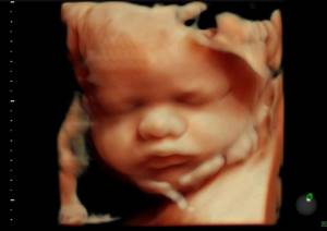
- A three-dimensional image is visible on the screen. You can even count the fingers and toes. This significantly increases information content and reduces the number of medical errors.
- The image obtained during such a procedure is understandable not only to the doctor, but also to the future parents, so the whole family often comes for diagnosis. Mothers say that after giving birth they recognized their baby because he looked the same as in the picture. This indicates the accuracy of the technique.
- The method reveals heart defects that are difficult to diagnose with a simple ultrasound. The study allows doctors to take measures in advance so that the baby not only survives, but is also operated on at an early age.
4-D ultrasound in pregnant women allows:
- Diagnose the skull and facial structure of the fetus.
- Examine the condition of the skeletal system and spine.
- See any disorders of the gastrointestinal tract, cardiovascular and respiratory systems.
- Detect Down's disease, as well as some other chromosomal diseases in the early stages.
- Identify any pathological abnormalities in fetal development.
- Re-examine.
- See the female organs and assess their state of health, since in the second half of pregnancy this is very important for normal oxygen supply to the fetus.
With a 4D ultrasound, the child can be viewed from different angles, obtaining a video image, which, at the request of the future parents, is recorded on a disk.
Indications for 4D ultrasound: family and medical
From the very beginning, 4D ultrasound has been in demand among parents who want to see their baby in action even before birth: watch him move his arms and legs, yawn, discuss who he looks more like, and then watch a film with him at home many times.
And, what is important for almost all families, to accurately find out the gender of the future heir. For this purpose, 4D ultrasound is recognized as the most accurate method, when such factors as “the baby sat down poorly”, “covered the most important place with his legs”, and so on are practically excluded.
You can discuss the emotional state of expectant mothers as much as you like, but it has been noted that when a pregnant woman is nervous, which can negatively affect the process of preparing for childbirth and the birth itself, she gets to know the unborn baby during a 4D ultrasound and receives visual information that the baby is fine , helps her to better survive the last weeks of pregnancy, worry less and be more conscious about motherhood.
Therefore, family indications for 4D ultrasound:
— getting to know the future family member before his birth,
- better preparation of the expectant mother for childbirth,
— obtaining reliable information about the baby’s gender.
But there are also very serious medical indications . Thus, 4D ultrasound is recommended in the following cases:
— if, during a screening 2D ultrasound, the doctor suspects that the baby has a pathology in the development of the bones of the face, skull, spine or other parts of the musculoskeletal system,
— if a questionable result is obtained during genetic screening during pregnancy or the mother, for some reason, does not consider further genetic research using invasive procedures, which in her opinion or the opinion of doctors may be unsafe for the baby,
- if the unborn baby is at risk of developing malformations of the heart or brain - for example, if such diseases exist in the child’s family, or the need for additional research was discussed by the parents during consultation with a geneticist, a doctor managing the pregnancy or any other medical specialist, observing this family - for example, with a heart surgeon.
For example, during a 4D ultrasound, a doctor can carefully and in detail evaluate the functioning of the baby’s heart chambers, the blood flow through them, and its distribution after leaving the heart cavity. No other non-invasive (that is, without violating the integrity of the skin and internal organs) technique during pregnancy can cope with this task.
Operating modes of 4D ultrasound
The technique includes several operating modes:
- Superficial
. This mode allows you to see the surface of the body of the unborn baby at 11-22 weeks. You can examine the face, limbs, genitals, etc. In certain cases, visualization becomes difficult, most often due to incorrect position of the embryo, placement of the umbilical cord, or the volume of amniotic fluid. - Volumetric negative
. This function is resorted to when the doctor suspects a pathology of the vascular system, in particular vascular and non-vascular neoplasms, disorders in the vessels and ventricles of the brain. Using this mode, the condition of the fetus is clarified in detail and the necessary organs are assessed. - Multiplane
. This method helps to obtain an image of the fetus in three planes located perpendicularly. The multiplane mode is the basis of this technique. - Multiplanar
. This is the most optimal mode, allowing you to quickly detect any fetal anomalies through a two-dimensional image of all organs. - Multiplanar real-time mode (STICS technology)
. This method is intended to assess the functioning of the heart and detect heart diseases in the unborn child.
4D diagnostic capabilities
The technique for performing 4D ultrasound is practically no different from 3D. The only and most important difference is the ability to see the baby in real time. 3D diagnostics differs in that parents receive a three-dimensional image of the fetus a few minutes after the procedure. 4D allows you to observe movements and even record the child on video.
4D ultrasound can only be used in conjunction with a standard examination in order to diagnose the baby’s condition and satisfy the interest of the parents. It is ideal to combine two-dimensional and four-dimensional examination.
In addition to standard two-dimensional ultrasound diagnostic data (fetal size, its position, condition of organs and limbs), 4D ultrasound significantly expands diagnostic capabilities in terms of determining the presence or absence of the following developmental anomalies in the fetus: skeletal malformations, space-occupying formations, facial defects (so-called clefts faces, etc.), quantitative pathologies of the fingers and other anomalies of the hands, defects of the anterior abdominal wall, anomalies of organ development. The 4D ultrasound mode provides an expanded overview, so the doctor can evaluate all external signs. This advantage of 4D ultrasound is especially relevant for studying facial structures (nasolabial triangle, lips, chin, ears, etc.). On the monitor of the device, the specialist monitors the movements of the fetus; their nature may be important for confirming pathologies of the development of the musculoskeletal system.
Decoding the results
The conclusion about a 4-D ultrasound is deciphered by a gynecologist. They also analyze the results obtained with normal indicators, which depend on the duration of pregnancy. Helps the doctor to correctly decipher a special photometric table, which contains all the parameters of the fetus, indicated weekly. The main parameters taken into account in the decoding are:
- Head circumference.
- Inspection of tubular bones and their length.
- Distance from crown to sacrum
- Biparietal size (width of the head from temple to temple).
In addition, the conclusion about a 4-D ultrasound also includes indications about the condition of the placenta and amniotic fluid (a decrease or increase in them often indicates the development of a pathological process). Great importance is also attached to the examination of the umbilical cord, which is the main connecting link between the mother and the fetus. Also, in the transcript of this ultrasound examination, the doctor must indicate the gender of the child, with almost one hundred percent certainty.
Equipment used for 4D ultrasound
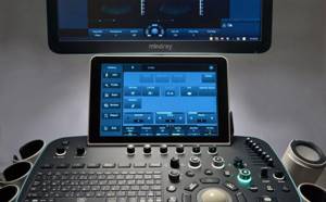
The quality of scanning depends entirely on the equipment. The ultrasound machines used to conduct research are classified as expert-class devices. Such scanners are distinguished by their great capabilities and excellent research quality. They are slightly larger than usual, this is due to the presence of a powerful sensor. They are also equipped with color monitors with high quality images and software.
The most popular models among modern ultrasound scanners used in medical practice are: Accuvix XG, EKO, Philips Epig 5, etc. Our medical center has installed a new 4D ultrasound machine with Doppler SonoAce X8. You can find out why we chose this particular ultrasound machine here ➡ Modern ultrasound 3D, 4D with Doppler from Samsung Medison

How safe is 4D ultrasound?
Despite the fact that the duration of a 4D scan is longer than a regular one, it is completely safe for the mother and the unborn child, and therefore can be performed as many times as the doctor deems necessary.
Of course, the 4-D ultrasound procedure is a unique chance for parents not only to see their future baby in real mode and capture this moment for life, but also to make sure that the child will be born completely healthy.
You can undergo a fetal ultrasound in the “4D + recording on disk” format at the Diana Clinic. We have installed new equipment that allows us to take high-quality photographs. Diagnostics and interpretation are carried out by a doctor of the highest category.
CLICK TO MAKE AN APPOINTMENT, TEST OR ULTRASOUND
If you find an error, please select a piece of text and press Ctrl+Enter
- Gallery
- News
- Reviews
- Vacancies
- Licenses
- Insurance partners
- Controlling organizations
- Schedule for receiving citizens for personal requests
- What you need to know about coronavirus infection?
- Rules for patients
- Important! Visiting a clinic during self-isolation
- Online consultation with a doctor
- to corporative clients
- Documentation
Recently, a new method of ultrasound diagnostics has become increasingly popular among both patients and doctors - three-dimensional ultrasound, which significantly expands diagnostic capabilities, while remaining the same safe and reliable method.
Over the history of using two-dimensional ultrasound in medical practice, doctors have developed a system for analyzing data obtained during such a procedure. For example, each period corresponds to certain sizes of the baby’s head, other parts of the body and organs. That is, 2D ultrasound allows you to fairly accurately determine the condition of the mother and child and identify possible deviations in the development of pregnancy. Three-dimensional research data complements and refines the resulting picture. With its help, you can obtain important information about certain developmental defects. If the doctor suspects something is wrong after a 2D examination, he can prescribe a 3D ultrasound session. This combination of two methods gives the clearest picture of the condition of the expectant mother and baby.
3D ultrasound is an ideal identification of intrauterine fetal anomalies, because Thanks to the three-dimensional image, doctors can evaluate different parts of the fetal body in three projections simultaneously.
It is obvious that with the help of such 3D ultrasound, small defects are diagnosed better than with traditional ultrasound. Three-dimensional examination data provides additional information for the diagnosis of certain fetal malformations: face, spinal column, limbs. These are polydactyly, spina bifida, cleft lip, cleft palate and other developmental anomalies. Genetic defects are better diagnosed with 3D ultrasound. The role of 3D ultrasound in the diagnosis of heart defects also deserves attention.
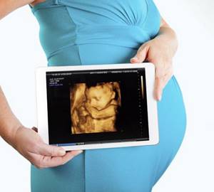
In addition to medical purposes, 3D ultrasound allows the expectant mother to see a (3D) photo-like image of her baby , and with 4D ultrasound you can see a 3D image in motion. It turns out that the 4th dimension is time. The result is a video recording of the child's movements, but with fewer frames per minute than we are used to. Carrying out a 4D ultrasound allows you to start creating a video archive even before the baby is born. This is of great importance in the formation of parental instinct before the baby is born.
The more the baby moves, the clearer the image and the more interesting the “frames” will be. If the baby is not active, you can drink some sweet drink. This usually “excites” the baby within 10-15 minutes.
By the way, not always the mother, and the doctor too, can clearly and distinctly see the child on the screen.
The image quality of 3D ultrasound also depends on several points:
- position of the baby in the womb;
- location of the umbilical cord and placenta;
- the amount of amniotic fluid (the less there is, the worse the image);
- overweight pregnant woman;
- the presence of scars on the abdomen after operations.
3D and 4D ultrasound reports are issued to patients on the day of the examination, both on paper with photographs attached, and on a CD.
Sign up for an examination by phone
Services and prices
Ultrasound of the fetus in 3D - 4D mode
RUB 5,040
Ultrasound of the fetus in 3D - 4D mode with recording on disk
6,000 rub.
Tyan Oksana Aleksandrovna Head of department, obstetrician-gynecologist Doctor of the highest category Work experience: 25 years
Zabolotnova Olga Valentinovna Obstetrician-gynecologist Doctor of the first category Work experience: 24 years
Moiseeva Alla Vitalievna Obstetrician-gynecologist, ultrasound diagnostics doctor First category doctor Work experience: 36 years
Volkova Polina Dmitrievna Obstetrician-gynecologist, ultrasound diagnostics doctor Doctor of the highest category Work experience: 34 years
Postnikova Nadezhda Anatolyevna Obstetrician-gynecologist, ultrasound diagnostics doctor Work experience: 34 years
Shchelokova Elena Nikolaevna Obstetrician-gynecologist Doctor of the highest category Work experience: 37 years
Kovalenko Elena Evgenievna Ultrasound diagnostics doctor Doctor of the highest category Work experience: 42 years