Friday, October 16, 2020
The lymph node system in the human body plays the most important role of a protective barrier against the spread of attacking harmful microorganisms, rods, helminths, viruses, cancer cells, and toxic substances in the body. Lymph nodes are among the organs of the immune system that are the first to take the hit and change due to infection. Therefore, the occurrence of the slightest painful symptoms in places where immune filters are concentrated requires their detailed examination to draw up a complete clinical picture of the disease.
The cost of ultrasound of lymph nodes in Moscow at the Men's and Women's Health Clinic is affordable and designed for patients of any income. Qualified doctors will conduct diagnostics, and doctors will assess your health based on the results obtained.
Ultrasound as a method for diagnosing cervical lymph nodes
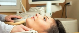
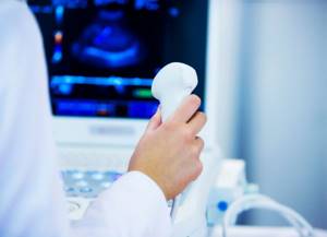
Ultrasound examination of the lymph nodes of the neck is carried out with a special device using high-frequency waves. The method is based on the ability of ultrasound to penetrate soft organs without consequences, be reflected from them and return. The scanner picks up the returning waves and converts the information onto the screen in the form of a “picture” of the internal organs.
Ultrasound of the soft tissues of the neck and lymph nodes will reveal pathologies, among which are often found:
- inflammatory changes;
- metastases;
- cysts;
- tumors, etc.
An ultrasound of the lymph nodes is completely safe and painless. The method does not cause discomfort or pain to the patient during the study, does not cause harmful effects to the body, and therefore is prescribed to patients regardless of condition, age and suspected disease. An exception may be damaged skin in the diagnosed area or the postoperative period.
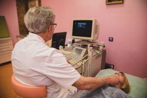
Research process
How the check is carried out:
- the patient is exposed to provide access to the examined area and takes a comfortable position on the couch;
- Universal ultrasound gel is applied to the treatment area;
- the device’s sensor is pressed tightly against the area being diagnosed;
- the image appears on the screen, the specialist takes measurements and analyzes the resulting image.
The results are interpreted based on the echogenicity of the tissues, the size, shape and clarity of the contour, and the condition of the vascular network. In the presence of metastases, everything changes - structure, echogenicity, size and shape (the node becomes more round). This phenomenon makes ultrasound a specific test for detecting metastases.
When is it necessary to perform an ultrasound examination of lymph nodes?
A qualified doctor can make an accurate diagnosis only based on the results of diagnosing the patient’s condition. Having the conclusions of a set of diagnostic procedures, the doctor will correctly determine the source and degree of development of the pathology and prescribe a course of effective treatment.
One of the informative instrumental methods for studying the health of the body is ultrasound of the lymph nodes. Using ultrasound equipment, you can determine any deviations from the norm in the condition of the lymph nodes:
- changes in shape and consistency;
- increase in contours and sizes;
- change of localization;
- compaction of the structure;
- the relationship with surrounding soft tissues changes;
- redness of the surface, etc.
These characteristics make it possible to identify and determine the nature of lymphadenopathy (deviations from the normal state).
Since the course of pathological processes leads to changes in the lymph nodes, the occurrence of any of these symptoms is a mandatory reason to sign up for an ultrasound scan of the lymph nodes.
An accurate study using modern premium equipment is carried out by specialists from the Clinic for Men’s and Women’s Health. Here you can do an ultrasound of the lymph nodes without queues or fuss, the price for services is affordable, and the results are reliable. Pre-registration is made by telephone based on a doctor’s referral.
Accuracy of the study
The accuracy of the study depends on three factors - the location (depth) of the nodes, the quality of the equipment, and the experience of the specialist. In hard-to-reach places, lymph nodes can be confused with cysts or old abscesses - a duplex study is required. Suspicions of hematomas, hernias or aneurysms may not be confirmed by ultrasound; in this case, an ultrasound-guided biopsy is performed. Since the method is safe, it can be used to regularly monitor the patient's condition.
To obtain an accurate diagnosis, you should choose a medical center with good doctors (on the NEARMEDIC website you will find biographies of doctors with photos) and the most modern equipment. You can make an appointment with us by phone or through the website without queuing or waiting. There are several branches in Moscow; choose the most convenient one and the one closest to your place of residence or work - each of them has high-quality ultrasound equipment.
Indications for the ultrasound procedure
The patient’s complaints of malaise are a reason to pay attention to the condition of the lymphatic vessels. Indications for prescribing an ultrasound of the lymph nodes of the neck, which shows the presence of minimal deviations from the norm, may include a number of accompanying symptoms:
- the appearance of a lump, bump or redness at the location of the cervical lymph nodes;
- pain in the area of clusters of lymphatic vessels;
- night sweats;
- general weakness;
- temperature, etc.
The presence of one or a complex of these disorders requires immediate contact with a specialist. At the Men's and Women's Health Clinic, distinguished doctors of the highest category will provide qualified medical care with care and understanding.
Hyperechogenicity of tissues - white spots on an ultrasound image
Dense organs and tissues reflect ultrasonic waves at high speed. This means that they are hyperechoic. Increased echogenicity is characteristic of bones, deposits of calcium salts (stones, sand), inflamed tissues, scars or accumulation of fat in tissue.
High echogenicity can also be detected when there is a change in the tissue parenchyma - the main tissue of non-hollow organs. Its hyperechogenicity indicates a decrease in cell saturation with fluid, which occurs as a result of:
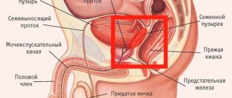
- imbalance of hormones in the body (for the mammary, thyroid, prostate glands are detected by ultrasound of the mammary glands, ultrasound of the prostate, ultrasound of the thyroid gland);
- failure of metabolic processes (metabolism);
- improper nutrition (especially for the pancreas - clearly visible on ultrasound of the pancreas);
- smoking, drinking alcohol or using drugs;
- pathological process in the parenchyma;
- inflammatory or traumatic swelling of the tissue.
What diseases can cause changes in the lymph nodes of the cervical region?
The cervical region is a place of accumulation of lymph nodes, which doctors especially often pay attention to. If they are soft, the body fights dangerous pathogens; if they become dense, this is a sign of inflammation, which is called lymphadenitis.
After palpation, the doctor prescribes an ultrasound of the cervical lymph nodes and a number of additional tests that will help clarify the cause of the ailment. Since the nodes in the neck are located very close to the mouth, nose, nasopharynx, throat and trachea, diseases of the lymphatic system can occur with the following diseases:
- caries;
- sinusitis;
- tonsillitis;
- pharyngitis;
- tracheitis;
- sinusitis, etc.
An ultrasound of the lymph nodes should be performed if there is slight compaction that persists for a long period. This may be a signal of a hidden inflammatory process. Therefore, ultrasound of the lymph nodes of the neck can identify a number of diseases that occur unnoticed in the body:
- HIV/AIDS;
- diabetes;
- monoculosis;
- tuberculosis;
- Sezary's disease;
- syphilis;
- leprosy;
- oncological diseases;
- diseases affecting the lymphatic system.
If external changes or pain symptoms occur, it is recommended to consult a doctor without taking self-medication measures. If necessary, the doctor will order an ultrasound of the lymph nodes of the neck and additional tests.
Ultrasound of lymph nodes in Moscow can be performed at the Clinic for Men's and Women's Health, where certified physicians, using innovative scanners, will make a diagnosis and coordinate further treatment tactics with clients.
Normal echo pattern
With age, the structure of the mammary gland changes. It consists of the following types of fabrics:
- fat;
- connecting;
- glandular.
Glandular tissue on ultrasound is visualized as a homogeneous cell of large or medium size, covered with a hyperechoic capsule. In women over 60 years of age, fibrofatty transformation is more pronounced. Lymph nodes have a hemogenic structure, a round shape, which does not exceed 10 mm in diameter. The capsule is hyperechoic, continuous, and smooth.
How to prepare for an ultrasound procedure
Depending on the symptoms, the doctor gives a referral for ultrasound diagnostics of one of the groups of nodes. The choice falls on the lymphatic vessels that are as close as possible to the location of the disease. In case of severe condition, it is possible to diagnose several groups of nodes.
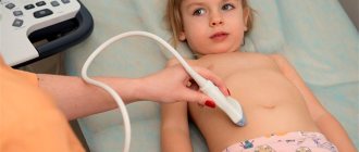
For effective diagnosis, the doctor may ask you to prepare for the examination in advance. Ultrasound of the lymph nodes in the cervical region is performed without special preparation, with the exception of cases when the patient needs to remove hair growing on the neck, special bandages or bandages.
You must arrive for the examination on time in agreement with the diagnostician. Before the procedure, you need to inform your doctor about taking medications, since a number of medications provoke changes in the lymph nodes.
Main ultrasonic parameters
When performing an ultrasound, the doctor evaluates several parameters, the main ones being echogenicity, structure and contours of the organ.
The image on the monitor of an ultrasound machine consists of dots - pixels, each of which is colored in one of 1024 shades of gray. The degree of color intensity will directly depend on the volume of the reflected ultrasound beams. Denser organs reflect waves very well because they absorb environmental vibrations and become intense secondary sources of sound. Therefore, the ultrasound returns to the sensor in almost its original state.
This phenomenon is very similar to the echo in the mountains. So, a strong reflection will color the image light gray or white, and a weak reflection will give a dark gray tint, close to black. Based on the color of the resulting image, the doctor determines the condition of organs and tissues.
Survey methodology
The diagnostic room has a specialist workplace, and a couch for the patient is required.
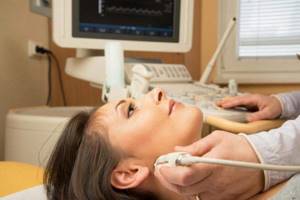
Examinations of the cervical spine are performed while sitting. The area being diagnosed is exposed. The doctor applies a gel to the area, which promotes soft gliding and effective passage of ultrasound pulses towards the tissues and back. The information received is displayed on the monitor and recorded in the protocol.
An ultrasound of the lymph nodes, which shows the slightest deviation from the norm, can diagnose the disease at an early stage and prevent death in the future.
The procedure lasts from 10 minutes to half an hour, depending on the complexity of the placement of the nodes and the thoroughness of the diagnosis.
The diagnostician enters the results obtained on a form and gives them to the patient. A specialized doctor deciphers the data and makes a diagnosis.
Rules for preparation before performing an ultrasound of the mammary glands with regional lymph nodes
Special preparation is not needed, however, there are general rules regarding the days of the menstrual cycle when the study will show the most informative and objective results. At the end of menstruation, the ducts are restored, and at the beginning of the cycle, glandular structures develop and branch. During ovulation, the gland begins to prepare for possible conception, so there is a noticeable swelling and increase in size. Experts recommend performing an ultrasound before ovulation. It is at this time that the mammary glands do not swell (not edematous), and the structure and tissues are best visible.
Optimal schedule for:
- 7-14 days with a menstrual cycle that lasts more than 28 days;
5-12 days with a 28-day cycle.
When there are urgent indications for diagnosis, ultrasound examination is performed regardless of the day of the menstrual cycle.
Are additional tests needed?
The accuracy of the diagnosis depends on the completeness of the information received about the patient’s condition. In addition to ultrasound, the doctor must prescribe additional tests that will complement the clinical picture of the disease. Current research methods:
- general blood analysis;
- blood chemistry;
- tests for tuberculosis;
- blood test for HIV/AIDS;
- X-ray;
- MRI;
- CT scan.
The functionality of the tomograph in our clinic allows us to most accurately perform ultrasound, which eliminates the need for many additional tests.
What is echogenicity
The content of the article
An inverted black and white image is what the sonographer sees during the examination. All human organs reflect ultrasound in their own way. The color depends on the density of the organ: the denser it is, the whiter the picture. For example, liquid is depicted in black. The ability of organ tissue to reflect ultrasound is called echogenicity.

Inaccuracies and preliminary diagnoses based on ultrasound diagnostics
Correct diagnosis and interpretation of ultrasound results determines the further outcome of treatment. Conducting ultrasound examinations by inexperienced specialists or under conditions of turmoil and nervous tension in public health facilities shows low effectiveness. In stressful environments, human error may occur more often than usual.
For an error-free diagnosis, it is recommended to contact a private diagnostic and treatment institution, the Women’s and Men’s Health Clinic, which offers ultrasound of the neck lymph nodes at a low price. The center does not create queues outside the office door, accepts patients by appointment, and pays due attention to patients.
What does increased echo density mean: norms and pathology
The concept of “increased echo density” is often encountered on ultrasound. This can be explained this way. Hyperechogenicity of different organs looks different on ultrasound and has a variable meaning:
| Organ | Features of the ultrasound image | What could it indicate? |
Uterus | Diffuse increase in echogenicity, increased organ size | Inflammatory process |
| A hyperechoic round object with sound enhancement is visible | Myoma (fibroma) | |
| Tumor (malignant or benign) | ||
| Blurred contours and heterogeneous echo structure, an echogenic round formation is visible | Endometriosis | |
Endometrium | Uniform thickening of the layer, uniform echogenicity, smooth contours | Hyperplasia |
| Uneven and unclear contours, heterogeneous echo structure | Endometrial cancer | |
Ovaries | An area of increased echo density is visible | Calcium salt deposition |
| Neoplasms of a malignant or benign nature | ||
Pancreas | Hyperdensity of parenchyma | Inflammatory process |
| Swelling | ||
| Increased gas formation | ||
| Malignant tumor | ||
| Increased pressure in the portal vein system | ||
| Stone or calcium deposits in tissue and ducts | ||
| Diffuse increase in echo density, decrease in gland size | Replacement of normal tissue with scar tissue. Occurs as a result of exacerbation of chronic pancreatitis | |
| Diffuse increase in echo density, the size of the gland is not changed | Replacement of normal tissue with adipose tissue. The phenomenon often occurs in diabetes mellitus or old age | |
| High echo density as a transient phenomenon | Poor nutrition (abuse of fatty foods) | |
| General disease – reactive pancreatitis | ||
| Irregular bowel movements | ||
| Wrong lifestyle | ||
Gallbladder | A hyperechoic object is detected | Deposition of stones in the organ |
| Diffuse increase in permeability, thickening of organ walls | Chronic inflammatory process | |
Thyroid | Increased echogenicity of the organ | Endemic goiter (lack of iodine in the body) |
| Toxic goiter (organ damage by poison) | ||
| Autoimmune thyroiditis | ||
| Subacute thyroiditis | ||
| An echogenic formation is visible | Oncology | |
| Areas of sclerosis | ||
Mammary gland | Increased echogenicity of the gland (except for pre-, post- and menopause periods) | Post-inflammatory changes in organ tissues |
| Hyperechoic formations detected | Abnormal cyst | |
| Calcification | ||
| Fibrous area | ||
Kidneys | Hyperechoic structure of the kidneys, their size is increased, the pyramids have reduced echogenicity | Diabetic nephropathy |
| Diffuse increased echogenicity of the parenchyma, organ layers are not differentiated, hypoechoic pyramids | Glomerulonephritis (severe) | |
| Hyperdense area visible | Malignant formation | |
| Myelomas | ||
| Kidney infarction | ||
| Parenchyma calcifications | ||
| Increased echogenicity of the renal sinus | Inflammatory, metabolic or endocrine disorders | |
Spleen | Increased echo density of the organ (excluding age factor) | Increased portal pressure |
| Konovalov-Wilson syndrome | ||
| Glycogenosis | ||
| Amyloidosis | ||
| Increased iron levels in the blood | ||
During pregnancy | Fetal intestinal hyperdensity (after 16 weeks) | Intrauterine TORCH infections |
| Intestinal ischemia | ||
| Cystic fibrosis | ||
| Fetal growth restriction | ||
| Intestinal perforation | ||
| Hyperechogenicity of the placenta | Infarction (hemorrhage) of the placenta | |
| Placental abruption | ||
| Presence of calcifications |
Increased echogenicity can be both normal and pathological, so the diagnosis must be clarified using other examination methods.
How much does an ultrasound scan of neck lymph nodes cost in Moscow?
The price of an ultrasound scan of lymph nodes depends on the area being examined. The men's and women's health clinic has a loyal pricing policy. The cost of medical care is acceptable for residents of Moscow and the regions. Specific figures for all types of services can be found on the website in the “Cost of Services” section.
Price of ultrasound of lymph nodes in the clinic The difference in cost is insignificant; it differs from the cost of the procedure in city multidisciplinary clinics. I can compare with the comfort, efficiency of examination and treatment provided in our center.
Benefits of breast ultrasound
Among the main advantages of this diagnostic method:
- affordable price;
- the ability to adjust and control the treatment process at any stage;
- no absolute contraindications;
- painlessness;
- high information content and safety;
- efficiency of carrying out and obtaining results.
The absence of restrictions on the number of tests (can be used an unlimited number of times) is another significant advantage of this diagnostic method. And due to the absence of radiation exposure, the procedure is indicated for use by people of absolutely any age.
What does a decrease in echo density mean: table of pathologies
A hypoechoic node can be found in many organs and does not always turn out to be a pathology. Let's take a closer look at what a formation with reduced echogenicity is in various organs, and what preliminary diagnosis can be made.
| Organ | Features of the ultrasound image | What could it indicate? |
Thyroid | Hypoechoic node | Cyst |
| Fluid formation | ||
| Vascular formation | ||
| Oncology (in 5% of cases) | ||
Uterus | Hypoechoic structure with a fuzzy shape, M-echo indicators are increased, the uterine cavity is expanded, heterogeneous endometrial structure | Carcinoma |
| Hypoechoic zone with internal echo structure | Inflammatory process | |
| An area on the wall of an organ with reduced echogenicity | Myoma | |
| Hypoechoic zone near the fertilized egg | Accumulation of blood under the embryo - an incipient miscarriage | |
Mammary gland | A formation that is heterogeneous in structure with reduced echo density, with a fuzzy and uneven contour, gives a shadow | Carcinoma |
| Hypoechoic formation (often irregular in shape) with unclear contours and boundaries | Adenosis | |
| Round hypoechoic inclusion with unclear contours | Typical cyst | |
| Formation with reduced echo density, thickened walls, internal growths and calcifications | Abnormal cyst | |
| Hypoechoic formation with smooth and clear contours | Fibroadenoma (similar to a malignant tumor) | |
Ovaries | Areas with reduced echo density | Follicle |
| Vascular formation | ||
| Corpus luteum | ||
| Cyst | ||
| Oncology (rather rare) | ||
| Heterogeneous structure of the organ during childbearing age and homogeneous during menopause | Normal, not pathological | |
Kidneys | Hypoechoic area with clear boundaries and homogeneous structure | Cyst |
| A formation of heterogeneous structure with reduced echogenicity and unclear contours | Tumor | |
| Hypoechoic formation, retroperitoneal lymph nodes are enlarged, blood flow is not visualized | Oncology | |
Pancreas | Hypoechoic formation with unclear contours, which does not occupy all the tissue of the organ | Metastases |
| A homogeneous formation with low echo density and a smooth contour, without inclusions | Cyst | |
| Several hypoechoic areas | Fibrolipomatous process | |
| Hemorrhagic pancreatitis | ||
| Hypoechoic formation with thin peripheral outgrowths, increased size of the gland, displacement of its large vessels, blood flow is not visualized | Gland cancer | |
Liver | Hypoechoic formations near the portal vein or gallbladder bed, often have a triangular or oval shape | Residual areas of normal tissue against the background of liver steatosis (accumulation of fat in the cells of the organ) |
| Hypoechoic round nodes, possible tuberosity along the periphery | Cirrhosis | |
| Formation with reduced echo density, smooth contours and internal echo signals | Cyst with hemorrhage | |
| Oval, round or elongated inclusion with a loose echostructure | Thrombosis | |
| Areas of varying echogenicity, presence of gas bubbles (not always), uneven contour, echogenic membrane | Abscesses | |
| The hypoechoic zone is round in shape with smooth contours, the presence of echogenic outgrowths extending to the periphery | Nodular hyperplasia | |
| Homogeneous formation with smooth contours, presence of internal signals (not always) | Adenoma | |
| An area with a heterogeneous structure, the presence of areas of hemorrhage and calcifications (not always), changes in local lymph nodes, possible definition of ascites | Liver cancer | |
| Formations with uneven borders and a hypoechoic contour (sometimes), the organ tissue has not changed | Metastases |
For the most accurate and informative study, the patient must properly prepare for the procedure, taking into account exactly how it will be carried out.
Anechoicity - a black spot on the health map
Areas that are unable to reflect ultrasound waves are called anechoic, or echo-negative. They are displayed on the monitor of the ultrasound machine in black. Liquids also lack echogenicity, so they are depicted in the same way.
Often detected anechoic areas indicate the presence of cystic formations. Small cysts (up to 5 cm in diameter) regress after a few months. Large formations are tolerant to special therapy.
In patients over 50 years of age, such a “finding” during an ultrasound usually indicates a malignant formation. Its untimely treatment is life-threatening. Immediate treatment is necessary for complications of a tumor in the kidneys: pyelonephritis, urolithiasis or arterial hypertension.
Echo-negative formation is a sign of normality. This is the cyclically formed ovarian gland - the corpus luteum. It is impenetrable to ultrasound, so it is displayed on the monitor in a dark color. After menstruation, the formation can be defined as the corpus luteum. If menstruation is delayed, then it indirectly indicates the onset of pregnancy.
What does anechoic formation mean: dangerous symptoms
Table of norms and pathologies when detecting anechoicity::
| Organ | Features of the ultrasound image | What could it indicate? |
Ovaries | Round anechoic formation (or several) up to 2.5 - 3 cm (the rest up to 0.7 - 1.2 cm) | It is not a pathology. These formations are growing follicles |
| An echo-negative round formation more than 3 cm in diameter with a homogeneous structure and surrounded by a thin capsule. Found in the second half of the cycle | Follicular cyst (as a rule, spontaneously disappears in 1 – 3 cycles) | |
| Anechoic inclusion from 3 cm. Formed after ovulation | Corpus luteum cyst (resolves spontaneously after several cycles) | |
| Two-chamber or multi-chamber echo-negative formation with heterogeneous contents, sometimes with various echo-positive inclusions or growths on the walls | Dermoid cyst | |
| Endometrioid cyst | ||
| Cysts that can develop into malignant ones | ||
| Anechoic structure next to the ovary | Luteal cyst | |
Mammary gland | Echo-negative structure (a cavity containing transparent contents) | Cyst |
| Galactocele is a cavity containing breast milk (during lactation) | ||
| Homogeneous anechoic formation | Simple cyst | |
| Anechoic structure with hyperechoic inclusions | Complex cyst | |
| Anechoic formation with uneven contours, various inclusions and deformations | Malignant tumor | |
Thyroid | Echo-negative formation of a round shape with smooth contours and a dorsal enhancement effect | True cyst |
| An anechoic inclusion with a flocculent structure, the walls of which are formed by glandular tissue | Pseudocyst | |
| Anechoic or hyperechoic formation (depending on internal composition) | Adenoma | |
| Avascular inclusion with extremely low density for ultrasound | Colloid cyst | |
Uterus | An echo-negative formation detected during ovulation or 1–2 days after it | Not a pathology. The formation is fluid from a ruptured follicle |
| Anechoic structure in the uterus | Leiomyoma | |
| Degenerative changes in myomatous nodes | ||
| Pregnancy or approaching menstruation | ||
| Echo-negative formation in the suture area | Hematoma formation | |
Cervix | Anechoic formation up to 5 mm | “Normal” occurrence in postpartum women |
| Anechoic formation more than 5 mm | Endocervical cyst | |
| Nabothian cyst (the result of self-healing of ectopia) | ||
| Echo-negative structure with a fine suspension inside or a thickened wall | Endometrial cyst | |
| Thickening and deformation of the organ, several structures with different echogenicity differing in their heterogeneity | Cervical cancer | |
During pregnancy | Anechoic formations are detected in the fetus | Cyst (usually not detected after childbirth) |
| Anechoic formation in the upper third of the uterus with a hyperechoic rim (at 5–6 weeks) | Fertilized egg (pregnancy) | |
| Echo-negative structures | Luteal or follicular cyst (if the inclusions are located in the ovaries) | |
| Paraovarian cysts or serosoceles | ||
Kidneys | Anechoic round inclusion with smooth contours and thin walls | Simple cyst |
| Multiple echo-negative formations in both kidneys | Polycystic | |
| Echo-negative non-round formation with heterogeneous echogenicity, localization - near scar tissue | Secondary cyst | |
| The contours of the kidney are not changed; an anechoic formation is visible near them, next to which there is usually an area of hypoechoic parenchyma | Perirenal hematoma | |
| Echo-negative structure with unclear contours and various inclusions | Cancer | |
| Inclusions of an anechoic nature with unclear contours, vessels are not visualized, the wall of the pelvis is more than 2 mm | Kidney abscesses | |
Liver | Round or oval anechoic formation with partitions (not always), gives a shadow | Simple cyst |
| Echo-negative formations communicating with the branches of the portal vein; the vessels are not visualized | Dilatation of the hepatic vein | |
| Pulsating echo-negative structure communicating with the artery | Hepatic artery aneurysm | |
| Round anechoic formation with echogenic walls and calnates inside | Hydatid cyst |
Thus, an anechoic formation is understood as a cystic, fluid structure. To accurately determine the nature of echo-negative areas, it is necessary to undergo additional examinations.