Antinuclear (antinuclear) antibodies, ANA - (from the English anti-nuclear antibodies; in Russia, a less successful synonym is widespread - ANF, antinuclear factor, which does not reflect the long-established biological nature of the phenomenon) - antibodies against the main components of the cell nucleus (nucleoproteins, proteins chromatin, etc.). In systemic autoimmune diseases, as well as in the development of many other pathological conditions, lymphocytes are sensitized to the body’s own proteins and autoantibodies are produced directed against antigens of the components of cell nuclei and cytoplasm. The resulting autoantigen-autoantibody immune complexes are deposited on the basement membranes of various tissues and organs (skin, kidneys, synovial, serous membranes, brain, etc.), as well as the vessels supplying them with blood, activating the complement system and causing inflammation and tissue damage. At the same time, lysosomal activity increases and inflammatory mediators are released. The cytotoxic effect is exerted by complement fixed by immune complexes and sensitized lymphocytes. Vascular damage at the level of the microvasculature underlies systemic damage to connective tissue and parenchymal organs. A test for antinuclear antibodies is carried out if there is a suspicion of an autoimmune disease or the addition of an autoimmune component that plays a significant role in the pathogenesis of many nosoforms. The ANA test is highly sensitive (up to 98%) for systemic lupus erythematosus, however, positive results can be observed in other collagenoses and immune complex diseases, as well as in other diseases that occur with the development of an autoimmune component. Test results obtained in different laboratories may differ due to the characteristics of the methods used. Therefore, a complete laboratory response when studying antinuclear antibodies must necessarily include a description of the method used. A very extensive list of antigens combined into the concept of “nuclear” also requires mandatory clarification; antibodies to which of them can be detected by the method used, and to which they cannot. Immunofluorescence methods, which use animal cells as a substrate, allow, in addition to antinuclear antibodies, to identify and differentiate antibodies to certain components of the cytoplasm, which expands their capabilities. Indications for the analysis: diagnosis and differential diagnosis of systemic connective tissue diseases (especially SLE), autoimmune hepatitis, viral hepatitis with an autoimmune component (induced by viruses or medical interventions), primary biliary cirrhosis, multiple sclerosis; identification of the autoimmune component in various diseases, in the pathogenesis of which autoaggression may play a role. Material for research: blood serum. To determine antinuclear antibodies, the technique of indirect immunofluorescence is used using cells of the cultured Hep-2 line as a substrate. The sample (tested blood serum) is incubated with the substrate; if autoantibodies to nuclear or cytoplasmic antigens are present in the serum, they bind to the corresponding components of the substrate to form an antigen-antibody complex, which is detected by a conjugate - antibodies against human immunoglobulins labeled with FITC. As a result, when studied using a fluorescent microscope, specific fluorescence of the corresponding nuclear or cytoplasmic structures of cells is revealed. The use of cells of a malignant cultured line as a substrate due to their characteristics (high nuclear-cytoplasmic ratio, large size, presence of a large number of mitoses) makes it possible to evaluate various types of luminescence characteristic of the presence in a blood sample of a wide range of antibodies to various antigens that have various clinical associations. It should be borne in mind that some of the detected types of luminescence are highly specific, clearly associated with certain diseases and have a definite diagnostic significance, especially when detected in high titers; however, some antibodies to different antigens may produce similar patterns of fluorescence, in which case further testing may be required to clarify the specific antigen to which the autoantibodies are directed. On the other hand, a certain type of antibody can occur in several nosological forms. There are also antibodies (they are rare) that react exclusively with components of the spindle apparatus of cells; they are usually not associated with autoimmune diseases, and their detection is regarded as a false positive result without clinical associations. All of the above features are necessarily reflected in the laboratory report, where, in addition to indicating the type of glow and the most likely type of antibody corresponding to it, recommendations are also given on the interpretation of the results in the sense of a possible association with specific nosoforms or syndromes, as well as the necessary additional laboratory tests. The advantage of the immunofluorescence method using cells of malignant cultured lines is also the possibility of detecting antibodies to some antigens of cytoplasmic localization: mitochondria, actin, lysosomes, etc. Some of the detected antinuclear and anticytoplasmic antibodies with their characteristic types of fluorescence and clinical associations are given below:
- nuclear homogeneous type - antibodies (AT) to DNA (double- and single-stranded), antibodies to DNP (deoxynucleoprotein - a complex of DNA with histones), antibodies to histones, antibodies to other components of chromatin (protamines, etc.); clinical associations - SLE (anti-DNA, antibodies to histones - in high titers); drug-induced lupus, rheumatoid arthritis (antibodies to histones);
- nuclear peripheral type - AT to double-stranded DNA, histones; clinical associations - SLE, drug-induced lupus);
- nuclear membrane lamin type - AT to lamins (fibrillar proteins of the nuclear membrane); clinical associations - SLE, linear scleroderma, rheumatoid arthritis, combined hepatitis/thrombopenia/anemia syndrome;
- nuclear-membrane ring-shaped type with a speckled glow of the cytoplasm - antimitochondrial antibodies to mitochondrial components M2 (pyruvate dehydrogenase complex), M3, M6; clinical associations see mitochondrial type;
- nuclear channels - AT to proteins of the nuclear membrane canaliculus complex; clinical associations - polymyositis;
- perichromine type - AT to perichromine; clinical associations - SLE;
- heterogeneous nuclear RNP (ribonucleoprotein) - AT to various components of the nuclear matrix; clinical associations—mixed connective tissue disease (MCTD);
- Sm/RNP —AT to nuclear RNP or Sm complex; clinical associations - SLE, CTD (antibodies to Sm); Sjogren's syndrome, scleroderma, rheumatoid arthritis, SLE, discoid lupus caused by procainamide (antibodies to RNP);
- centromeric type - AT to the centromere (kinetochore); clinical associations - CREST syndrome (calcinosis, Raynaud's phenomenon, esophageal dysfunction, sclerodactyly, telangiectasia), scleroderma, primary biliary cirrhosis
- nuclear speckled type (NSP1) - antibodies against poorly characterized proteins, often combined with antibodies to actin or mitochondria; clinical associations - primary biliary cirrhosis (NSP1 + mitochondrial type), chronic active hepatitis (NSP1 + actin);
- nucleolar homogeneous type - AT to nucleolin and other components of nucleoli; clinical associations - combined polymyositis/scleroderma syndrome, scleroderma mainly with kidney damage;
- nucleolar-cluster type - antibodies to fibrillarin (7-2-RNA), antibodies to ribonucleoprotein U3RNP; clinical associations - scleroderma, found mainly in young men;
- nucleolar-speckled type - AT to Scl-70 (AT to topoisomerase 1); clinical associations - scleroderma;
- mitochondrial (cytoplasmic) type - antibodies to M2 (pyruvate dehydrogenase complex), M3, M6 components of mitochondria (antibodies to M1 and M5 are not detected on Hep-2 cells); clinical associations - primary biliary cirrhosis, drug-induced SLE (antibodies to M3, M6), chronic active hepatitis, Reynolds syndrome, Sjögren's syndrome, rheumatoid arthritis;
- finely speckled (cytoplasmic) type - AT to various cytoplasmic proteins, for example, SPR (signal recognition particle); clinical associations - myositis;
- perinuclear type - antibodies to antigens Jo-7, PL1, PL7, PL12; clinical associations - polymyositis, mainly in combination with connective tissue disease of the lungs; dermatomyositis;
- ribosomal type - AT to ribosomal antigens; clinical associations - SLE, mainly with neurological symptoms, rheumatoid arthritis, Sharpe's syndrome;
- actin - AT to actin (antibodies to smooth muscles); clinical associations - chronic active hepatitis, primary biliary cirrhosis;
- tubulin - AT to tubulin; clinical associations - alcoholic cirrhosis, Hashimoto's thyroiditis, scleroderma, CREST syndrome, Raynaud's phenomenon;
- cytokeratin - AT to cytokeratin; clinical associations - rheumatoid arthritis, scleroderma, SLE.
Detailed description of the study
Anti-double-stranded DNA antibodies (anti-dsDNA) are a type of autoantibody called antinuclear antibody (ANA). Antibodies usually protect against infection, but autoantibodies are produced when a person's immune system loses tolerance to its own cells.
Double-stranded DNA antibodies specifically target the genetic material (DNA) found in the cell nucleus, which explains their name. These antibodies may be present in low concentrations in a number of diseases (infectious mononucleosis, primary biliary cholangitis, etc.), however, they are most often associated with lupus. The double-stranded DNA antibody test, among others, can be used to help establish the diagnosis of SLE and distinguish it from other pathologies.
Systemic lupus erythematosus (SLE) is an autoimmune disease in which the human immune system produces antibodies against its own cells, similar to what happens when defending against viruses or bacteria. As a result of the autoimmune process, organs and tissues are damaged.
It is believed that the development of SLE is promoted by a combination of certain genetic, immune, endocrine factors, along with unfavorable environmental influences. There is a certain hereditary risk, although there is no compulsory inheritance. Female gender and hormonal influences increase the risk of developing SLE.
Several environmental triggers for SLE have been identified, such as ultraviolet rays or sun exposure. Infection with certain infectious diseases (Epstein-Barr virus and others), smoking and vitamin D deficiency can also affect the development of this disease.
The clinical signs of SLE are very varied. Symptoms may come on suddenly or develop slowly, and may be temporary or permanent. Most people with this disease experience periods of exacerbation, when symptoms worsen for a while, then improve.
Signs of SLE depend on which body systems are affected by the disease. The most common symptoms include: a butterfly-shaped rash on the face covering the cheeks and bridge of the nose, joint pain and swelling, fever, and constant fatigue.
Diagnosis of SLE is based on evaluation of symptoms, examination, and laboratory findings. Detection of antibodies to certain cell components serves as the basis for laboratory diagnosis of this disease. In most cases, there is a relationship between the amount of anti-dsDNA and the course of the disease. Thus, during the period of remission, a low level of antibodies is determined, which is a prognostically favorable sign. An increase in the concentration of anti-dsDNA in the blood indicates a period of exacerbation.
A serious complication of SLE includes lupus nephritis, a condition characterized by inflammation of the kidneys that can cause kidney failure. Studying the level of antibodies to double-stranded DNA helps to assess kidney damage in this disease and determine the activity of the autoimmune process. Increased levels of anti-dsDNA precede exacerbation of lupus nephritis.
The anti-dsDNA test is used to diagnose SLE in a person who has clinical signs of the disease and a positive antinuclear antibody (ANA) test result. The study is also used in the diagnosis of other connective tissue diseases. The analysis is carried out using an immunochemiluminescence method, which is highly specific and allows one to determine the presence of even a small amount of anti-dsDNA, and also helps in the differential diagnosis of systemic connective tissue diseases, especially when several types of autoantibodies are present in the blood.
ANA OR ANTI-CYTOPLASMIC ANTIBODIES
- Systemic connective tissue lesions:
- systemic lupus erythematosus (SLE), - rheumatoid arthritis, - Sjogren's syndrome, - scleroderma, - dermatomyositis, - periarteritis nodosa, etc.;
- Infections (tuberculosis, infectious mononucleosis, acute and especially chronic viral hepatitis, subacute infective endocarditis, HIV infection, etc.);
- Chronic autoimmune hepatitis, primary biliary cirrhosis of the liver;
- Diabetes mellitus (insulin dependent);
- Multiple sclerosis;
- Pulmonary fibrosis;
- Systemic vasculitis.
Antinuclear antibodies can also be detected in acute and chronic leukemia, acquired hemolytic anemia, Waldenström's disease, malaria, chronic renal failure, thrombocytopenia, lymphoproliferative diseases, myasthenia gravis and thymomas. Antinuclear antibody titers in periarteritis nodosa can reach 1:100, in dermatomyositis - 1:500, in SLE - 1:1000 and higher. In SLE, a test for detecting antinuclear antibodies using indirect immunofluorescence has a high degree of sensitivity (up to 98% according to various authors), but moderate specificity (78%), since antinuclear antibodies can also occur in other diseases mentioned above. For comparison, the test for determining antibodies to native DNA using ELISA in the diagnosis of SLE has a significantly lower sensitivity - 38% (since not only antibodies to DNA, but also antibodies to histones, deoxyribonucleoprotein and many others can play a pathogenetic role in the development of SLE nuclear antigens); however, the specificity of anti-native DNA antibodies for the diagnosis of SLE is almost absolute (98%). The clinical interpretation of these differences in the sensitivity and specificity of different laboratory tests in the diagnosis of SLE is as follows: a negative result of detection of antibodies to native DNA using ELISA does not exclude the diagnosis of SLE, but the detection of these antibodies confirms the diagnosis with almost 100% probability. There is a different situation with regard to ANF: a negative (especially multiple) result of detection of antinuclear antibodies by indirect immunofluorescence makes the diagnosis of SLE (at least the active form) very doubtful (since the method detects almost all autoantibodies found in SLE), but the detection antinuclear antibodies will not always be a sufficient basis for making a diagnosis of SLE; it confirms the presence of the phenomenon of autoimmunity and usually requires further clinical and laboratory differentiation to establish a final diagnosis, taking into account the type of luminescence, antibody titer, additional clarifying or confirmatory tests recommended by the laboratory, comparison of results with clinical picture. In SLE, there is usually no correlation between ANA titres and clinical status, although persistence of high titers over a long period of time is an unfavorable prognostic sign. A decrease in the level of antibodies portends remission, but sometimes death (the phenomenon of antibody consumption: with increasing autoimmune tissue damage, the rate of release of nuclear antigens, to which accumulated antinuclear antibodies bind, exceeds the rate of synthesis of new autoantibodies). In scleroderma, the frequency of detection of antibodies to nuclear antigens is 60-80%, their titer is usually lower than in SLE. In rheumatoid arthritis, SLE-like forms of the course often occur, so ANAs are detected quite often. With dermatomyositis, antibodies to nuclear antigens in the blood occur in 20-60% of cases, with periarteritis nodosa - in 17% (titer up to 1:100), with Sjögren's disease - in 56% in combination with arthritis and in 88% of cases - in combination with Gougerot-Sjögren syndrome. In discoid (cutaneous) lupus erythematosus, antinuclear factor is detected in 50% of patients. Frequency of detection of antinuclear antibodies in rheumatic diseases and in healthy individuals (Nasonova V.A. et al., 1997)
| Disease | ANA detection rate, % | Captions |
| SLE - active form | 98-100 | +++ |
| Discoid lupus erythematosus | 40 | ++, +++ |
| Drug-induced lupus | 100 | ++ |
| Systemic scleroderma | 70 | ++, +++ |
| Sjögren's syndrome | 60 | ++, +++ |
| Mixed connective tissue disease | 100 | ++, +++ |
| Raynaud's disease | 60 | ++, +++ |
| Rheumatoid arthritis | 40 | +, ++ |
| Juvenile chronic arthritis | 20 | +, ++ |
| Polymyositis and dermatomyositis | 30 | + |
| Periarteritis nodosa | 17 | + |
| Healthy individuals under 40 years of age | 3 | + |
| Healthy faces after 40 years | 25 | + |
Antibodies to nuclear antigens (ANA), screening
General information about the study
Antibodies to nuclear antigens (ANA) are a heterogeneous group of autoantibodies directed against components of the body's own nuclei. They are detected in the blood of patients with a variety of autoimmune diseases, such as systemic connective tissue diseases, autoimmune pancreatitis and primary biliary cirrhosis, as well as some malignancies. The ANA study is used as a screening for autoimmune diseases in a patient with clinical signs of an autoimmune process (prolonged fever of unknown origin, articular syndrome, skin rashes, weakness, etc.). Such patients, if the test result is positive, require further laboratory evaluation, including tests more specific for each autoimmune disease (for example, anti-Scl-70 for suspected systemic scleroderma, anti-mitochondrial antibodies for suspected primary biliary cirrhosis). It should be noted that a negative ANA test result does not exclude the presence of an autoimmune disease.
ANAs are most common in patients with systemic lupus erythematosus (SLE). They are found in 98% of patients with it, which allows us to consider this study as the main test for diagnosing SLE. The high sensitivity of ANA for SLE means that repeated negative results make the diagnosis of SLE questionable. However, the absence of ANA does not completely exclude the disease. In a small proportion of patients, ANAs are absent at the onset of SLE symptoms but occur during the first year of the disease. In 2% of patients, antibodies to nuclear antigens are never detected. If the test result is negative in a patient with symptoms of SLE, it is advisable to perform more SLE-specific laboratory tests, primarily anti-double-stranded DNA antibodies (anti-dsDNA). Detection of anti-dsDNA in a patient with clinical signs of SLE is interpreted in favor of the diagnosis of SLE, even in the absence of ANA.
SLE occurs as a result of a complex of immunological disorders that develop over a long period of time. The degree of imbalance of the immune system gradually increases over the course of the disease, which is reflected in an increase in the spectrum of autoantibodies. The first stage of the autoimmune process is characterized by the presence of genetic features of the immune response (for example, certain alleles of the major histocompatibility complex, HLA) in the absence of abnormal laboratory tests. At the second stage, autoantibodies can be detected in the blood, but there are no clinical signs of SLE. Antibodies to nuclear antigens, as well as anti-Ro-, anti-La-, antiphospholipid antibodies are most often detected at this stage. Detection of ANA is associated with a 40-fold increase in the risk of SLE. The period between the onset of ANA and the development of clinical symptoms varies, averaging 3.3 years. Patients with a positive ANA test result are at risk for developing SLE and require periodic follow-up with a rheumatologist and laboratory testing. The third stage of the autoimmune process is characterized by the appearance of symptoms of the disease, while the widest range of autoantibodies can be detected in the blood, including anti-Sm antibodies, antibodies to double-stranded DNA and ribonucleoprotein. Thus, to obtain complete information about the degree of immunological disorders in SLE, the ANA test must be supplemented with an analysis for other autoantibodies.
The course of SLE varies from persistent remission to fulminant lupus nephritis. To predict the disease, assess its activity and the effectiveness of treatment, various clinical and laboratory criteria are used. Since none of the tests can clearly predict exacerbation or damage to internal organs, monitoring of SLE is always a comprehensive assessment, including the study of ANA, as well as other autoantibodies and some general clinical indicators. In practice, the doctor independently determines a set of tests that most accurately reflect how the course of the disease changes in each patient.
Drug-induced lupus represents a special clinical syndrome. It develops while taking certain medications (most often procainamide, hydralazine, some ACE inhibitors and beta blockers, isoniazid, minocycline, sulfasalazine, hydrochlorothiazide, etc.) and is characterized by symptoms reminiscent of SLE. ANA can also be detected in the blood of most patients with drug-induced lupus. If there are symptoms of an autoimmune process in a patient taking these drugs, an ANA test is recommended to rule out drug-induced lupus. The peculiarity of drug-induced lupus is the disappearance of immunological disorders and symptoms of the disease after complete discontinuation of the drug - at this time, a control ANA study is recommended, and a negative result confirms the diagnosis of drug-induced lupus.
ANAs are detected in 3-5% of healthy people (in the group of patients over 65 years of age, this figure can reach 10-37%). A positive result in a patient without symptoms of an autoimmune process must be interpreted taking into account additional anamnestic, clinical and laboratory data.
What is the research used for?
- For screening autoimmune diseases such as systemic connective tissue diseases, autoimmune hepatitis, primary biliary cirrhosis, etc.
- For diagnosing systemic lupus erythematosus, assessing its activity, making a prognosis and monitoring its treatment.
- For the diagnosis of drug-induced lupus.
When is the study scheduled?
- For symptoms of an autoimmune process: prolonged fever of unknown origin, joint pain, skin rash, unmotivated fatigue, etc.
- For symptoms of systemic lupus erythematosus (fever, skin lesions), arthralgia/arthritis, pneumonitis, pericarditis, epilepsy, kidney damage.
- Every 6 months or more frequently when evaluating a patient diagnosed with SLE.
- When prescribing procainamide, disopyramide, propafenone, hydralazine and other drugs associated with the development of drug-induced lupus.
References
- Alexandrova, E.N., Novikov, A.A., Nasonov, E.L. Recommendations for laboratory diagnosis of rheumatic diseases of the All-Russian public organization “Association of Rheumatologists of Russia” - 2015. Modern rheumatology, 2015. - V. 9(4). — P. 25-36.
- Kishkun, A.A. Guide to laboratory diagnostic methods. - M.: GEOTAR-Media, 2007. - 800 p.
- Kavanaugh, A., Solomon, D. American College of Rheumatology Ad Hoc Committee on Immunologic Testing Guidelines. Guidelines for immunologic laboratory testing in the rheumatic diseases: anti-DNA antibody tests. Arthritis Rheum., 2002. - Vol. 47(5). — P. 546-55.
- Padalko, E., Bossuyt, X. Anti-dsDNA antibodies associated with acute EBV infection in Sjögren's syndrome. - AnnRheumDis., 2001. - Vol. 60(10). — P. 992.
- Tsuchiya, K., Kiyosawa, K., Imai, H. et al. Detection of anti-double and anti-single stranded DNA antibodies in chronic liver disease: Significance of anti-double stranded DNA antibodies in autoimmune hepatitis. - J Gastroenterol, 1994. - Vol. 29. - P. 152–158.
- Severin, S.E., Aleynikova, E.V., Osipov, S.A. and others. Biological chemistry: Textbook. - M.: Publishing House "Medical Information Agency", 2021. - 496 p.
Systemic lupus erythematosus. Antibodies (IgG) to double-stranded (native) DNA
Anti-double-stranded DNA antibodies are autoantibodies directed against one's own double-stranded DNA, seen in systemic lupus erythematosus. Are being studied for the diagnosis, assessment of activity and monitoring of treatment of this disease.
Units
U/ml (unit per milliliter).
What biomaterial can be used for research?
Venous blood.
How to properly prepare for research?
Do not smoke for 30 minutes before donating blood.
General information about the study
Anti-double-stranded DNA antibodies (anti-dsDNA) belong to the group of antinuclear antibodies, that is, autoantibodies directed by the body against components of its own nuclei. While antinuclear antibodies are characteristic of many diffuse connective tissue diseases, anti-dsDNA is considered specific for systemic lupus erythematosus (SLE). Detection of anti-dsDNA is one of the criteria for diagnosing SLE.
The concentration of anti-dsDNA varies depending on the characteristics of the disease. As a rule, a high indicator indicates high activity of SLE, and a low indicator indicates the achievement of remission of the disease. Therefore, measurement of anti-dsDNA concentration is used to monitor treatment and prognosis of the disease. An increase in concentration indicates insufficient control of the disease, its progression, as well as the possibility of developing lupus nephritis. On the contrary, a consistently low antibody concentration is a good prognostic sign. It should be noted that such a dependence is not observed in all cases. Anti-dsDNA levels are measured regularly, every 3-6 months, in cases of mild SLE and at shorter intervals in the absence of disease control, when selecting therapy, during pregnancy or the postpartum period.
Although high levels of anti-dsDNA are characteristic of SLE, low concentrations are also found in the blood of patients with some other diffuse connective tissue diseases (Sjögren's syndrome, mixed connective tissue disease). In addition, the test may be positive in patients with chronic hepatitis B and C, primary biliary cirrhosis and infectious mononucleosis.
When is the study scheduled?
For symptoms of systemic lupus erythematosus: fever, skin lesions (butterfly erythema or red rashes on the face, forearms, chest), arthralgia/arthritis, pneumonitis, pericarditis, epilepsy, kidney damage;
- when antinuclear antibodies are detected in serum, especially if a homogeneous or granular (speckled) type of immunofluorescence of the nucleus is obtained;
- regularly, every 3-6 months, for mild SLE, or more often if the disease is not controlled.
What do the results mean?
Reference values:
Positive result:
- systemic lupus erythematosus;
- effective therapy, remission of systemic lupus erythematosus;
- Sjögren's syndrome;
- mixed connective tissue disease;
- chronic hepatitis B and C;
- primary biliary cirrhosis;
- Infectious mononucleosis.
Negative result:
- absence of systemic lupus erythematosus;
What can influence the result?
- Effective therapy and achievement of disease remission are associated with low anti-dsDNA levels;
- lack of disease control, exacerbation of the disease, lupus nephritis are associated with high levels of anti-dsDNA.
AT to 2-stranded DNA

No special preparation is required before collection; the conditions are standard:
- the analysis is taken on an empty stomach;
- Eliminate smoking and physical activity within half an hour;
- avoid taking any medications as much as possible.
Diagnostic significance of testing autoantibodies to double-stranded DNA
These specific immunoglobulin proteins belong to the group of antinuclear antibodies and are a highly specific marker in the diagnosis and clinical differentiation of diffuse connective tissue diseases (rheumatoid arthritis, Sjogren's syndrome, scleroderma, thyroiditis), chronic hepatitis, biliary cirrhosis, cholestasis and infectious diseases caused by cytomegaloviruses and Epstein-Barr, as well as determining the active phase of a serious autoimmune pathology - systemic lupus erythematosus (SLE) . Its clinical picture is characterized by:
- prolonged fever;
- damage to the skin - the appearance of an erythematous “butterfly” on the face (specific hyperemia) and red rashes on the forearms and chest;
- arthralgia , expressed by painful sensations in bone joints (however, not accompanied by the development of an inflammatory process in them and a change in mobility), often accompanied by myalgia (pain in muscles of various types);
pulmonitis - inflammation of the alveoli and distal bronchioles caused by an immune reaction;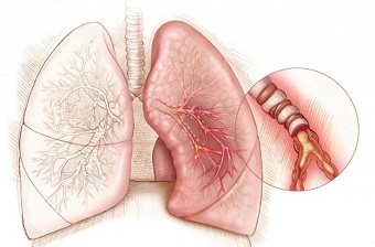
- repeated epileptic seizures caused by short-term abnormal activity of brain neurons, which is provoked by excess charges arising in them;
- pericarditis - inflammation of the serous membrane of the heart muscle, often accompanied by the accumulation of fluid in its cavity;
- damage to the glomeruli of the kidneys , in which primary urine is formed during the filtration of blood plasma.
The concentration of immunoglobulins acting against double-stranded DNA (deoxyribonucleic acid - a macromolecule that ensures the storage and transmission of genetic information) directly correlates with the level of circulating immune complexes formed in the glomerular apparatus during the development of a severe pathological process in the kidneys and containing class G antibodies. Accumulation of auto -antibodies provoke activation of the complement system and the occurrence of inflammation, accompanied by tissue damage.
The specificity and sensitivity of the immunological test is almost 90%; its diagnostic value if a patient is suspected of having SLE increases when a comprehensive study of antinuclear antibodies and antibodies to 2-sDNA is carried out. Regular monitoring of their quantity is used to monitor ongoing therapeutic measures and the patient’s condition, as well as predict the course of the disease - antibody titers vary according to changes in the activity of the pathological process and correlate with the severity of inflammation of the glomerular kidney tissue of autoimmune origin (glomerulonephritis). A pronounced increase in the number of anti-2sDNA isotype G and a decrease in the level of complement is considered a harbinger of reactivation of the disease.
What might influence the final data of the study?
In some cases, with an exacerbation of glomerular nephritis, a decrease in antibody titers is observed - this phenomenon means that a negative test result cannot completely exclude pathology. Sometimes a small amount of antibodies to double-stranded DNA can occur in individuals who do not have any clinical signs of autoimmune pathology.
A positive immunological test result is typical for:

Such a clinical syndrome as drug-induced lupus, provoked by taking medications - Lithium, Procainamide, Chlorpromazine, Hydralazine, Propylthiouracil. This condition goes away completely after stopping the use of these medications. The pathological process rarely involves internal organs; its course is characterized by a favorable prognosis and is rarely combined with the presence of antinuclear antibodies in the blood.- Mixed connective tissue pathologies.
- Chronic hepatitis B and C are anthroponotic viral diseases that affect hepatocytes.
- Infectious mononucleosis - or Filatov's disease, caused by human herpes virus type IV.
- Primary biliary cirrhosis is a chronic liver disease associated with impaired immune regulation. Accompanied by the gradual destruction of intrahepatic bile ducts, which are perceived by the immune system as foreign antigens.
The information contained in the study conclusion cannot be used for self-diagnosis and treatment! The interpretation of the test results is carried out by the attending physician, who takes into account data obtained from other sources - medical history, instrumental examination, general clinical and biochemical tests.

