Transvaginal ultrasound (TVUS) is performed to diagnose the condition of the pelvic organs in women. The study is prescribed to identify urological and gynecological diseases, monitor their course and treatment results, as well as to determine the duration of pregnancy and exclude pathologies in the first trimester. TVUS today is the most accurate and informative method for examining the pelvic organs in women.
CLICK TO MAKE AN APPOINTMENT, TEST OR ULTRASOUND
COST OF TRANSVAGINAL ULTRASOUND 1000 RUB. CONSULTATION WITH A GYNECOLOGIST ON THE RESULTS OF AN ULTRASOUND OR ANALYSIS - 500 rubles
This type of examination has a minimum of contraindications, is absolutely painless and inexpensive.
Features of the diagnostic technique
The content of the article
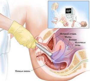
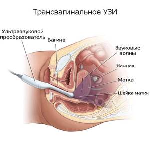
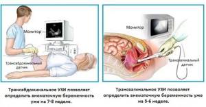
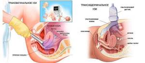
Transvaginal examination differs from transabdominal examination only in that in the first case, ultrasound waves are supplied through a sensor that is located in close proximity to the organs being examined or the fertilized egg: they are separated only by a thin vaginal wall. With transabdominal ultrasound, the sensor is moved along the lower abdomen, but this technique does not provide a clear overview if the patient is obese or suffers from increased gas formation in the intestines.
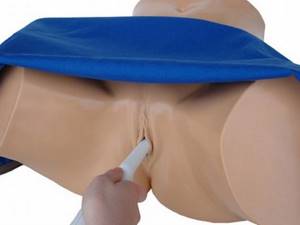
The intracavitary sensor (transducer) looks like a rod 12 cm long, and its diameter does not exceed 3 cm, so its penetration during the study does not cause unpleasant sensations. The sensor has an oblique view relative to its axis, which is necessary for better visualization of the uterus, taking into account its anatomical location. At the end there is a channel with a needle, which may be needed in case of tissue biopsy.
Intravaginal ultrasound examination is absolutely safe, so it can be done multiple times. Sometimes TVUS is prescribed several times during one menstrual cycle.
WE DO ALL TYPES OF ULTRASOUNDS 2D, 3D, 4D with DOPPLER / We do all kinds of ultrasound - 2D, 3D, 4D diagnostics with Doppler
We use one of the best ultrasound scanners with Doppler SonoAce X8 from SAMSUNG MEDISON. This device combines a maximum range of functions and a wide range of diagnostic capabilities - it includes all types of sensitive Doppler, it has an excellent degree of visualization, the ability to panoramic scanning of organs and constructing images in real time in 2D, 3D and 4D formats. The kit contains all the sensors necessary for performing any type of ultrasound - transavaginal, transabdominal and transrectal. We use one of the best ultrasound scanners with the SonoAce X8 doppler from the company SAMSUNG MEDISON. The device for the diagnostic combines the maximum set of functions and a wide range of diagnostic capabilities. SonoAce represents all kinds of sensitive Doppler, has an excellent degree of visualization, the possibility of panoramic scanning and the construction of images in real time in 2D, 3D and 4D formats. The kit includes all necessary ultrasound sensors of any type – transvaginal, transabdominal and transrectal.
ATTENTION! IN THE CLINIC IS A DOCTOR SPEAKING IN ENGLISH LANGUAGE!
Modern ultrasound 3D, 4D with Doppler from Samsung Medison
Contraindications
There are no contraindications for performing transabdominal ultrasound. This study can be carried out for absolutely any condition in patients of different ages and with such frequency as the situation requires. But if possible, it is better to postpone the procedure for some time if there are lesions of various origins on the skin of the lower abdomen, pustular rashes, allergic rashes, etc., so as not to injure the skin even more and not to provoke the spread of the pathological process over a larger area.

Transvaginal ultrasound still has some limitations. Its execution is not shown:
- children and virgin girls;
- people with psychological disabilities;
- in some cases, older women;
- within 2-5 days after performing any interventions on the pelvic organs from the vagina, in particular after surgical treatment of cervical erosion, hysteroscopy, hysterosalpingography, abortion).
Also, it does not make sense to conduct a transvaginal examination if a large formation is detected during a gynecological examination, which can be easily felt with the hands.
Indications for TVUS
Transvaginal ultrasound examination is prescribed for suspected diseases of the pelvic organs, emergency conditions, and also to assess the results of treatment. Indications for this diagnostic technique are:
- Severe pain during menstruation.
- Lack of menstruation or irregularity.
- Suspicion of hormonal imbalance in the body.
- The appearance of bleeding outside the menstrual cycle.
- Presence of symptoms of inflammation of the uterus or ovaries.
- Pathological changes in the endometrium: hyperplasia, polyps, chronic endometritis, submucosal nodes.
- Diagnosis of endometriosis of the uterus or adjacent organs.
- Determination of the presence of pathological fluids in the fallopian tubes.
- Suspicions of underdevelopment of the pelvic organs.
- Identification of neoplasms suspected during a gynecological examination.
- Diagnosis of neoplasms and tumor processes of the uterus and bladder.
- Detection of ovarian tumors and cysts.
- Suspicion of ruptured ovarian cysts.
- Infectious diseases of the urinary tract.
- Suspicion of uterine fibroids.
- Diagnosis of the causes of urological diseases, urinary incontinence and other urinary disorders.
- Determining the location of the intrauterine device.
- Monitoring the effectiveness of treatment.
- Monitoring the condition of the endometrium with an intrauterine device installed or taking hormonal drugs.
- Inability to conceive for six months.
- Preparation for IVF and support of the procedure.
Transvaginal ultrasound allows a woman to know when she is ready to conceive a child. To do this, during a TVUS, a special substance is injected into the fallopian tubes, which, as a contrast, shows the patency of the tubes on the day of the study.
No other method, except TVUS, is capable of recording the baby’s heartbeat as early as the 5th obstetric week of pregnancy. Therefore, transvaginal ultrasound in early pregnancy can confirm the fact of successful conception.
Is it possible to eat before an ultrasound of the pelvic organs?
You may eat before undergoing a transvaginal ultrasound examination. But a few days before the procedure, you should avoid eating foods that contribute to increased gas formation:
- flour products;
- legumes;
- milk and fermented milk drinks;
- cabbage, radishes, turnips;
- grapes, apples, pears;
- carbonated drinks, etc.
The large amount of gases in the intestines makes it difficult to obtain an accurate image of the organs being examined. When experiencing flatulence, a woman needs to take a drug that normalizes gas formation processes.
TVUS during pregnancy
Since the intravaginal examination procedure is associated with a certain irritation of the walls of the vagina and pelvic organs (including the uterus), it is allowed only in the first trimester - up to 13 weeks of pregnancy. TVUS makes it possible to detect the very fact of pregnancy and determine its exact date. But there are other indications for conducting such a study:
- monitoring the fetus and its development over time;
- determination of intrauterine and ectopic pregnancy;
- early diagnosis of fetal and pregnancy pathologies;
- identification of threats of miscarriage;
- suspicion of detachment of the ovum;
- distinguishing the fertilized egg from a pathological formation in the uterus;
- assessment of the condition of the scar that remained on the uterus after a previous cesarean section;
- selection of the method of delivery based on the data obtained;
- complete and partial hydatidiform mole;
- determination of the position of the fetal egg in the uterus, the state of its attachment (presentation, low location).
TVUS during pregnancy allows you to determine the number of fetuses in the uterus, heart rate, and possible malformations. This type of study accurately determines the gender of the unborn child.
During pregnancy, transvaginal ultrasound can be done on demand as many times as needed. The sensor and ultrasound radiation are not able to affect the condition of the fetus and the pregnant woman herself.
Which ultrasound method is better?
Most often, doctors prescribe ultrasound of the abdominal cavity, mammary glands, pelvis, kidneys, thyroid gland, bladder and liver. For men, one of the most important diagnostic methods is ultrasound of the prostate gland. Separately, it is worth mentioning ultrasound during pregnancy. This examination is carried out on pregnant women regardless of their health status, because its purpose is to examine the fetus.
Diagnostics are performed using different types of sensors. The choice of method and type of sensor depends on which organs need to be checked.
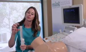
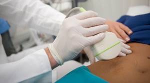
How is a transvaginal examination done?
To undergo an intravaginal examination, the patient needs to undress from the waist down and lie down on the couch, spreading her legs slightly to the sides. The diagnostician usually asks why the woman was sent for an ultrasound and what complaints she has. This information is necessary in order to pay special attention and examine the most interesting pelvic organs at multiple magnification. In this case, the examination involves a complete and thorough examination of the condition of the patient’s entire genitourinary system.
The procedure is carried out as follows:
1. The doctor places a medical condom on the ultrasound sensor and lubricates it with a special gel, which facilitates its penetration into the vagina and eliminates the air space between the transducer and the organs being examined.
2. The doctor performs all movements slowly and carefully, so there is no discomfort during the procedure. A woman can feel pain only if there is an inflammatory focus in the area being examined. If any discomfort occurs, you should immediately report this to your diagnostician.
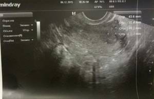
3. An exact image of the organs being examined is visualized on the monitor of the ultrasound machine; the doctor can enlarge the resulting image many times and view the elements of interest in great detail.
4. Having recorded all the necessary data, the doctor removes the sensor from the vagina. The woman can get dressed and pick up the ultrasound results, which are later deciphered by the doctor who referred for the study.
The effect of ultrasound on a child
Thermal index
When the technology operates, ultrasound interacts with tissues, heats them, and increases the temperature of cells that contain a lot of water. Such heating can cause harm, especially for the fetus, whose dimensions reach only millimeters, but only if equipment with such high-power radiation is used.
The largest amount of water is contained in brain cells. This tissue is subject to the greatest heating during ultrasound.
In accordance with scientific developments, the thermal index should not be higher than 20C. Ultrasound heats tissue by 1.0 degrees. But different fabrics have different heating abilities. Doctors have not yet recorded the consequences of this effect on the health of the embryo, since modern ultrasound machines cannot create waves of harmful power.
At the same time, doctors study the effect of ultrasound only on born children; it is impossible to determine anything in utero, so the average person still has reasons to doubt the complete safety of the technique.
Women often say that it is not without reason that foreign doctors send expectant mothers for an ultrasound scan only after 11-12 weeks. Previously, an ultrasound was performed only if the patient complains of something (bloody discharge, pain in the lower abdomen). But we must not forget about the insurance model of medicine in Western countries: the less research, the more profitable it is for them.
Mechanical index
Ultrasound examination is also called echolocation because ultrasonic waves that are reflected from tissues provide doctors with information about the size and location of the fetus, similar to an echo.
Ultrasound also affects fetal tissue mechanically. This lies in the pressure of the waves. Experts believe that the sound amplitude should not be higher than 1.9. When diagnosed in the early stages, it should be from 0.05 or more.
During ultrasound, the effect on tissue of a sound wave less than the above is observed.
Preparation for TVUS
Intravaginal ultrasound is distinguished not only by its ease of implementation with high information content, but also by the lack of preparation for this examination. There is no need to drink water to fill the bladder; the transvaginal sensor clearly visualizes the uterus, ovaries, fallopian tubes, ligaments and adjacent organs without additional manipulation.
Only patients suffering from severe flatulence need specific preparation:
1. 2 days before the test, you should limit the consumption of foods that increase gas formation in the intestines: baked goods, fresh vegetables and fruits, dairy products.2. On the day of the TVUS, the doctor may prescribe you to take Enzistal, Espumisan, activated charcoal or Smecta. The drug is selected by the doctor based on the patient’s tolerance to the active ingredients of the drug and cannot be chosen at his own discretion. Transvaginal ultrasound examination for pregnant women is recommended to be carried out on a moderately full bladder, so for such patients a preliminary visit to the toilet is not required.
Advantages of ultrasound with a vaginal sensor
Ultrasound scanning through the vagina has several advantages:
- has accessibility;
- performed with an empty bladder;
- has high information content;
- there is no special preparation (full bladder, special diet for bloating);
- makes it possible to examine the cervix (ultrasound through the anterior abdominal wall does not allow detailing the cervix);
- used in women with a pronounced subcutaneous fat layer on the abdomen;
- does no harm.
On what day of the menstrual cycle is TVUS performed?
Women often ask whether it is possible to do a transvaginal ultrasound during menstruation. For the most accurate assessment of the condition of the female genitourinary system (outside the period of pregnancy), it is worth adhering to a certain dependence of TVUS on the day of the menstrual cycle:
1. A planned transvaginal ultrasound should be done on the 5th, 6th or 7th day of the cycle, while the endometrium of the uterus is not yet in the secretory phase. If you ignore this recommendation, the doctor may incorrectly interpret the results of ultrasound diagnostics.2. To determine the presence of a fertilized egg in the uterus and the exact duration of pregnancy, a woman needs to come for a transvaginal ultrasound diagnosis no earlier than the 10th day of the delay.3. If a patient is suspected of having endometriosis, then the study should be planned after ovulation - no earlier than the 14th day of the cycle.4. If uterine fibroids are suspected, a transvaginal ultrasound is performed on days 18-24 of the cycle5. To monitor the patient’s reproductive function, monitor the ovulation process, or determine the cause of hormonal imbalance in the body, transvaginal ultrasound is recommended to be performed on the 10th day of the cycle, and then every 3 days until its end.
However, there are situations when an emergency procedure is necessary, regardless of the day of the cycle:
- sudden disruption of menstrual function: the next menstruation turned out to be more abundant and with clots, longer (longer than 7 days) or did not occur at all within 1 month;
- spotting that the patient does not associate with the onset of menstruation, i.e. they can appear before, after the next menstruation or in the middle of the cycle;
- discharge with blood in pregnant women;
- discharge mixed with blood during or after sexual intercourse;
- the appearance of bleeding in a woman during the menopausal period;
- pain in the lower abdomen.
The appearance of such symptoms should prompt a woman of any age to contact a gynecologist and further undergo a TVUS scan.
How is the procedure done?
The examination itself does not take more than 10 minutes. After the doctor examines the image displayed on the screen, he writes a conclusion. During this time, the patient may be asked to wait outside the office. The conclusion is handed over to you and you can take it to your doctor. It is worth knowing that this procedure is not prescribed for girls who are not sexually active. In this situation, the examination is performed rectally or using an abdominal ultrasound.
On what day of the cycle is a vaginal ultrasound performed?
If you do not know what a vaginal ultrasound is and when it is best to do it, it is worth knowing that the most suitable time for examination is the 5-8th day of the cycle, that is, immediately after menstruation. However, this is only suitable if a routine inspection is being carried out. If the gynecologist suspects endometriosis of the uterus, the examination is postponed to the second half of the cycle. Several studies in one cycle allow you to monitor the dynamics of the disease. An emergency procedure can also be performed on any day of the cycle if bleeding begins that is not related to menstruation.
Transvaginal ultrasound: photo
Transvaginal ultrasound
Pelvic ultrasound in St. Petersburg
transvaginal ultrasound during pregnancy
transvaginal ultrasound
transvaginal ultrasound
Transvaginal ultrasound at the Diana clinic
Gynecological ultrasound: vaginal or transabdominal?
The main advantage of transvaginal ultrasound in gynecology is its high accuracy in displaying the structure of the tissues of the pelvic organs, giving the doctor the necessary information about their functional state. Intravaginal sonography allows you to obtain detailed data on the condition of the tissues of the cervix and body of the uterus, ovaries, and visualize both fallopian tubes, while transabdominal examination allows you to examine only the left one.
Unlike transabdominal ultrasound, vaginal ultrasound makes it possible to detect and study areas of altered endometrium, which in many cases makes it possible to recognize endometriosis without performing a diagnostic operation - laparoscopy.
Despite the high information content of vaginal ultrasound, its use is not always possible. Transvaginal ultrasound is not prescribed for virgins due to the likelihood of damage to the integrity of the hymen. Vaginal ultrasound examination also cannot be used:
- if the patient is allergic to latex;
- in the later stages of diagnosed cervical cancer (due to the risk of bleeding);
- in the acute phase of diseases accompanied by inflammation, tissue swelling and severe pain (the study should be postponed until recovery or alternative methods should be used);
- in the 2nd and 3rd trimesters of pregnancy (due to the risk of premature contractions).
If there are contraindications to vaginal ultrasound, the doctor may prescribe transabdominal sonography or other diagnostic methods. Also, both types of examinations can be prescribed simultaneously to obtain the maximum amount of data on the condition of the pelvic organs through examination in different planes.
The results obtained during the study are interpreted by a sonologist, after which he gives his opinion and refers to the appropriate doctor to prescribe the necessary treatment.
1
0
2
Article rating:
5 out of 5 based on 6 ratings
Author: Shulga Tatyana Alexandrovna
Ultrasound diagnostics (USD) doctor. First category. Work experience 27 years.
How often should a transvaginal ultrasound be done?
You need to contact a gynecologist and undergo a transvaginal ultrasound examination every time a woman develops the symptoms and conditions described above in the indications for the procedure. A gynecologist or urologist may prescribe this type of examination if the development of a disease of the genitourinary system is suspected.
If there are no complaints and the woman assesses her condition as satisfactory, she needs to undergo TVUS for preventive purposes at the following frequency:
1. For women with the onset of sexual activity – once every 2 years.2. Women over 40 years old – annually.
If endometriosis is suspected, the procedure is performed in the second half of the cycle. If bleeding occurs in the middle of the cycle, a transvaginal ultrasound is performed urgently to determine the cause of this phenomenon.
Transrectal ultrasound (TRUS)
TRUS is the most informative method for diagnosing the prostate gland in men. The ultrasound wave is reflected from the tissues, thereby forming an image of the organ on the monitor; in this case, the doctor observes the image of the prostate gland.
Indications for transrectal diagnostics
All men over 40 years of age should undergo a routine ultrasound of the prostate gland in order to prevent diseases of the genital area. In addition, this research method is indicated for:
- The appearance of pain in the perineal area.
- Signs of prostate adenoma.
- Decreased potency.
- Feelings of discomfort when urinating.
- Frequent urination at night, accompanied by pain and discomfort.
- Inability to conceive a child (in the absence of health problems in the woman).
- Deviation from normal values in spermogram and urine analysis.
- When pathologies are detected in the development of the genital organs.
How to prepare for TRUS?
Transrectal diagnosis of the prostate gland requires mandatory preliminary preparation. To ensure that the results are as accurate as possible, the patient should perform a number of actions:
- A couple of hours before the examination procedure, you need to empty your bowels. To facilitate the process, you can use Microlax or place a glycerin suppository rectally.
- Fill your bladder. If an ultrasound of the prostate gland is prescribed for suspected infertility or decreased potency, then an hour before the diagnosis you need to drink 0.5 liters of water and not empty your bladder.
If a test is ordered to identify problems with urination, the patient comes to the doctor with a bottle of still water and starts drinking it. As soon as he feels the urge to urinate, he informs the doctor and goes for an ultrasound procedure.
How is transrectal ultrasound performed?
The patient comes to the doctor, lies down on the couch on his left side, bends his knees and presses them as tightly as possible to his chest. A special sensor with a condom previously put on it is inserted through the anus into the rectum to a shallow depth (up to 6 cm). The diameter of the sensor is about 2 cm, so when it is inserted the patient does not experience pain - only some discomfort is felt.
If it is necessary to study the prostate and seminal vesicles, the sensor is inserted to a greater depth. If it is necessary to determine bladder emptying, then the ultrasound is repeated after the patient goes to the toilet. This is how the amount of residual urine is assessed and possible pathologies are determined.
Decoding the research results
TRUS of the prostate gland evaluates indicators such as the shape and size of the gland, its contours, structure, and echogenicity. Normally, the indicators should be as follows:
- Shape: triangular, semicircular or oval;
- Contours: clear, even;
- Echogenicity: fine-grained;
- Structure: heterogeneous (gland tissue and capsule have different echo densities);
- Dimensions: length – 2.4-4 cm, width – 2.7-4.3 cm, thickness – 1.5-2.3 cm;
- Volume: up to 200 cm³.
TRUS of the prostate shows a hypoechoic seminal tubercle in the shape of a triangle and up to 2 mm in size. The bladder, rectum and ejaculatory ducts are also clearly visualized.
According to numerous reviews from specialists, transrectal ultrasound is a highly informative study, and the urologist receives accurate data. If no echo signs of pathology were detected during the ultrasound, then the patient can undergo an ultrasound again only after a year if there are no complaints about men’s health.
Evaluation of TVUS results
Having transvaginal ultrasound data in hand, an experienced specialist can accurately diagnose and select the appropriate treatment for the identified pathology. The results are interpreted by a gynecologist.
About consultation with a gynecologist / Consultation of a gynecologist
Our clinic employs gynecologists of the highest and first certification categories. All doctors have certificates confirming their qualifications, issued in St. Petersburg and Moscow. The cost of an initial appointment with a gynecologist is 1000 rubles, a consultation based on test results or ultrasound is 500 rubles. Appointment with a gynecologist You can make an appointment with a gynecologist without an insurance policy, registration in St. Petersburg and Russian citizenship. We have gynecologists and ultrasound specialists who speak English. You can apply to us without having an insurance policy, registration in St. Petersburg and Russian citizenship. ATTENTION! IN THE CLINIC IS A DOCTOR SPEAKING IN ENGLISH LANGUAGE! Sign up for an appointment with the doctor
During a TVUS, the diagnostician evaluates the following indicators:
Uterus size and position
The size of the organ depends on the individual characteristics of the anatomical structure, the number of pregnancies, childbirths and some other factors. In women who have given birth, the dimensions of this organ are approximately 70 x 60 x 50 mm. If these sizes are much larger than acceptable, the doctor looks for the cause of this pathology.
When performing a transvaginal ultrasound, the position of the uterus is also taken into account. If it is slightly tilted forward, this is considered normal. If its position is shifted back, they speak of a pathological location of the organ.
Echogenicity of the uterus
Here, the structure of the walls and cavity of the organ itself is important; the clarity of the boundaries of the uterus also determines. The presence of hyperechoic areas in the picture may indicate the presence of neoplasms in the uterus.
Endometrial thickness, cervix and endocervix
The inner layer of the uterus changes its thickness depending on the day of the menstrual cycle: at the beginning the endometrium has a thickness of about 3 mm, and at the end it can reach 2 cm. The data obtained on the thickness of the endometrium is compared with the day of the patient’s cycle: if there are discrepancies with the norm, inflammatory process.
Cervix and endocervix . What matters is the structure of the cervix and its size, which normally should correspond to 40 x 30 mm. The cervical canal should have a diameter of less than 3 mm and be filled with mucus.
Sizes and contours of the ovaries
Transvaginal ultrasound perfectly shows the size of the right and left ovaries. At the same time, they may differ slightly, but their total volume should not exceed 10 cubic cm. If this indicator is exceeded, polycystic syndrome or an inflammatory process may be suspected. Their contours can be either smooth or irregular due to the formation of follicles.
Presence of free fluid in the pelvis
A small amount of it can normally be seen in this area, but only immediately after ovulation, which is associated with rupture of the follicle. Otherwise, the fluid may be a sign of ovarian inflammation resulting from an infectious lesion.
Fallopian tubes
They are usually visualized in the presence of an ectopic pregnancy or an inflammatory process in them. To diagnose tubal patency, a contrast agent is used. Already at the stage of ultrasound diagnostics - transvaginal ultrasound, the doctor can determine the following pathological conditions:
- inflammation of the fallopian tubes, ovaries or endometrium of the uterus;
- polycystic ovary syndrome;
- ovarian cyst;
- endometriosis of the uterus and nearby organs;
- polyps or uterine fibroids;
- cancer of the ovary, uterus or cervix.
What is the frequency range of the sensor?
The frequency range of the sensors used for transvaginal scanning is in most cases 4-7 MHz. Higher frequencies, as a rule, are not used to study the uterine cavity.
You may be interested in: Sodium barbital: use and dosage
The fact is that the depth of the uterus is easily detected by a gynecologist, so there is no need to purchase sensors with a higher frequency.
The scanning angle of such sensors varies from 120 to 140 degrees. This angle is sufficient to completely examine the uterus. There are also special sensors, thanks to which they receive a 4D image and simultaneously display the image on the screen.
Such equipment is capable of studying the structure of small parts of the fetus and its cardiovascular system, as a result of which doctors can identify abnormalities at an early stage.
Interpretation of transvaginal ultrasound: table of indicators and diseases
| Genitals | Normal indicators | Deviations |
| Uterus | Anteflexio (anterior tilt); The contours of the uterus are smooth and clear; Dimensions of the uterus: length - 7 cm, width - 6 cm, diameter - about 4 cm; Homogeneous echogenicity of the walls; Homogeneous structure; Crisp and smooth edges | Retroflexio is a variant of the norm that prevents conception; Uneven contours indicate inflammation in the uterus and tissues or tumors of various origins; Heterogeneous echogenicity is a sign of a tumor; Hyperechoic formations (polyps, tumors, myomatous nodes); Heterogeneous structure: a sign of endometritis |
| Cervix | Dimensions: anteroposterior size - 2-3 mm, length - 3-4 cm. Diameter of the cervical canal: 2-3 mm. Homogeneous echostructure. Mucus of homogeneous echostructure. | Increased size of the cervix and cervical canal Heterogeneous structure |
| Free liquid | Normally, its amount is several mm. | A large amount of fluid is a sign of infection |
| Ovaries | Dimensions: width - 2.5 cm, length - 3 cm, thickness - 1.5 cm. Normal volume: 2-8 cm³; Fuzzy, lumpy contours. Homogeneous structure, small areas of fibrosis; In the middle of the cycle, follicles 4-6 mm in size and one dominant follicle up to 2 cm in size are formed | Increase in size (inflammation, polycystic disease) Large fibrous foci (inflammatory process). The size of the dominant follicle is greater than 2.5 cm (follicular cyst) |
| The fallopian tubes | Almost invisible | Clearly visualized (a sign of ectopic pregnancy or inflammation) |
Echo signs of uterine pathologies
| Pathology | Features |
| Uterine cancer | Changes in the contours of the uterus, swelling |
| Myoma | Increased size of the uterine body, altered contours, “hyperechoic zone” (nodule in the muscle) |
| Polyps | Fuzzy contours of the uterus, space-occupying formations in the uterus |
| Endometriosis | “Bubbles” on the cervix, in the tubes and in the uterus itself |
| Adnexit | Thickening of the fallopian tubes, enlarged ovaries, unclear and blurry contours |
| Ovarian cancer | Increase in size with deformation of contours |
| Ovarian cyst | Accumulation of fluid with a diameter greater than 25 mm |
| Endometritis | Swelling of the endometrial layer, increased size of the uterus, thickening of the walls |
| Cervical tumor | Deformation and enlargement of the cervix |
Timely diagnosis and identification of pathological processes in the genitourinary system allows, in most cases, to manage only with drug therapy. Only in rare cases are identified problems solved exclusively by surgery. This speaks to the importance of an annual preventive examination by a gynecologist and undergoing a transvaginal ultrasound by a good specialist.
CLICK TO MAKE AN APPOINTMENT, TEST OR ULTRASOUND
If you find an error, please select a piece of text and press Ctrl+Enter
What diseases does the examination reveal?
Transvaginal gynecological ultrasound easily diagnoses the following diseases and conditions of the female body:
- Cystic formations (cysts) of the ovaries.
- Inflammation in the fallopian tubes (pus, blood).
- Endometriosis of the cervix.
- Uterine fibroids.
- Pregnancy.
- Malignant formations in the uterus.
- Polyposis of the endometrial layer (polyps).
- Ovarian cancer.
- Cyst rupture.
- Abnormal development of the organs of the genitourinary system.
The method of transvaginal ultrasound of the uterus is also successfully used to determine the quality of the follicle maturation process. Such a study allows timely detection of obstruction of the fallopian tubes and the degree of this pathology. This is very important when searching for a suitable treatment method for female infertility.
The transvaginal sensor inserted into the vagina is very sensitive, so the method is applicable in diagnosing pathologies in fetal development during pregnancy and frozen pregnancy.
Advantages of this device
Thanks to ultrasound examination of the pelvic organs, the diagnosis can be significantly simplified. Because of this, this procedure has become widely accepted in medical practice. Moreover, this method is safe for the health of the woman and child, so ultrasound can be done multiple times.
Research with a transvaginal sensor provides the opportunity for timely detection of the development of many pathologies of the female genital organs that have an oncological or inflammatory etiology.
If Doppler sonography is used in conjunction with this method of examination, it is possible to timely identify the likelihood of developing thrombosis, examine blood flow in the pelvic organs and detect atherosclerosis.
Also, ultrasound of the pelvic organs with a transvaginal sensor is more meaningful and informative than a conventional examination through the abdominal wall.