Full text of the article:
Working with diseases of the internal organs is especially difficult because they cannot be seen. Previously, doctors had to treat patients literally “blindly,” since it was possible to examine a person’s internal organs only during surgery. Nowadays, doctors don’t have to pick up a scalpel; various types of scans help make a diagnosis. However, patients are wary of this type of examination. This is due to the high cost of some procedures and the fear of radiation. Let's try to figure out what the features of certain scans are and when to resort to them.
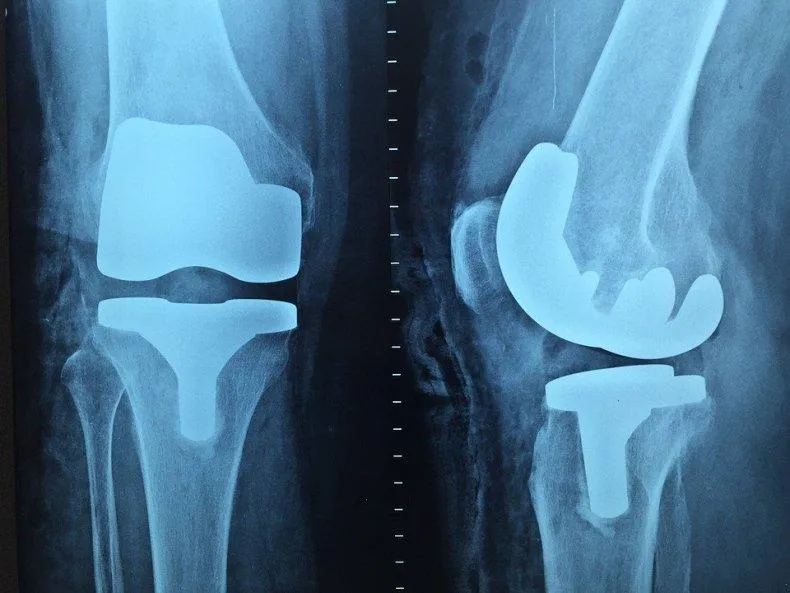
X-ray
The oldest and most common method of visualizing the human body. X-rays are used everywhere, from surgery to dentistry. The method is simple and clear: a person is irradiated with special rays that easily pass through soft tissues and linger in hard ones. Thanks to this principle, an image is transmitted to a photographic film or sensor located on the side opposite the ray source, and radiography or fluoroscopy is available to the doctor.
Main advantages
such an examination: speed and cost. Almost all hospitals are equipped with X-ray machines; the procedure is quick and inexpensive.
Main disadvantages:
exposure and image quality. When performing radiography, the patient is irradiated, and the picture is two-dimensional. The doctor can hardly see the internal organs individually, since their shadows overlap each other. It is also impossible to see the cartilage tissue and brain in detail. The cartilage practically does not block the rays, the brain is securely closed by the skull. X-rays are not suitable for their examination.
It will be most effective to carry out radiography for damage to bones, joints and teeth.
X-ray of the cervical spine is prescribed for:
- spinal injury in the neck area;
- limited mobility – it is difficult for a person to look around;
- pain that gets worse when moving your head;
- frequent dizziness, headaches;
- ringing in the ears;
- crunching when moving the neck;
- discomfort in the upper back;
- feeling of weakness, tingling, goosebumps in the hands.
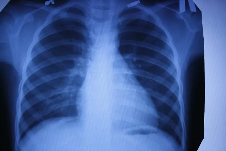
Fluorography
Another type of examination that all residents of our country regularly undergo. Fluorography was “invented” almost a hundred years ago. This is a kind of accelerated radiography. Scientists proposed photographing a screen with an image obtained from radiography. This made the procedure faster and more widespread. Screening tests began to be administered to everyone in order to detect latent pulmonary tuberculosis.
Main plus
The procedure is fast,
the main disadvantage
is the image quality. The patient also receives a dose of radiation, and the doctor has a rather blurry picture, so fluorography is recommended to be supplemented with questionnaires and laboratory tests for the presence of tuberculosis.
Features of X-ray of the thoracic spine
In order to exclude such a phenomenon as tissue layering in the images, X-ray images of the thoracic region are taken in different projections. Thus, it is possible to achieve their clarity and visualize existing pathologies. The most commonly used projections are:
- Straight - the patient takes a standing position in front of the X-ray screen and emitter, his chin is fixed so that it is strictly parallel to the floor, and the spinal table takes the required position. Thus, rib deformations, as well as spinal curvatures, are well visualized;
- Lateral - the patient takes a standing position, placing his hands behind his head and pressing the diagnosed side of the chest tightly against the screen. The images clearly show the lower vertebrae and intervertebral discs, and it is possible to obtain a different projection of the ribs.
The shooting itself lasts a few seconds, during which the patient needs to hold his breath, and the entire procedure will require no more than 10 to 15 minutes.
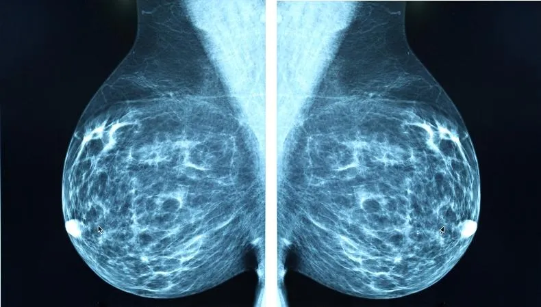
Mammography
A separate type of radiography designed to diagnose breast diseases, which is why women undergo mammography. There is no consensus on the recommended age for the procedure. Mammography helps ensure the absence of a malignant tumor with an accuracy of 89%. It is believed that women should be screened regularly starting at age 39, although some cancer societies recommend screening at a younger age.
Mammography is prescribed to diagnose breast cancer, the procedure is quick
, this is a plus, but the patient
is irradiated
, and the risk of an incorrect diagnosis remains, this is a minus. Mammography can be digital or film; digital mammography provides a clearer image.
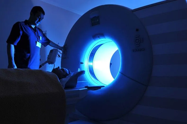
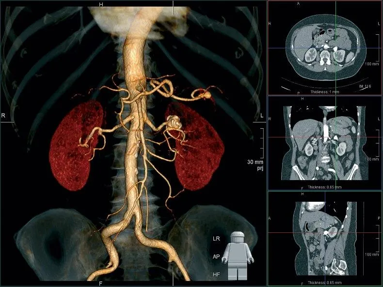
Computed tomography (CT)
Computed tomography is also carried out on the principle of radiography, but as a result the doctor receives not a flat two-dimensional picture, but a three-dimensional image. This is achieved by simultaneously taking a large number of images, which are assembled into a single image. CT scanner sensors are highly sensitive and distinguish a huge number of shades, so the doctor can examine in detail all the bones and organs of the patient. The image quality can be further improved by injecting the patient with a special substance, the so-called “contrast”. The contrast helps to distinguish healthy tissues from altered ones and detect abnormal structures in the body, and also makes it possible to study the condition of blood vessels in detail. CT with contrast is not prescribed in every case; often a simple computed tomography is sufficient.
CT scan is done quickly
, with its help to screen for lung cancer. You can also use computed tomography directly during surgery.
Disadvantages of CT
can be considered a high radiation dose to the patient. Therefore, CT is not prescribed to pregnant women, children and overweight patients (more than 200 kilograms).
Evgeny Avsharov: what's wrong in the kingdom of radiology?

Evgeniy Mikhailovich Avsharov – head of the development and implementation of the PACS system for receiving, storing and transmitting medical DICOM images AS_VIMeN, processing and visualization on 2D workstations Real-time processing and visualization AS_GSV “Michelangelo”, DICOM servers and AS series printers, chief architect of the Microsecond Radiology technology ”, based on new physical and technological principles, to create a fundamentally new class of x-ray diagnostic medical and industrial equipment. Certified engineer from Siemens in X-ray technology and from General Electric in computed tomography; developer of the ideology and technology for creating digital adaptive systems for receiving, storing and pipeline 2D processing and real-time visualization of a stream of high-resolution dynamic medical images. Technical Director of LLC “KURS-AS1” , Moscow.
What technical difficulties prevent large manufacturers of X-ray equipment from significantly reducing the dose of harmful radiation?
The X-ray dose required to create a correct diagnostic image is directly dependent on the initial quantum efficiency DQE(0) of Flat Panel Detector (FPD).
Many FPD manufacturers are not very fond of providing characteristics of the initial dependence of the quantum DQE(0) efficiency on the value of the incoming X-ray dose, and here's why: even for direct conversion FPDs performed on a CsI scintillator with an amorphous selenium a-Se amplifier with a gain of 1.0, in the region of the low dose range 1.0-10nGy (0.1-1.0µR), the change in the DQE(0) value is in the range from 0.10 to 0.30, therefore it is necessary to set the gain value in the a-Se layer from ~50 and higher, which leads to a proportional increase in quantum noise.
Thus, leading manufacturers of X-ray equipment have reached the limit of increasing the power of X-ray radiation and have reached the limits of converting X-ray detectors using traditional technologies, both in medicine and in industry. Currently, existing approaches to the construction of X-ray diagnostic systems do not allow combining in one product such characteristics as: increasing the resolution of dynamic X-ray detectors with a simultaneous increase in the shooting speed in frames per second, which directly affects the diagnostic process; reduction of the X-ray dose by an order of magnitude or more, because to increase the resolution of X-ray images by 4 times, it is necessary to increase the dose per pixel of the image by 16 times; reduction of the focal spot of an X-ray tube to a value of 0.1 * 0.1 mm or less, with the shooting time of an X-ray image frame being several milliseconds (even the best processing allows achieving a real resolution of no more than 0.7-:-0.5 of the size of the focal spot, even if the resolution of the X-ray detector is very high exceeds this value); reduction of the integral radiation power per x-ray image frame without increasing the instantaneous power of the x-ray tube.
On what principles is it possible to increase the resolution of radiology with a decrease in the X-ray dose?
The quality and diagnostic value of images obtained in radiology depend on four main components: X-ray emitters (X-ray tubes), high-voltage X-ray generators, X-ray detectors and image processing and visualization systems.
We solved the problem of leveling the influence of secondary (parasitic) X-ray radiation on image quality. Currently available mathematical methods for partial leveling of secondary spurious radiation arising in the object of study allow processing both a stream of 16-bit frames in real time and a stream of subtraction images, format up to 1024 * 1024 (i.e. 1 megapixel = 1.0Mpix) at 30 frames per second.
We achieve this by creating a multi-stage 16-bit flexible multifunctional pipeline for mathematical processing of an image stream with the sequential use of sets of functional elements - matrix filters, nonlinear and spectral converters, the parameters of which are available to the operator of the processing process. In this case, the physical data stream can reach 120MB/s at 60 frames per second (for the 1024*1024*16b format), and the required for visualization, without display delay and the ability to be perceived by the human eye, is 60MB/s at 30 frames per second. The quality of partial leveling of secondary parasitic radiation depends on the processing technology and the capabilities of the functional elements of the processing pipeline.
Stages of image processing from the original to obtaining a subtraction image in which even the smallest blood vessels are clearly visible
The video shows the stages of pipeline processing from the primary source image to the subtraction one, which makes it possible to partially level out the secondary (stray) radiation, but not completely; the degree of leveling characterizes the quality of mathematical processing. The request to increase the dynamic display of large format images 9-12Mpix at 30-60 frames per second requires an increase in the power of the computing pipeline by an order of magnitude or more, due to an increase in the input stream to 1440MB/s (12Mpix at 60 frames per second), which is for processing a very non-trivial task of creating a dynamic pipeline for streaming image processing.
Thus, a way out of the systemic limitations of traditional radiology to greatly increase the efficiency and safety of the method is possible through the creation of a new class of X-ray diagnostic systems within the framework of the Microsecond Radiology technology. The solution has no analogues in world radiological practice.
What are the main results of microsecond radiology?
- A radical reduction in the x-ray dose by more than 20 times or more for any type of x-ray examination, such as in medical examinations, especially in mammography and angiography, in x-ray examinations based on the principles of “tomosynthesis” and computed tomography; and in X-ray non-destructive testing systems, in X-ray inspection and security systems.
- At the same time, the resolution of dynamic images will increase by 3-4 times, i.e. up to (100-:-50)µm versus (400-200)µm at 30/60 frames per second, compared with modern X-ray medical systems, which will require the creation of new standards for the diagnostic process in general radiology, in the field of computed tomography, with angiographic studies, mammography, etc.
- An additional effect is a decrease of more than 20 times in the integral power of the X-ray generator from (50-:-120)kW to (2-:-4)kW at the instantaneous power of the X-ray tube - from (30-:-100)kW to (15 -:-30)kW, will ensure virtually no heating of X-ray tubes and generators, and will lead to a manifold increase in their life cycle.
- Real-time streaming pipeline processing (more than 1500MB/s), to reduce X-ray noise of dynamic images, based on a unique real-time parallel computing tool, while increasing the resolution and clarity of the resulting images.
- Increasing the resolution of a computed tomograph to (100-:-50) microns with an X-ray tube focus ranging from 0.15×0.15mm to 0.1×0.1mm (in the medical field).
- Multimodality – combining a high-resolution X-ray diagnostic device and a computed tomograph in one device.
- Reducing the cost of a microsecond X-ray tube, microsecond X-ray generator and microsecond X-ray detector, associated with the design and technical features of the latter and the high manufacturability of their manufacture, which will lead to a reduction in the “total cost of ownership” (CTO) of the entire X-ray complex by up to 50%.
Where were your developments implemented?
Currently, medical image processing systems operate in Moscow at the MEDSI clinic, at the children's clinic of the Presidential Administration, at the Gazprom clinic, at the 12th city hospital and more than 47 clinics across the country.
Is it possible to use the developed methods in astronomy for in-depth space exploration, microscopy, photo processing and digital processing of arbitrary signals?
Restoring images from projections is one of the functions of practical astronomy and many other practical fields (geology, biology, etc.). The technology can be applied to any processes involving the absorption and reflection of signals inside the object under study - for example, for computed tomography it significantly improves images obtained by reconstructing images from projections.
It must be emphasized that although this technology was developed for medicine, primarily for radiology, it is also applicable for microscopy and processing of high-resolution photo images, has proven itself to be excellent for processing MRT images, is relevant for optical images, and in general for any two-dimensional high-resolution images resolution – both in size and in pixels.
Interview: Ivan Stepanyan
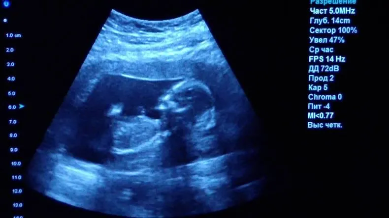
Ultrasound examination (ultrasound)
X-ray is not the only way to “look inside” the human body; another technology is ultrasound. Some animals, such as bats, use sound waves to orient themselves in space. People have also learned to use waves to solve some problems, including in medicine. A picture of the internal organs can be obtained by sending a sound wave into the human body and monitoring its return. The computer helps process the results and present them in the form of a three-dimensional picture.
The main advantage of this research method is safety.
. Ultrasound can be performed even on pregnant women; in addition, ultrasound devices are mobile and can easily be placed in the patient’s room to monitor the condition of organs and blood flow in real time.
However, ultrasound cannot provide a high-definition image, so the use of this method of research is limited, for example, gastrointestinal diseases cannot be diagnosed using ultrasound.
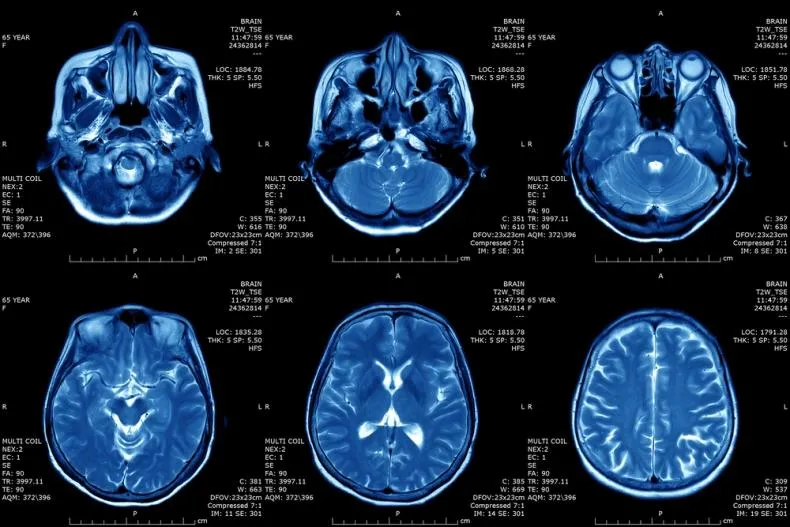
Magnetic resonance imaging (MRI)
The principle of MRI is based on the property of atomic nuclei to respond to a strong magnetic field. The calculation is based on the reaction of hydrogen nuclei, of which there are many in the composition of water molecules, and the human body, as is known, consists of 60% water. When entering a magnetic field, the nuclei of atoms are oriented along it; they can be excited and the energy that they will give off when the influence weakens can be recorded, i.e. “relaxation”. Computer analysis allows you to convert the information received and determine the location, density and structure of tissues in the body.
MRI allows you to “see” cartilage, soft tissue and the human brain without causing harmful effects
, so the procedure can be performed by anyone and as many times as desired.
However, the examination takes a long time
, and closed-type tomographs can cause attacks of claustrophobia. True, there are open-type devices. MRI procedures should not be performed on people who have electrical devices (such as pacemakers) or metal implants implanted in their bodies.
MRI will be effective in studying tumors, brain and vascular abnormalities.
What is more harmful: MRI or X-ray?
The difference is significant. From the point of view of the effect on the body, x-rays are more dangerous. Ionizing radiation stimulates the formation of free radicals. The latter damage healthy cells and provoke their uncontrolled division with the risk of malignant degeneration. High doses can cause radiation sickness. That is why the procedure is contraindicated for children and pregnant women.
The doses used are minimal and the risk of adverse effects on the body is low, provided that the recommendations for the frequency of examination are followed. X-ray diagnostic methods can be used no more than once every six months. If dynamic monitoring of the development of pathology is necessary, in many cases tomography is better than x-rays.
The magnetic field of MRI is completely harmless to the body. Risks arise when contraindications are ignored, which include the first trimester of pregnancy and the presence of metal implants in the body.
If there are titanium structures in the body, there will be no harm. Even in the presence of ferromagnetic implants outside the examination area, life-threatening consequences are excluded. If the patient has a pacemaker, insulin pump, defibrillator, or metal hemostatic clips, tomography is strictly contraindicated for him. In this case, X-ray is better than MRI. Ionizing radiation has no effect on the operation of electronics and does not change the properties of metals.
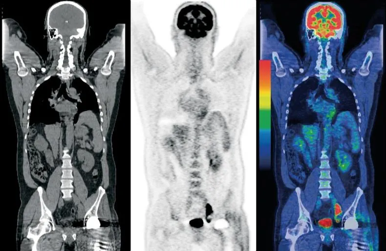
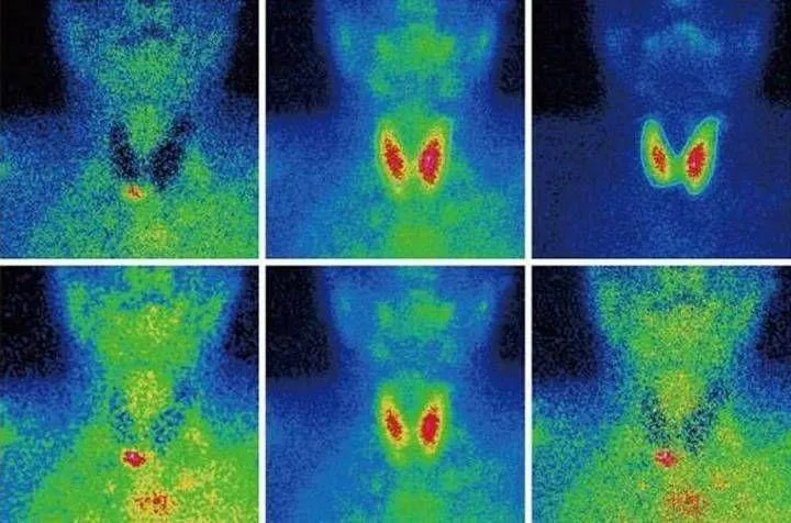
Scintigraphy, SPECT, PET
Perhaps these are one of the rarest procedures on our list. These examination methods are based on radiation diagnostics, but it is used in reverse. The patient is not irradiated from the outside, but a special radioactive drug is injected into him to make him “glow from the inside.” First, scientists invented and tested scintigraphy. With its help, it was possible to obtain two-dimensional images. Then research went further and single photon emission computed tomography ( SPECT)
), followed by positron emission tomography (
PET
). The difference between these methods is rather technical; they use different radiopharmaceuticals and different types of detectors that record radiation from the patient’s body.
The question arises: “Why such difficulties?” The fact is that thanks to these procedures, you can see formations in the pictures that are not visible in pictures obtained by external irradiation. Metastases and tumors can appear inside bones or organs and remain silent for a long time. The radiopharmaceutical is injected into the body and accumulates in the tissues, which allows it to “illuminate” certain areas.
Main disadvantage
of this examination method is the cost. The radiopharmaceutical is developed individually for each patient; in addition, the patient receives radiation exposure, and the procedure itself is more complex than those we described earlier. However, in some cases it cannot be avoided, for example, in cases of oncological and neurological diseases, diagnosis of heart and thyroid diseases.
Indications for x-ray of the thoracic spine
X-ray diagnostics of the thoracic spine is carried out if pathological conditions of the lungs, heart and bone structures are suspected. It is also prescribed if the patient has suffered injuries. There are a considerable number of symptoms that are signs of certain conditions and serve as indications in this case:
| Clinical manifestations | Diseases they indicate |
| Pathologies of the spinal column:
|
| Heart pathologies:
Congenital and acquired heart defects. |
| Lung pathologies:
|
|
|
Hybrid Imaging Techniques
Probably, science would not be science if it did not constantly move forward and try to create something new from the old. So, doctors began to combine different scanning methods to obtain even more detailed and high-quality images. PET and SPECT are combined with CT, MRI complements PET. Such experiments are not cheap, but can sometimes help make decisions about further treatment for the patient.