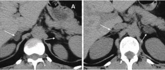
In the 21st century, undergoing a computer scan is a common process for patients and doctors. A CT scan is a clinical screening of a specific area of the body that is aimed at detecting abnormalities. It is used to establish a correct conclusion before surgery and to monitor the effectiveness of previously prescribed therapy. The device operates by generating minimal amounts of radioactive radiation, so it does not harm the patient.
What can you see on a CT scan?
This relatively new method of human condition testing, which dates back to the early 1970s, continues to be highly effective in a variety of situations. With the help of clinical equipment, doctors receive highly accurate and informative information about the patient’s current health. Radiation screening helps to diagnose a dangerous disease in the initial stages, which ensures timely administration of therapy and a speedy recovery. The concept of the abdominal cavity includes many sections. Using diagnostics, you can study the condition of the following areas of the body:
- gastrointestinal tract structures;
- stomach;
- intestines;
- liver and biliary tract;
- pancreas;
- spleen.
Additionally, CT is used to examine the retroperitoneal region, which includes:
- kidneys;
- ureter;
- adrenal glands;
- lymph nodes;
- urinary canals.
Using diagnostic technology, doctors can find out the condition of the bone structures, as well as the lower thoracic region of the spinal column.
When is a CT scan prescribed?
Typically, a computer examination of the presented department is prescribed by doctors in the following situations:
- in case of damage;
- in severe conditions;
- for painful sensations without obvious reasons;
- for chronic problems with the urinary system or digestion;
- in case of illness, jaundice;
- with the likelihood of the formation of neoplasms of a different nature.
Additionally, screening of the abdominal cavity is done before preparing for surgery, if it is necessary to decide on the need for surgical intervention, and to check the effectiveness of prescribed drugs for treatment.
On the search portal “Unified Appointment Center” you can book a place in any clinic in the capital in a few minutes. All you need to do is call the hotline to instantly request an appointment for an abdominal CT scan. The manager will inform you about the number of available places in the coming days at the desired medical center, conduct an informative consultation and inform you about current promotional offers. On the portal you can find all the necessary information about the city’s clinics, for example, prices, work schedules, information about doctors, as well as reviews from visitors.
Indications for examination
You can get a referral from your doctor for a computed tomography scan of the uterus and ovaries in the following cases::
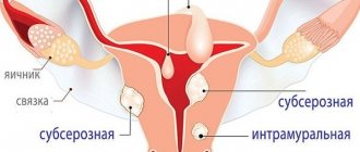
- benign and malignant neoplasms of the pelvic organs;
- injuries of the uterus, ovaries, pelvic bones;
- inflammatory diseases of the female reproductive system;
- sharp pain in the lower abdomen or pelvis, the cause of which cannot be established without additional research;
- examination of the pelvic organs before surgery;
- monitoring the effectiveness of the treatment and the persistence of remission (MRI after hysterectomy, after a course of radiation or chemotherapy).
Abdominal CT scan in women: what shows the disease
Using computer scanning, you can obtain verified information about the size and location of a specific organ, the condition of its structures, blood vessels and nerves. Doctors receive information about the ribs, sternum, lumbar region, and lower thoracic region of the spine. When performing a CT scan, the following changes can be seen in the images:
- the presence of internal hemorrhages;
- accumulation of fluid;
- foreign objects;
- parasitic cysts;
- inflammatory reactions;
- oncology;
- benign neoplasms;
- blood diseases;
- deformations in blood vessels such as thrombosis;
- disturbances in the functioning of the lymph nodes;
- the presence of hepatitis, cirrhosis and hepatosis;
- deformations of the structure of congenital organs;
- appearance of stones;
- formation of abscesses and other purulent anomalies;
- pinched nerve.
Using a tomogram, the doctor will be able to immediately study the problem, draw up a conclusion and the necessary treatment plan.
Lead tactics
1. Scope of surgery for a malignant ovarian tumor: extirpation of the uterus with appendages and removal of the greater omentum, i.e. removal of the cervix, uterus, appendages. The greater omentum is removed because in 18-20% of cases micrometastases are detected; the omentum is actively involved in the accumulation and production of ascitic fluid (especially in advanced stages).
2. Scope of surgical intervention for a benign process: adnexectomy (removal of appendages). During the operation, a careful examination of the internal lining of the cyst is performed (there may be malignant growths). During the operation, a rapid histological examination is performed.
Abdominal CT scan: what it shows in women and how it goes
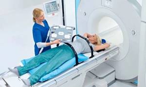
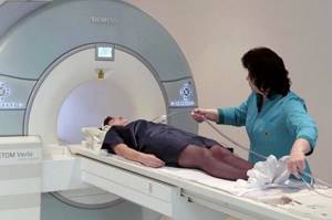
It is clear what can be seen using a diagnostic technique. Let's look at the stages of a scanning session. No special preparation is required before diagnosis. It is enough to remove metal jewelry and other metal objects. If there is a foreign object like an implant inside the body, be sure to inform the diagnostician about it. The step-by-step procedure for screening the abdominal cavity is as follows:
- The patient is placed on a mobile couch.
- If necessary, doctors fix the limbs to prevent accidental movements during the test.
- If prescribed, the patient is given a contrast agent.
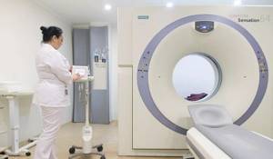
- The transforming table is pushed into the tomograph tunnel.
- Scanning begins.

The medical staff will carefully study the resulting images of a specific area in the adjacent room using a PC. The results are printed on a special film or sent by email. Also, answers describing the state of organs can be obtained by dumping them onto a portable storage device. On average, preparing the results takes about a couple of hours.
Transcript of research
The tomograph takes images of the area under study in series. Each of these images is a thin slice of the area under study, on which all contours and anatomical formations are clearly visible.
Decoding is carried out only by experienced specialists who determine from the images received:
- the presence of pathologies and its types;
- the organ in which the pathological process occurs;
- neoplasm with abnormalities.
As a result of the resulting description, a diagnosis is established. Sometimes, in order to accurately determine the diagnosis, a doctor needs to familiarize himself with the previous results of instrumental examinations, treatment performed and the opinions of other doctors.
Abdominal CT scan: contraindications
Based on reviews from patients who have previously undergone similar diagnostics, it becomes clear that the studies practically do not provoke any complications or other adverse consequences. However, doctors warn that testing is not suitable for everyone. CT scans of various organs have specific limitations:
- carrying a child to avoid radiation exposure to the unborn child;
- the patient’s inability to control his own movements, as the quality of the images will decrease;
- acute pain syndrome that will not allow the patient to remain motionless;
- obesity, since the devices are designed for a weight not exceeding 150 kg.
Children under five years of age are not allowed to be diagnosed, so as not to expose the growing body to radiation.
What can you eat before an abdominal CT scan?
Before visiting the diagnostician’s office, it is forbidden to eat for several hours before the appointment. The abdominal cavity should be examined on an empty stomach to prevent digestion of food from giving a false result. Doctors advise starting to adhere to a certain diet in a couple of days and reducing portion sizes. It is recommended to avoid foods that cause flatulence, as well as alcoholic beverages. It is forbidden to eat hard-to-digest foods that cause constipation.
Additionally, the timing of the appointment plays an important role when organs need to be examined:
- morning session - dinner should be low-calorie dishes;
- daytime - a light snack is allowed 5 hours before the diagnosis;
- evening - you can have breakfast, but you should skip lunch.
The doctor may prescribe a bowel cleanse to get more reliable information after the tomogram. In this case, the day before your appointment, you need to take a laxative prescribed by your doctor. You can book a place in any metropolitan clinic using the portal. Just call the hotline. The company manager will conduct an informative consultation for free, answer your questions, inform you about current promotions and make an appointment at a discount. Don’t put off the examination until later, find out about your health status in the coming days!
Abdominal CT with additional intensifier
Standard screening, where it is necessary to examine the organs of the represented section, is carried out without the use of a contrast agent. In some cases, doctors may write referrals for research using an additional amplifier, which improves the reliability of the results. The substance enters the bloodstream and colors inflamed areas, making them more visible in images.
This technique is prescribed to detect oncology or the process of metastasis, as well as a detailed study of blood flow. Angiographic tomography can detect the following disorders:
- aneurysms;
- blood clot formation;
- development of atherosclerosis;
- disorders that cause narrowing of vascular walls;
- tumors of various types;
- vascular damage;
- dissection of the aortic walls.
Keep in mind that CT scans can cause side effects, for example:
- increased body temperature;
- dizziness;
- nausea;
- general weakness;
- irritation in the injection area.
.These symptoms go away without the need for medication. If complications such as shortness of breath, urticaria, itching, swelling, or decreased blood pressure occur, which is evidence of an allergy to contrast, the patient is provided with appropriate assistance.
Lead tactics
If after 2 months the cyst does not disappear, then surgical intervention is necessary, which is explained primarily by oncological alertness. When formed on the ovary, it is much higher than with other tumor processes of the female genital area, for example, uterine fibroids.
The task of general gynecologists is to prevent the development of an oncological process at any cost, and this task today has been greatly facilitated by the advent of ultrasound and laparoscopy. If a cyst and not a tumor is clearly identified, then the operation is limited to cystectomy - removal of the cyst with a capsule (to prevent relapse). Laparoscopy makes it possible to remove the cyst without damaging healthy ovarian tissue, and with minimal intervention to remove the paraovarian cyst. If complications develop, surgical intervention is also indicated.
CT scan of the abdomen with contrast: contraindications
Due to the potential complications caused by the additional booster, there are specific restrictions for a certain group of people:
- allergy sufferers who cannot tolerate iodine;
- persons suffering from severe renal failure;
- patients with hyperthyroidism and other thyroid diseases.
Children who have not yet turned 12 years old are diagnosed only for certain indications upon referral from the attending physician. Breastfeeding mothers are also prohibited from screening, as the contrast can pass into breast milk and harm the baby.
If there are no restrictions, book your appointment now using the One-Stop Appointment Center. Ask all your questions by calling the hotline. The manager will conduct a consultation and help you choose a suitable clinic for a computer examination. Get a scan discount to get tested on a budget!

