Braces are prescribed to straighten the teeth and correct the bite. They have to be worn for quite a long time: from several months to 3 years. During this time, the person may become ill or need to undergo preventive testing. Many people believe that MRIs cannot be done with braces. Let's find out if this is so.
In this article
- What are braces and what are they?
- MRI and braces: which metals do not affect diagnosis
- Myths about MRI and braces
- Alternative to MRI with braces
Magnetic resonance imaging (MRI) is one of the safest and most effective diagnostic methods, which allows many diseases to be identified at an early stage. Its essence is to scan a particular part of the body by exposing it to a magnetic field. The cells of the body react to it in a certain way, which is read by the device and transmitted to the computer. Based on the images obtained, the doctor can not only identify the pathology, but also determine its location, degree and other data that is needed to make an accurate diagnosis and prescribe treatment. An MRI is prescribed during a routine examination or if the following diseases are suspected:
- malignant neoplasms;
- inflammatory processes;
- CNS disorders;
- pathologies of the spine, joints and internal organs.
In other words, using MRI you can scan the entire human body and detect almost any disorder. This is the main advantage of this method. However, its advantage is not only that it is informative, but also that it is safe. There is no ionizing radiation during the examination. MRI does not cause complications, even if performed with the introduction of a contrast agent.
Despite the effectiveness and safety of the technique, there are a number of contraindications to it, for example, the presence of a pacemaker, joint prostheses and other foreign objects in the patient’s body. This especially applies to metal products, which cause distortion of diagnostic data. Let's find out whether orthodontic braces are included in the list of such items, and whether it is possible to do an MRI with braces.
What are braces and what are they?
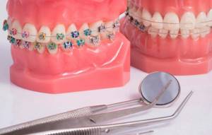
Braces are orthodontic structures that help correct bite pathologies. Treatment of dental diseases with their help is a rather lengthy process. Depending on the condition of the teeth, the patient’s age and other factors, it may take several years. During this time, the patient is not insured against other diseases that may require hardware diagnostics, including MRI.
Braces are made from various materials: metal, sapphire, ceramic or plastic. The arch of the structure, which passes through the clasps and exerts the main pressure on the dentition, is usually made of metal. As already noted, it can distort the results of the examination. For this reason, the doctor may prohibit an MRI if the patient wears braces. There is no direct contraindication to the study, but the information obtained during it cannot be trusted 100%.
However, refusal to conduct an MRI is justified only in cases where the head area is being examined. Other parts of the body can be scanned whether or not you have braces.
There are also types of metal that do not affect diagnostic results. Therefore, tomography of the brain or dentofacial apparatus can be carried out even if there are orthodontic structures in the patient’s oral cavity.
The influence of metal on the result
One of the main limitations to MRI is the presence of medical devices containing metal inside the body: for example, metal implants, crowns, pacemakers, artificial limbs and staples for fastening their fragments.
Metal elements cause a “magnetization” effect, which leads to distortion of the examination result, breakdown of devices, and the development of negative reactions from the body.
This contraindication does not apply to braces. You can undergo magnetic resonance imaging with this corrective device if you are examined:
- spine;
- limbs;
- abdominal or pelvic organs.
The presence of braces and other metal devices in the mouth contributes to the formation of interference in the operation of the device, which causes changes in diagnostic data. The tomograph's response is as follows:
- spots may appear on the photo;
- the image is unclear, blurry;
- the research result is distorted.
Similar results occur in cases where metal objects are exposed to a magnetic field during a diagnostic procedure. When studying parts of the body that do not contain any metal objects, the measurement accuracy will be high.
Important: undergoing an MRI does not pose a health risk. Patients with metal braces can undergo an examination of the entire body, except the head.
Therefore, if you have braces with metal parts, before undergoing the examination you need to find out from the orthodontist exactly what type of metal they are made of.
What should a person do if he accidentally swallows a brace or other structural element?
Read here whether you need to remove teeth when installing braces.
At this address https://orto-info.ru/sistemyi-vyiravnivaniya-zubov/breketyi/nalogovyiy-vyichet.html we will tell you how to get a tax deduction for installing braces.
MRI and braces: which metals do not affect diagnosis
Before your MRI procedure, tell your doctor what material your braces are made of. This information can be obtained from the orthodontist who installed the structure. The physicochemical properties of various metals differ significantly. Moreover, the reaction to the magnetic field of a particular metal product may be different.
Some of them, for example, titanium structures, do not react at all to the MRI machine and the magnetic waves it creates. Also, braces containing ferromagnets - substances that have spontaneous magnetization - will not interfere with the examination.
In any case, the procedure can be interrupted if the patient feels any discomfort. Before the examination, the doctor gives him a special device with which he can signal that he is not feeling well.
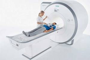
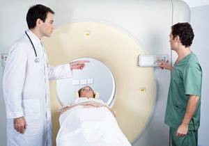
MRI machine
The device is equipped with a camera, which is placed in a special pipe. The patient is placed on a retractable table before he is immersed in a chamber - a tube. As soon as the examination begins, the image obtained by irradiating the patient with electromagnetic waves is immediately displayed on the monitor screen, which is located near the device itself. To obtain a clear image in the photograph, the patient is urged to lie still during the examination period (approximately 15-20 minutes). Using an MRI machine, tumors, pathologies and anomalies of internal organs are detected.
Myths about MRI and braces
Some people believe that MRI scans performed while there are metal objects in the body can cause electric shocks. Others believe that metal structures, such as braces, become red hot and leave severe burns on (or inside) the body. There is also an opinion that orthodontic brackets lose their functionality or begin to deteriorate under the influence of the magnetic field created by the MRI machine.

All these ideas about tomography are erroneous. The examination does not cause any complications, even if it is carried out while the patient has braces. It’s just that the exposed images obtained during such a study can be treated with skepticism. The doctor will not be able to make an accurate diagnosis and prescribe treatment. However, MRI cannot cause any harm to health. Moreover, it is not capable of damaging the orthodontic structure.
Common Misconceptions
Among patients themselves and on Internet forums, you can come across a lot of statements regarding the compatibility of MRI examination and braces.
Many of them are shocking with negative consequences for human health, others talk about obtaining a 100% inaccurate result. Much of the information is inaccurate and unsubstantiated.
The most common misconceptions are:
- During examination, the metal becomes quite hot and magnetized , causing the structure of the braces to collapse. In reality, nothing like this happens. Magnetic radiation does not heat up the correction device, and the magnetization effect is so small that it is not felt.
- Metal parts may damage tooth enamel during examination .
Modern braces are small in size and cannot move and injure the oral cavity during the diagnostic process. Only large structural elements made entirely of metal can become very hot, move and cause injury to the oral cavity. But today, in the manufacture of large parts, paramagnetic materials, which are passive in relation to the magnetic field, are usually used. - The result of the study will be greatly distorted .
This statement is only half accurate. Braces can affect the results and interfere with the diagnosis of only the part of the body in which they are installed. Therefore, if you perform an MRI of the head with an orthodontic system installed, the results will indeed be incorrect. In this case, radiography or angiography will be more informative. - During diagnosis, the patient feels electric shocks . In addition, they say that hot braces leave burns on the mucous membrane and soft tissues. In practice, not a single similar case has been recorded. The examination is absolutely painless.
Sometimes patients feel slight discomfort from lying on a hard surface for a long time, fatigue, and numbness due to being in one position for a long time. When a contrast agent is injected to confirm the diagnosis, you may feel cold during its administration.
Important! In isolated cases, there is a slight tingling sensation in the body, warmth in the examined area, slight dizziness, nausea, vomiting, and headache. These phenomena are considered in medicine to be a normal reaction of the body to the procedure.
The only negative consequence of MRI is a distortion of the actual picture of the patient’s health condition.
A physician will not be able to make an accurate diagnosis based on tomography data or select an acceptable treatment for a particular patient. The procedure does not pose any risk to the health, much less the life of the person being examined!
In the video, a specialist will express his opinion on the possibility of performing an MRI in the presence of braces.
Alternative to MRI with braces
So, are MRIs done with braces? This method for orthodontic braces is prescribed in the following cases:
- Tomography is performed below the head area;
- braces are made of titanium or contain ferromagnets;
- The orthodontic system does not contain metal parts.
If the patient needs to examine the brain or the dental system, and he has metal braces in his mouth that are not made of titanium and without ferromagnets, the doctor prescribes alternative examination methods, for example, CT - computed tomography.
Unlike MRI, which operates on the basis of magnetic waves, CT provides scanning by exposing the body to x-rays. Computed tomography is prescribed for bruises and fractures, dental injuries, bone diseases, internal bleeding and other pathologies. The effectiveness of MRI and CT may vary depending on which organ is being examined and what disease is suspected.
When a patient requires a detailed examination of the jaw, he is usually prescribed a CT scan, since the images obtained using MRI will be overexposed in the places where the braces are. Computed tomography, no less effective than magnetic resonance imaging, allows us to examine the hard tissues of the oral cavity. However, if it is necessary to assess the condition of the soft tissues in the area of the dental system, the doctor may suggest temporarily removing the orthodontic structure. In this case, additional consultation with an orthodontist will be required.
The essence of the method: what are the concerns of patients
The diagnostic method is based on the phenomenon of nuclear magnetic resonance: a living organism consists of cells, cells are made of molecules, and they are made of atoms of chemical elements. Inside every atom there is a positively charged nucleus. If you place it in a high-intensity magnetic field, it reacts to it, which is manifested by the transition of the atom to an excited state.
All human organs consist of different tissues, and they are made of different molecules and atoms. And each type of chemical element reacts to a magnetic field in a unique way. Reading their reaction to the influence of the magnetic field of the tomograph, the device scans the area under study. The computer helps process data by converting the received information into a two-dimensional or three-dimensional picture.
The concern of orthodontic patients is that some metals can become magnetized, and the magnetic field inside the scanner is very strong. What can happen if you perform a diagnostic procedure with braces or other metal objects?
Reviews
People's reactions to treatment and diagnostic procedures vary greatly from person to person. The same procedure can leave different impressions and sensations in different people.
If you had to undergo an MRI while wearing braces, tell us which organ was diagnosed and what your impressions of this procedure were. This can be done by leaving your review at the bottom of the page.
If you find an error, please select a piece of text and press Ctrl+Enter.
Tags braces diagnostics
Did you like the article? stay tuned
Previous article
Restoring chewing function using transosseous implantation
Next article
Tactics for correcting bite defects using ALF technology
What happens if TMJ dysfunction is not treated?
If the dysfunction is not treated, the compensatory capabilities of the body may sooner or later be exhausted, the symptoms will worsen, the pathology will begin to progress, causing greater discomfort (sometimes for several years), thereby affecting the deterioration of the function of the dental system.
In order to try to prevent this and carry out treatment taking into account the individual characteristics of the structure and functioning of the temporomandibular joints, patients are usually offered the following approach.
Can someone with braces have a CT scan of their teeth, head or neck?
Here, again, it all depends on the material of your structure. There will be no problems with a polymer alloy, sapphire or ceramic, but metal is again undesirable. X-rays themselves have no effect on it. It’s just that any metal objects degrade the quality of the pictures. For example, a dental CT scan will most likely be done in vain. Neither the upper nor the lower jaw can be seen. The images will show blurry spots and streaks that may interfere with diagnosis. As a result, time and money will be wasted (and the price of a dental CT scan in some clinics is quite high). However, the diagnostician will be able to discern the maxillary sinuses or neck vessels. Therefore, in some cases, computed tomography is still performed.
Well, if there is no metal in the braces, there are no obstacles at all. Feel free to sign up for a CT screening. And for those who have problems with their teeth while wearing braces, even a panoramic photo of the jaw is possible. Of course, the brackets will slightly obscure the view of the dental tissue, but otherwise the picture will be objective.
Will I have to remove my braces for the examination?
Most likely you won't have to. Doctors will try to do everything so as not to damage the orthodontist’s work and not disrupt the progress of dental treatment. Removing the structure is allowed only in emergency situations when it comes to saving life and health. In other cases, they will simply select the appropriate diagnosis for you. The doctor will weigh all examination options and choose the best one.
As you can see, the owner of braces on his teeth, in fact, has nothing to fear. The best option is to learn everything about the properties of their material before installing braces.
If for health reasons you often go to the hospital or undergo examinations, it makes sense to immediately order a brace system that will not cause problems during future procedures. For example, ceramic braces are completely inert to the magnetic field and X-radiation, and at the same time cope excellently with the tasks of correcting the bite. And here’s another fact to note for everyone who is going to correct their bite: metals can be ferromagnetic and paramagnetic. The first - steel, nickel or mixtures thereof - can distort diagnostic images. The latter - for example, titanium - do not affect the images in any way. Therefore, if it is absolutely impossible to do without metal in braces or retainers, then let it be titanium.
Occlusive therapy for TMJ dysfunction
After diagnosis, the patient is scheduled for an appointment with the orthodontist to determine the central relationship of the jaws (“true” position of the lower jaw, the position in which your joint and chewing muscles will be most comfortable).
In order to more accurately establish and fix this position, an occlusal splint (splint) will be individually made for the patient from a special plastic, which is erased as it is worn. The splint must be worn constantly (sleeping, talking, eating in it if possible) - this is the meaning of occlusion therapy, which will help the joint and masticatory muscles rebuild into the most comfortable functional state.
Cleaning and caring for the splint is very simple - after eating (as well as while brushing your teeth), brush with a soft brush with toothpaste or soap.
What may be the symptoms of TMJ dysfunction?
- Tenderness or pain in the area of one or both TMJs at rest or when opening the mouth.
- Crunching, clicking, crepitation and other noises in the area of one or both TMJs when opening the mouth.
- History of TMJ injuries (previous), incl. dislocation, subluxation, chronic subluxation.
- Restrictions in the mobility of the TMJ, restrictions in opening the mouth.
- Excessive tone of the masticatory muscles, bruxism (“grinding” of teeth in sleep, at rest).
- Asymmetry of the chin, lips, lip frenulum, asymmetry of mouth opening, S-shaped opening.
- Suspicion of a forced position of the lower jaw.
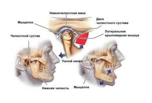
Structure of the TMJ
The presence of one or more of the above symptoms may indicate TMJ dysfunction.
Traditional orthodontic treatment does not address TMJ dysfunction. During orthodontic treatment, the severity of dysfunction may not change, decrease or increase. At the moment, in the world scientific orthodontic literature there is no convincing data on the connection between orthodontic treatment and the condition of TMJ. Deterioration of the joint after treatment may have nothing to do with this treatment.
Note! Even in the absence of visible clinical manifestations of joint dysfunction, hidden disorders may occur that require special diagnostics to identify them.
If there is a forced incorrect position of the lower jaw, its position may change during the treatment process with changes and complication of the treatment plan (the need to remove individual teeth, increasing the duration of treatment). A reliably forced position cannot be diagnosed by traditional orthodontic methods; to verify its presence, as a rule, a special analysis is required (manual functional analysis, determination of the central relationship of the jaws), the use of a special articular splint for a period of several months, which, however, does not give 100 % guarantees.
To conduct a detailed articular diagnosis, explain the specifics of your case, and further manufacture an articular splint, you can make an appointment with an orthodontist who deals with the issue of TMJ dysfunction.
TMJ dysfunction is a chronic condition that can be compensated, but not cured (i.e., it is possible to eliminate symptoms, however, pathological changes in the joints, if they have already occurred, will most likely persist).