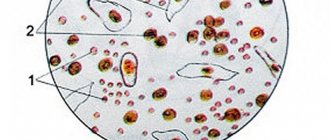
Classification
Three types of epithelial cells can be found in urine:
- flat;
- transitional;
- renal.
Flat
The most common type of epithelium found in urine tests. Its cells line the urethra, and an excess of this value usually occurs with various types of inflammation of this organ (urethritis).
Flat epithelium
Transition
This epithelium lines the walls of the bladder, renal pelvis, and ureters. May be present in urine, but only in a single form. An increase in its content indicates dangerous diseases such as pyelonephritis, cystitis, and urolithiasis.
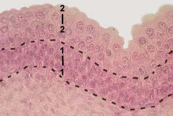
Stratified transitional epithelium
Renal
This epithelium lines the kidney tubules and should not be present at all in the urine of a healthy adult. However, it can be found in newborns - in quantities from one to five units, no more. An excess is considered an indication of an inflammatory process in the renal parenchyma.
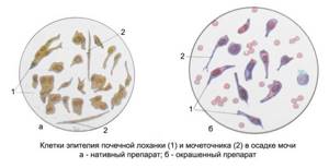
Renal epithelium
Types of epithelial cells
There are several types of epithelial tissue:
- Single layer
. It can be divided into several more: flat, edged, cylindrical and cubic; - Multilayer
. This type includes flat keratinized or non-keratinized, glandular, transitional and, quite rarely, cubic and cylindrical.
Doctors and various laboratories most often use the term “squamous epithelium”. Using an analysis of the quantitative content of epithelial tissue in a smear or urine, the doctor can draw appropriate conclusions about the patient’s health and give the necessary recommendations for treatment. There are certain standards; in a healthy person, the content of squamous epithelium is within certain permitted values. A significant increase in indicators indicates a progressive course of the inflammatory process, which means that prompt therapeutic intervention is required.
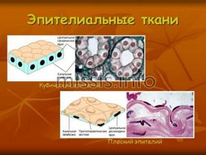
If squamous epithelium is found in the urine, what does it mean?
If squamous epithelial cells are found in large quantities in urine during analysis, there may be several reasons for this phenomenon. But one way or another, all causes are usually associated with inflammatory pathologies in the body.
Next, we will consider the most common causes of the appearance of squamous epithelium in the urine.
- Cystitis . This pathology often occurs in both adults and children. Inflammation is caused by bacteria, viruses or fungi - and you should get rid of the disease only after an accurate diagnosis.
- Kidney problems . Various inflammatory diseases of the kidneys are a very likely cause of the appearance of squamous epithelium in the urine. The most common diseases in this case include: bilateral kidney damage;
- renal failure at various stages.
If an excess of the indicator is associated with inflammatory diseases of the genitourinary system, the following symptoms will most likely be present:
- decreased amount of urine and decreased frequency of urination;
- cloudy urine with a specific pungent odor;
- lower abdominal pain;
- painful urination with frequent urge;
- bloody spots in the urine.
The presence of squamous epithelium in the urine can sometimes mean inappropriate (excessive dosage) taking medications. And in men, this fact can mean inflammation of the prostate gland.
Important: if squamous epithelium is found in minimal quantities, this fact can be considered normal. Therefore, the presence of epithelial cells in the urine does not always mean illness.
Flat epithelium in urine: norms and causes of increase
3.1 Leukoplakia of the bladder
Leukoplakia of the bladder and vesical triangle (squamous metaplasia) in many cases is practically asymptomatic for several years. In such patients, sexually transmitted infections (chlamydia, ureaplasma, gonococci, Trichomonas, herpes, mycoplasma, Escherichia, Gardnerella) are often detected in smears. In addition to high values of squamous epithelium and horny scales in the urine, there may be the following signs of the disease:
- the occurrence of a sudden strong urge to urinate;
- prolonged pain in the urethra or pelvis;
- frequent urination;
- abnormal location of the external urethral opening;
- increased symptoms upon initiation of sexual activity or change of sexual partner.
Often sick women are diagnosed with cystitis, although the cause of these disorders is the presence of another pathology. In 60-96% of patients, cystoscopy reveals leukoplakia located in the bladder neck and bladder triangle. This disease is a pathological process in the mucosa, characterized by dysfunction of the squamous epithelium and the occurrence of keratinization, which is absent normally. Many experts consider this condition as precancerous. The reasons for the development of leukoplakia are the following:
- chronic infectious process in the urogenital tract;
- prolonged irritation of the mucous membrane with chemicals, medications, catheters;
- lack of vitamin A;
- genitourinary schistosomiasis (irritation of the mucous membrane from contact with sharp spines on the eggs of a parasitic worm), a complication of which is bladder cancer.
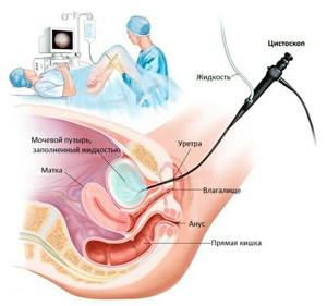
Cystoscopy
Diagnosis of the disease is carried out using cystoscopy (insertion of an endoscope into the bladder through the urethra) and biopsy of epithelial tissue. Cytological examination of urine sediment reveals degenerative squamous epithelial cells. In later stages, red blood cells and a significant number of leukocytes appear. Complications of leukoplakia are:
- malignancy of the process;
- the development of renal failure due to loss of elasticity of the bladder walls;
- formation of ulcers and polyps;
- development of chronic urethritis and cystitis.
Areas of stratified squamous epithelium without keratinization are also detected in healthy women (up to 80% of cases), especially during childbearing age. Cells in lesions with altered tissue resemble vaginal epithelium. This condition in medicine is considered a variant of the norm, and it is associated with hormonal changes. An acceleration of this process can occur in a pregnant woman due to the increased secretion of estrogen during gestation. Therefore, it is important to recognize this disease in time and carry out its treatment, which consists of the following:
- use of antibacterial and antiparasitic agents;
- antiviral therapy (if herpes is detected);
- local treatment by introducing natural or synthetic analogues of glycosaminoglycans into the bladder (Heparin, hyaluronic acid, chondroitin sulfate) to restore the mucin layer;
- transurethral surgery in the absence of effect from conservative therapy (electrovaporization or electroresection).
Normal squamous epithelium in urine
To find out whether a specific indicator of the content of epithelial cells in urine is normal, special tables have been created.
Sometimes it is necessary to slightly underestimate/overestimate the norms, based on the characteristics of a specific diagnosis and the patient’s age; however, in most cases, the indicators fall within the established norms.

Table
Normal epithelium in men
As for men, in this case squamous epithelium in the urine is undesirable in quantities of more than 5 units within the foreseeable limit of the microscope eyepiece.
Normal squamous epithelium in urine in women
When examining female biological material, it is necessary to make sure that no epithelial cells are noticed at all. Only single cells are allowed.
However, when a woman is expecting a child, the norms of epithelium in her urine can be increased to 5 units. As for children, the number of squamous epithelial cells in their urine should not be more than three units.
If a urine test shows the presence of squamous epithelium, then in most cases the number of cells is insignificant. If up to three cells are present, this is always considered the norm: for women, men, and children.
Norms of squamous epithelium in urine by age:
- for children in infancy, the norm is from 0 to 5 units, a pathological indicator is considered to be from 6 to 12 units;
- for a child in the first year of life, the norm is 0.3 units, pathology is 4-6 units;
- for a child from one to 18 years of age, the norm is 0.3 units, pathology is 4-10 units;
- for adults from 18 to 50 years old, the norm is 0-3 units, pathology - 4-10 units;
- after 50 years of age, the norm is 0-6 units, pathology - 7-12 units;
- for women during pregnancy, the norm is 0-6 units, pathology 7-12 units;
- during antibiotic treatment, the norm is 0-5 units, pathology is 6-12 units.
As can be seen from the table below, the norm of squamous epithelium for all patients is acceptable up to 5 units. As for other types - renal and transitional epithelium, ideally they should not be in the urine at all.

Click on the table to enlarge
Normal amount of epithelium in urine
Even completely healthy people have squamous epithelium in their urine. The norm for women is slightly higher than for men, but, regardless of gender, the normal number of cells is calculated in the units.
The same indicator of the number of squamous epithelial cells in urine may indicate the healthy condition of a woman and developing prostatitis or urethritis in a man.
In a person who does not have any diseases, up to 10 units
.
Exceeding this norm indicates an inflammatory process occurring in the genitourinary system. Significant excesses of indicators can be detected in the following diseases:
- cystitis;

- prostatitis;
- urethritis;
- nephrosis;
- acute nephritis.

To determine the number of cells, a general analysis is taken, in which several parameters of urine are studied at once, which include the following: leukocytes, erythrocytes, casts, bacteria, protein, glucose. However, it is not advisable to conduct a study only for the presence of epithelium in the urine, since the indicator is rather weak and does not always completely indicate the presence of the disease. Therefore, the analysis is carried out comprehensively, with the study of various characteristics.
Reference values for general urine analysis.
| Color | Yellow-straw |
| Smell | Unsharp |
| Transparency | Transparent, not cloudy |
| Reaction | pH 4 to 7 |
| Protein | Maximum 0.033g/l |
| Density | From 1012 to 1022 g/l |
| Glucose | Maximum 0.8 mmol/l |
| Ketone bodies | — |
| Hemoglobin | — |
| Leukocytes | Until 6 |
| Red blood cells | Until 3 |
| Epithelium | Within 10 |
| Cylinders | Single |
| Mushrooms | — |
| Bacteria | — |
| salt | — |
What to do if there is increased squamous epithelium in the urine?

Excessive levels of squamous epithelium in urine almost always mean the presence of inflammatory disease of the kidneys or urinary tract.
Therefore, in most cases, discomfort occurs when urinating, and other negative symptoms appear.
If such alarming symptoms appear, you should definitely visit a doctor and get the necessary tests.
Treatment should be prescribed only by a doctor after a thorough examination, diagnosis of the biomaterial and the body as a whole.
Usually, if squamous epithelium is detected in the urine in quantities exceeding the norm, antibiotics are prescribed.
Treatment with antibiotics
The following drugs are often used:
Cefazolin

Clarithromycin Ceftriaxone

Azithromycin
In addition, it is necessary to take immunomodulators and vitamins, since most cases of this symptom are accompanied by reduced immunity and weakening of the body as a whole.
If inflammation of the bladder occurs, antibiotics are also used:
Ofloxacin Levoflaxocin Bactrim

Furadonin
In addition, non-steroidal anti-inflammatory medications are added:
Canephron

Cyston

Phytolysin
The standard course of treatment is 10 days. However, depending on the severity of the disease and the age of the patient, other important factors, the duration of the course can be changed by the attending physician in one direction or another.
If squamous epithelium appears in the urine as a result of chronic urethritis, local antiseptic therapy is necessary. In this case, the drugs are administered directly into the urethra using the drip method.
If the problem is due to renal nephropathy, you should stop taking medications that may cause this disease. However, only a doctor should prescribe or exclude any medications.
For renal nephropathy - and the disease is quite serious - treatment is carried out with the following drugs:
Prednisone
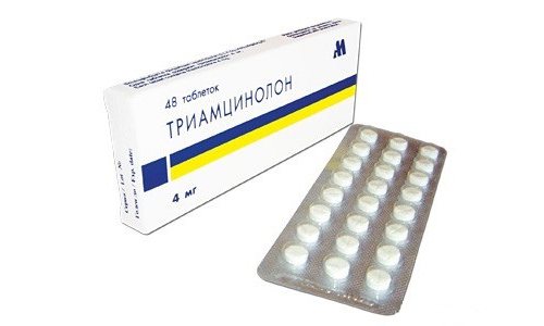
Triacinolone
For the period of treatment, a gentle diet is recommended, excluding fried and spicy foods. It is highly advisable to drink plenty of fluids, but with the exception of alcohol and coffee.
Physical therapy will also be useful - exercises should also be prescribed by a doctor. And simple home methods to strengthen the immune system will benefit not only the genitourinary system, but the entire body.
Attention: treatment will be most successful if squamous epithelium in the urine is detected as early as possible.
Video
Therapeutic procedures
The most commonly diagnosed disease of the female reproductive system is vaginitis.
. It is caused by various fungi, bacteria, parasites, and a decrease in natural hormonal support. Its treatment is carried out using antibacterial therapy, antifungal agents, depending on the type of causative agent of the inflammatory process.
Failure to go to the clinic in a timely manner can aggravate the problem, because as the infection progresses, it rises higher and affects the uterus.

Urethritis is one of the most common diseases of the genitourinary system. The cause of inflammation can be chlamydia, herpes, cytomegalovirus.
It is unacceptable to use any medications without medical supervision.
Treatment is most often carried out with the help of antibacterial drugs if the pathogen is infectious in nature. Non-infectious urethritis is treated with special substances that relieve irritation of the mucous membranes. After restoration of the normal amount of epithelium and other indicators, the patient’s condition stabilizes.
Transitional epithelium in urine
The epithelium, called transitional, lines the mucous membrane of the following organs:
- renal pelvis;
- bladder;
- ureters;
- in men - prostate ducts (large);
- the upper portion of the urethra.
In principle, transitional epithelial cells should not be found in urine. Only rare single copies are allowed, maximum 3 units.

Transitional epithelium
If the norm is exceeded, we can talk about the following diseases:
- pyelonephritis;
- cystitis;
- kidney stone disease.
Renal epithelium in urine
According to medical standards, renal epithelium should, in principle, be absent in urine. If cells are still found, this may indicate the development of kidney diseases such as:
- damage to the parenchyma;
- pyelonephritis;
- glomerulonephritis, etc.
Note that glomerulonephritis develops slowly and may not manifest itself for many years. And it is possible to detect renal epithelium in the urine, indicating this disease, only when the disease is already severely expressed.
The following symptoms are likely to occur with this pathology:
- dark and foamy urine;
- swelling of the extremities (especially legs);
- arterial hypertension;
- pain in the kidney area.
- Often the norms of renal epithelium in the urine are exceeded after kidney transplant operations, and if the excess is significant, we can talk about rejection of the transplanted organ by the patient's body.
- And sometimes the presence of renal epithelium in the urine can be evidence of such a serious disease as diabetes mellitus - with ischemic kidney damage associated with this disease.
- A problem also arises in the case of poisoning or intoxication of the body.
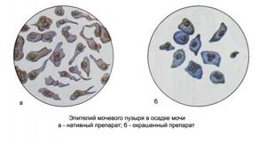
In addition to the above, the renal type of epithelium can be found in urine with congenital anomalies in the structure of the excretory organs. But in this case, the problem is diagnosed immediately after the birth of the child, and it cannot arise out of the blue during life.
Excess of squamous epithelium in urine during pregnancy
While expecting a child, a woman may well have squamous epithelium in her urine - and in most cases this fact is the norm (if it does not exceed 5 units).
The fact is that during pregnancy the genitourinary organs work in an enhanced mode, so frequent urination against the background of active exfoliation of the epithelial mucous layer is common.
In addition, the growing uterus compresses the genitourinary organs, displacing them, which also leads to more active desquamation of the epithelium.

The normal level of squamous epithelium during pregnancy is no more than 5 units
If the level of epithelium in the urine is more than 5 units, the woman is then under medical supervision: it is important to protect the health of the patient herself, and also not to forget about the health of the fetus.
Flat epithelium in the urine of a child
Cells of desquamated epithelium are sometimes found even in the urine of newborn babies. And this is considered quite normal, since when a child is born, the child’s body adapts to new conditions.
Attention: in almost all healthy newborns in the first two weeks of life, the level of epithelium in the urine is twice as high.
Analysis for squamous epithelium in a child.
The norm is no more than 3 units. On average, the presence of epithelium in a child’s urine should not exceed three units - and this is the norm for children of any age.
If the amount is exceeded, this may indicate the same problems with the kidneys and genitourinary organs as in adults.
A sharp excess of this indicator is especially dangerous for a child.
The most common causes of an excess of epithelial cells in the urine of children are the following diseases:
- inflammatory pathologies - cystitis and urethritis;
- inflammatory processes in the genital organs;
- kidney diseases;
- slow blood circulation in the pelvis;
- urolithiasis disease;
- neurological diseases accompanying urinary retention;
- reflux of urine (reverse movement);
- spasmodic pathologies of the genitourinary organs;
- salt deposits;
- purulent inflammatory processes, poisoning, intoxication.
Factors provocateurs
The appearance of an increased amount of epithelium in children does not happen out of nowhere.
The following factors provoke this phenomenon.
- Genetic. Occurs if one of the parents (usually the mother) suffered from similar genitourinary ailments.
- A pathologically passing, complicated pregnancy is also one of the powerful negative factors.
- If a child was born premature, this significantly increases the risk of various pathologies, including those associated with the kidneys and genitourinary system.
- With asphyxia (suffocation by the umbilical cord) during childbirth, the child is born with poor health, which also provokes various abnormalities.
- Developmental defects, especially congenital ones, greatly influence the occurrence of health problems throughout the body.
In addition to the above, infections that the child has suffered or that have not been treated can provoke the appearance of an increased amount of epithelium in the urine:
- furunculosis;
- angina;
- tonsillitis, etc.
Sometimes (and in the best case), an excess level of epithelium in children is observed due to incorrect, not very thorough preparation for the test: lack of disinfection of the container, hygiene of the genitals.
Normal amount of epithelium in the smear
To conduct a general clinical examination, material is taken from three zones
: from the outside of the cervix, from the urethra and cervical canal.
The purpose of this study is to identify and prevent inflammatory processes in the organs of the woman’s reproductive system. Indications for taking material are the following symptoms:
- suspicious, specific discharge;
- painful cramps in the lower abdomen not associated with menstruation;
- change in the color of the external genitalia;
- severe itching of the genitals.

A smear of the vaginal secretion flora must be taken from all women planning pregnancy in the near future, pregnant women and taking antibacterial drugs, antitumor agents and corticosteroids for a long time, as they reduce the protective functions of the immune system.
An ovarian cyst is a small, fluid-filled formation. It appears from the follicle. How dangerous cysts are, whether they need to be treated, and if so, how exactly, you will find out further. Read more in the article: “ovarian cysts in women.”
To obtain more objective results, it is necessary to exclude the use of any drugs used vaginally for 3 days, and to exclude sexual intercourse for 24 hours, otherwise the epithelium in the smear may be significantly overestimated.
The norm for women of epithelial cells in a smear on different days may differ, depending on the menstrual cycle.
Detection of a higher than normal number of cells in a smear may indicate a possible pathological process of an inflammatory nature (vaginitis, cervicitis, urethritis). While its insignificant amount indicates insufficient production of the necessary sex hormones.
Unpleasant sensations in an intimate place in women are a fairly common phenomenon that many ladies try to deal with on their own. Such signs may indicate an allergic reaction to the food product. Read more in the article: “discomfort in intimate places in women.”

Table for visual interpretation of analyses.
| Indicators | V | C | U |
| Epithelium is flat | From 5 to 10 | From 5 to 10 | From 5 to 10 |
| Leukocytes | 0 to 10 | From 0 to 30 | 0 to 5 |
| Gonococci | — | — | — |
| Trichomonas vaginalis | — | — | — |
| Key cells | — | — | — |
| Yeast-like fungi | — | — | — |
| Slime | Moderate content | Moderate content |
The pregnancy period is associated with hormonal changes that increase the risk of developing various pathological processes in the vagina. During this period, an analysis of the microflora in the birth canal is required at least three times throughout the pregnancy. If there are complaints and other indications, additional research is carried out.

Urinalysis to detect squamous epithelium
To determine the content of squamous epithelium, you will have to undergo a general urine test. It is necessary to approach this procedure competently and knowledgeably - the reliability of the analyzes is extremely important.

A general urine test for squamous epithelium should be taken in the morning and on an empty stomach.
The first step is to purchase a urine container. You should not use baby food jars and other household containers: even thoroughly washed and boiled, these containers can retain harmful microorganisms on their surface.
We advise you to purchase a special container at the pharmacy: this container is already properly sterilized and completely sealed. The cost is on average 100-200 Russian rubles.
- Two days before the donation procedure, you should not take diuretic medications, as well as medications containing nitrofuran compounds. It is highly undesirable to consume foods with coloring properties the day before testing: beets, citrus fruits, carrots.
- Before collecting urine, it is necessary to carry out hygienic procedures so that particles of mucus and fluid from the external genitalia do not get into the container. Women are advised to use a tampon to block vaginal discharge. And if a woman is menstruating, it is generally better not to donate urine during this period. The likelihood of the presence of excess impurities is too high.
- You need to collect urine immediately before you donate it, that is, usually in the morning. You should not carry out this procedure in the evening, since by the morning the biomaterial will no longer be fresh, and the results of the study will not be able to fully reflect the actual state of affairs. It is optimal to transfer the container to the laboratory no later than an hour or two after collecting the biomaterial.
Attention: there is no need to strain a whole jar/container of urine for analysis. The optimal amount is 100 ml, which is about half a glass.
Modern pharmacology can offer an independent method of examining urine. In pharmacies you can find express strips for sale, with which you can easily and quickly do a general urine test at home.
But in any case, only a doctor should make a diagnosis and prescribe treatment.
Diagnostic features
Determining which epithelium is present in urine and in what quantity is not an easy task, and requires attention, perseverance, certain knowledge, and experience from the laboratory assistant. And if previously cell counting was done “manually”, now there are instruments and programs that allow analysis to be done without any human intervention at all. This significantly improves the accuracy of the results.
If the indicator is significantly exceeded, a repeat analysis is usually prescribed before making a final diagnosis. The fact is that the first result sometimes turns out to be incorrect due to the patient’s failure to comply with hygiene rules.
So, the content of squamous epithelium in urine should not exceed 3-5 units at any age and condition - the norm applies to both men and women. If the analysis shows that this indicator is exceeded, it is necessary, under the guidance of a doctor, to identify the cause and carry out a course of treatment.
The sooner the excess is established and, accordingly, the diagnosis is made, the greater the chances for a speedy and successful recovery.
“Flora smear”: what will the analysis tell you?
The “Flora Smear” study refers to a microscopic type of analysis. The test is prescribed if an infection of the reproductive organs is suspected. To carry out the analysis, you need to obtain a smear. In men, the biotope is the urethra, in women - the cervical canal, vagina, urethra. Based on the location of the pathological process, the doctor can direct the woman to take a smear only from the vagina or cervical canal. However, more often all three loci are required at once.
| Taking a smear is a painless and safe procedure. All pregnant women also undergo a smear test at least three times: the first visit, 30 and 36 weeks, subject to a normal pregnancy. The resulting smear is transferred to glass, dried and stained, after which a laboratory doctor examines the preparation under a microscope and describes it. The result form contains information about microflora, leukocytes, epithelial cells and pathological agents. |
Microflora
During the analysis, the doctor describes its type and abundance. Normally, rod flora should predominate. The rods are representatives of lactobacilli and maintain a certain pH level. The main role of the sticks is to protect the vagina from pathogens and stimulate local immune reactions. When other bacteria, such as cocci, are present in the smear, the number of rods decreases. This condition indicates dysbiosis and requires mandatory correction.
Leukocytes
Leukocytes in a smear indicate the presence of an inflammatory reaction. The number of leukocytes in the “results” column is indicated with a hyphen. The first and second digits correspond to the minimum and maximum numbers found in the microscope's fields of view. Each locus has its own reference indicators. In the cervical canal no more than 25 cells per field of view are allowed, in the urethra - no more than 5, in the vagina - no more than 10.
An increase in the absolute number of leukocytes indicates inflammation, so it is necessary to consult a gynecologist or urologist.
Epithelium
Epithelial cells in the preparation are arranged in layers or scattered in the form of individual cells. In a smear their number should be single. In the case of an increase in the number of epithelial cells, the doctor indicates: moderately means that there are up to 10 epithelial cells in one field of view, a lot - up to 20, abundantly - over 25.
Changes in the size and morphology of epithelial cells may indirectly indicate an inflammatory reaction or hormonal problems. Epithelial cells covered over the entire surface with cocci are called “key cells.” Their detection in a smear is a morphological criterion of bacterial vaginosis.
Slime _
The values “little”, “moderate”, “absent” are a variant of the norm. If mucus is present in larger quantities, then this is a sign of dysbiotic disorders in the vagina.
Pathological elements
In a smear, the doctor can identify the mycelium and spores of yeast-like fungi, trichomonas, actinomycetes, sperm, etc. But he will not be able to determine the exact species of the pathogen due to the limitations of the method. For example, in the human urogenital tract there are several representatives of the genus Neisseriaceae, which are similar to each other, so it is not possible to distinguish them in a standard microscope.
The diagnostic sensitivity of microscopy in diagnosing gonococcal infection in Russia is 30%, and trichomonas infection - 44–68%. Microscopy is descriptive; to accurately determine the type of pathogen, it is necessary to use molecular biological methods (PCR, NASBA) and the cultural method.
