Endoscopy of the ENT organs is one of the most effective examination methods, which allows the doctor to examine hard-to-reach areas of the nasopharynx, larynx, and ear.
An endoscopic examination is carried out by an ENT specialist if it is necessary to clarify the diagnosis. The following symptoms may be indications for examination using an endoscope:
- difficulty breathing through the nose,
- nosebleeds,
- purulent discharge,
- headache in the face,
- decreased sense of smell,
- hoarseness, sore throat,
- itching/burning in the ear,
- snore,
- the possibility of having a foreign object in the throat, sinuses, or ear.
The appointment is conducted by otolaryngologists
Liliya Sagidullovna Lankina is an otorhinolaryngologist (ENT), of the highest qualification category. She graduated with honors from the Krasnoyarsk State Medical Academy in 2003.
Ask a question Read more...
Sergeev Fedor Yuryevich is an otorhinolaryngologist with 17 years of experience, the highest category. Graduated from Krasnoyarsk State Medical University named after. prof. V. F. Voino-Yasenetsky Ministry of Health of Russia.
Ask a question Read more...
Malashkovets Alexander Sergeevich is an otorhinolaryngologist (ENT), graduated from Krasnoyarsk State Medical University named after Professor V.F. Voino-Yasenetsky in 2017.
Ask a question Read more...
How is endoscopy performed?
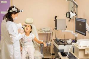
Endoscopic examination is not a complicated or painful procedure. It only takes a few minutes. To make the child comfortable and not afraid of the upcoming manipulations, we first explain to the little patient what will happen now. Our doctors have extensive experience working with children and know how to find an approach to each child.
Endoscopic examination proceeds as follows:
- The child is placed in a chair, he must throw his head back.
- The doctor applies a painkiller to the device and injects it into the throat or nasal passage. The endoscope is equipped with a video camera, lighting, and a thin tube. The monitor displays a picture of the area being examined. The doctor can move the instrument and direct it to the desired area.
- If necessary, simultaneously with a visual examination, a sample is taken and sent for analysis.
- At the end of the procedure, the endoscope is carefully removed from the cavity, the data obtained is recorded and printed.
Endoscopy can be performed on a child at any age. If the baby requires it, the procedure is carried out in a lying position with the head firmly fixed. If the child is small, then an adult can hold him.
What is an endoscope?
The endoscope is a thin optical tube, which the doctor carefully inserts into the nasal cavity, ear canal or leads to the entrance to the larynx; if necessary, local anesthesia is used - vasoconstrictor drugs in the form of drops or spray.
With the help of optics, the image is magnified several times. The video camera transmits the resulting image to the monitor. Thus, the patient himself or the parents of the child being examined can, together with the doctor, observe the diagnostic process
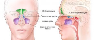
Endoscopic examination of the sinuses
Nasal endoscopy is recommended for the following symptoms:
- rhinitis, adenoiditis, other diseases of the nasal cavity,
- persistent runny nose,
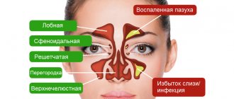
- frontal sinusitis, sinusitis, other inflammatory phenomena in the sinuses,
- complete/partial loss of smell, hearing, taste,
- frequent headaches for an unknown reason,
- frequent nosebleeds,
- trauma to the face, nose,
- suspicion of a deviated nasal septum or other abnormal development,
- heavy snoring,
- allergy.
The examination helps to identify developmental abnormalities, thinning of the mucosa, any neoplasm, polyps, enlarged adenoids, and ulcers. No special preparation is required for the procedure. The day before, it is better to avoid inhalations, ointments, and nasal drops.
Using a video endoscope, an otolaryngologist can:
• examine the nasal cavity and nasopharynx;
- consider adenoid vegetations in children, which is extremely important for determining indications for further treatment;
- determine the presence of small polyps that are invisible during normal examination;
- determine the presence of pathological secretion;
- evaluate the shape of the nasal turbinates, their altered middle and posterior sections;
- conduct an examination of the oral cavity and pharynx in places that are difficult to reach during normal examination;
- identify or suspect neoplasms in the initial stages;
- examine all elements of the larynx, assess the integrity and motor activity of the vocal apparatus.
For both adults and children. Children are allowed to undergo ENT endoscopy from the age of three.
Endoscopic examination of the larynx
The examination is carried out in the same way, only the instrument is inserted not into the nasal passage, but into the patient’s throat. The doctor can examine any part of the larynx and identify the disease at an early stage.
Endoscopy of the larynx in a child is performed for the following indications:
- discomfort, sore throat,
- difficulty swallowing,
- coughing up blood
- hoarseness, lack of voice,
- sensation of a foreign body inside.
To avoid gagging, it is better not to feed the child 4 hours before the procedure. You will be able to eat within an hour after visiting the doctor.
University
Currently, methods for rapid diagnosis of streptococcal antigen in smears from the surface of the tonsils and/or posterior pharyngeal wall (Streptatest, STREP-A-CHECK-1, Binax-Strep A) have become widespread, which allow obtaining results within 15–20 minutes . However, despite their high specificity (95–100%), they are characterized by lower sensitivity (60–80%) than culture tests.
However, neither cultural examination nor rapid tests make it possible to differentiate patients with ATP of streptococcal etiology from asymptomatic carriers of GABHS with intercurrent viral infection.
According to the 2012 ESCMID (European Society for Clinical Microbiology and Infectious Diseases) guidelines, smear culture for GABHS is not recommended routinely in all patients with acute sore throat. If there is a high probability of GABHS based on the assessment on clinical scales, rapid diagnostics should be carried out whenever possible; in the case of a negative result of the rapid test, bacteriological examination is not performed.
The IDSA (Infectious Diseases Society of America) recommends increased use of the rapid test and/or culture of the posterior pharyngeal swab because clinical symptoms are not considered reliable in differentiating ATF caused by viruses from GABHS unless severe symptoms of a viral infection (runny nose, cough, hoarseness and/or ulcerative lesions of the oral mucosa).
In children and adolescents, negative results of the rapid test should be confirmed by culture (in adults, this is not necessary due to the low prevalence of GABHS infection and the extremely low risk of developing rheumatic fever in the future). If the rapid test is positive, culture testing is not advisable. In children under 3 years of age, classic streptococcal ATP practically does not occur, and testing for GABHS in this age group is indicated only if it is diagnosed in a brother or sister.
Laboratory data also do not always allow one to distinguish a bacterial infection from a viral one. Markers of bacterial infection can be considered leukocytosis more than 15 x 109/l, neutrophilia more than 10 x 109/l and/or band forms more than 10% or more than 1.5 x 10⁹/l, CRP more than 70 mg/l. Lower numbers or a significant increase in only one of these indicators are often found with viral infections. Thus, in 1/3 of children with tonsillitis caused by adenoviruses and the Epstein-Barr virus, leukocytosis exceeds 15 x 109/l, and CRP exceeds 60 mg/l. On the other hand, low marker numbers do not necessarily rule out a bacterial infection. Thus, laboratory markers of inflammation do not always help in the diagnosis of streptococcal infection, their diagnostic value is lower than that of catarrhal symptoms, and routine biochemical blood tests are not recommended for ATP.
Serological diagnosis of GABHS infection has become widespread. Most often in general clinical laboratories it is limited to determining the titer of antibodies to streptolysin O (ASL-O). True GABHS infections always cause a specific immune response—an increase in ASL-O titer. The determination of ASL-O in venous blood is a highly sensitive and specific (about 80%) reaction; the determination of ASL-O in capillary blood is much less reliable.
The synthesis of antibodies in general and ASL-O in particular depends on the individual characteristics of the immune response: in some it is pronounced and long-lasting, in others it is slow. Antibody synthesis starts 1–2 weeks after the onset of streptococcal infection, but even 8 weeks after GABHS tonsillopharyngitis, the ASL-O titer may not reach its maximum. ASL-O can remain at very high levels for up to a year.
The value of ASL-O in the acute period does not give any indication of the risk of developing immune complications and does not depend on the severity of the disease. On the other hand, if the patient has an increased ASL-O already in the first three days from the onset of tonsillopharyngitis, this excludes an acute streptococcal infection and indicates that the patient suffered from it within a period of 2 weeks to 6-12 months before the examination or suffers from chronic tonsillitis . Immune complications (glomerulonephritis, ARF) develop 3–8 weeks after GABHS infection; if they did not arise within these periods, then it is hardly possible to assume their development in the future (even despite the increased level of ASL-O).
It is necessary to clearly understand that ASL-O are antibodies of antigen recognition, and not of its elimination, and not treat the patient with antibiotics in order to normalize this indicator. The immune process has a certain stage, which develops despite the death of the pathogen. It is the infection that needs to be treated, not the elevated ASL-O level! Determination of the ASL-O titer is recommended only in cardiorheumatology and nephrology to prove the streptococcal etiology of heart and kidney damage.
To diagnose the streptococcal etiology of ATF, determination of the level of antibodies is not used; the diagnosis is verified by cultural methods. This test can be used to differentiate active infection from a carrier state. (The meaning of the definition of ASL-O was described in sufficient detail by Alexander Lavrinovich in “MV” dated January 5, 2021).
There are early and late (immune) complications of GABHS infection.
Early complications are represented by cervical lymphadenitis, paratonsillitis, parapharyngitis, retropharyngeal and peritonsillar abscesses. The etiology of these complications is associated with a combined flora (GABHS and anaerobes) penetrating into the peritonsillar tissue and lymph nodes from the depths of the lacunae in severe tonsillitis or as a result of injury, for example, during processing or shading of the tonsils. In this case, the patient, despite the treatment, continues to have fever, pain when swallowing, turning the head, and opening the mouth. Upon examination, asymmetry of the pharynx and bulging of the pharyngeal wall are revealed.
Immune complications of GABHS infection, which include post-streptococcal acute glomerulonephritis and acute rheumatic fever, are well known to clinicians. The latter disease develops in 0.3% of patients with untreated GABHS tonsillitis, of which 40% develop chronic rheumatic heart disease. Adequate ABT reduces the risk of developing acute rheumatic fever (ARF) by 80% and peritonsillar abscess by 85%.
The so-called rheumatogenous GABHS strains have a number of specific properties. Of particular importance is M-protein, a type-specific antigen located in the outer layer of the cell wall. Antibodies to M-protein cross-react with various tissues of the host body according to the principle of molecular mimicry as the main pathogenetic mechanism for the development of immune complications of GABHS infection. In addition, protein M has the properties of a superantigen, causing polyclonal activation of lymphocytes and the formation of low-affinity antibodies, which can lead to impaired tolerance to self-tissue antigens and the development of autoaggression, especially in genetically predisposed patients. These properties were noted in serotypes M-3, M-5 and M-18.
Immunopathological complications of GABHS infection develop in the convalescence stage. Acute glomerulonephritis manifests itself on the 7th–10th day of illness, and adequate therapy for angina does not reduce the risk of its development. The clinical manifestations of ARF appear 2–3 weeks after the symptoms of ARF are relieved, and fever occurs in no more than 30% of patients. However, even in the acute period of ATF, disorders of the internal organs are detected. Changes in the cardiovascular system are characterized by tachycardia, muffled or weakened heart sounds.
When body temperature normalizes, tachycardia gives way to bradycardia, and the dullness of heart sounds becomes even more pronounced. In 1/3 of patients in the acute stage of the disease, microhematuria is detected, which disappears when the intoxication syndrome is relieved. These pathological changes are associated with the direct effect of streptococcal toxins (exotoxins, streptokinase, hyaluronidase, etc.) on organs, are not related to immunopathological reactions and do not require any anti-inflammatory or immunosuppressive therapy.
Endoscopic examination of the ear
Ear endoscopy allows you to identify a variety of pathologies and diseases. Endoscopy may be required in the following cases:
- with complete/partial hearing loss,
- for pain, ear irritation,
- in case of mechanical, chemical damage, burns,
- with otitis media, inflammation,
- when you feel fluid, purulent discharge,
- if you suspect the presence of foreign objects, insects, in the ear,
- if necessary, remove plugs, polyps,
- with prolonged tinnitus.
The child sits in a chair, the doctor is located on the side of him. The device is carefully inserted into the ear, and the doctor performs manipulations. If necessary, medicine is immediately injected into the ear, the cavity is washed or numbed. At the end of the procedure, the instrument is carefully removed. The study allows you to identify the presence of inflammation and pathologies, prescribe treatment, and eliminate the cyst/polyp.
Clinical manifestations
Inflammation of the adenoid tissue is called adenoiditis. Its course can be acute, subacute and chronic. Let's briefly touch on the main symptoms that parents should pay attention to:
1. Runny nose, most often it has a protracted course.
2. Predominant breathing through the mouth. Caused by difficult nasal breathing. The degree of difficulty directly depends on the degree of hypertrophy of the adenoid tissue. Nasality often appears. With a long course of chronic adenoiditis and breathing through the mouth, changes in the facial skeleton are possible, which later manifests itself as a persistent violation of speech pronunciation.
3. Night snoring, restless sleep.
4. Morning cough caused by choking on mucus draining from the nasopharynx overnight.
5. Hearing loss, recurrent otitis due to mechanical obstruction of the auditory tubes by adenoid vegetations. In this case, hypertrophy can be 1-2 degrees, when the adenoids are located near the mouths of the auditory tubes, which are responsible for ventilation of the middle ear through the auditory tube. The child begins to constantly ask questions or watch cartoons too loudly.
6. Fatigue, apathy. Caused by constant oxygen starvation of the brain, especially in chronic adenoiditis. There may be a lag behind peers in mental and physical development.
Endoscopic examination of adenoids
Adenoid hypertrophy is one of the most common phenomena in children under 12 years of age. Enlarged tonsils make breathing difficult, reduce the child’s immunity, cause snoring at night, lead to deformation of the facial skeleton and a number of diseases of the internal organs. Often, parents do not pay attention to the first signs of adenoiditis and bring their child to the doctor when conservative treatment no longer helps.
Adenoid endoscopy is considered one of the most effective and painless methods for examining the adenoids. The procedure is carried out quite quickly, allowing the ENT specialist to determine the degree of pathology and decide on the treatment method. Adenoids of grades 1 and 2 can still be treated conservatively; for grades 3 and 4, surgical removal of the tonsils is recommended.
What are adenoids and where do they come from?
Adenoid vegetations (nasopharyngeal tonsil) are lymphoid tissue in the vault of the nasopharynx. It is present in all children without exception and is a peripheral organ of the immune system, part of the lymphoid pharyngeal ring. The main function of this anatomical formation is to fight bacteria or viruses that enter the child’s body. Its main difference from other tonsils is that the surface is covered with a special epithelium that produces mucus. An increase (hypertrophy) of adenoid tissue is provoked by frequent allergic and respiratory diseases of viral or bacterial etiology. Therefore, the peak of hypertrophy of adenoid tissue occurs precisely at the age of 3-7 years. Then the lymphoid tissue is gradually reduced at the age of 10–12 years. By the age of 17, only fragments of tissue often remain; in healthy adults, adenoid tissue is absent. Hypertrophy of adenoid tissue is usually divided into several degrees according to its volume in the nasopharynx, from the first, where the adenoids close the nasal passages (choanae) by 1/3, to the third or fourth degree, when complete obstruction of the nasopharynx occurs with the impossibility of nasal breathing.

Contraindications to endoscopy
The endoscopic method of examining ENT organs is used everywhere and is considered today the most effective and safest . It is suitable for children of all ages, does not cause discomfort, and does not require special preparation. However, there are exceptions in which we do not perform endoscopy:
- Allergy to anesthetic . When the tube is inserted into the nose or throat, the patient may experience mild discomfort. To avoid pain and discomfort, we lubricate the endoscope with an anesthetic drug. If your child is allergic to such medications, you should tell your doctor before starting the procedure.
- Large swelling of the mucous membrane , pathologically narrow nasal passage. If it is impossible to use the thinnest endoscope, the doctor will decide to abandon endoscopy and use other methods to make a diagnosis.
- Frequent nosebleeds . In most of these cases, we use other examination methods.
- Mental illnesses . Endoscopy is not performed on a child with autism, dementia, or ADHD, as there is a risk of organ damage during the procedure. Also, the doctor may refuse to conduct an examination if the patient has heart disease, inflammatory processes in the larynx, or nasal cavity.
Indications for use
Endoscopy is indicated for all children with ENT diseases, and is also prescribed by a doctor if there are symptoms indicating the possibility of developing pathology:
- discharge of mucus and blood from the nose;
- problems with breathing, smell, hearing, speech;
- injuries of the skull, face;
- deviated nasal septum;
- headache, fever covering the face;
- snore.
The study makes it possible to detect foreign bodies in the nasal cavity in children, neoplasms in the nasopharynx, polyps, tumors of various types, foci of inflammation, post-traumatic curvature of the nasal septum, and disturbances in the structure of the mucous membrane. The Clinic’s specialists use endoscope examinations to evaluate the effectiveness of the therapy and develop a scheme for further treatment of diseases of the ENT organs.
Basic methods of endoscopy in children
Endoscopic examination methods are successfully used in pediatrics for the diagnosis and, in some cases, treatment of diseases of the gastrointestinal tract, ENT organs, and respiratory system. There are several main research methods:
- endoscopy of the nasopharynx: used in otolaryngology to assess the condition of the middle ear, nasal passages, vocal cords, tonsils, adenoids and other structures;
- bronchoscopy: necessary for examining parts of the respiratory system;
- colonoscopy: allows you to identify pathological changes in the colon;
- gastroscopy: used in gastroenterology to study the digestive tract, stomach, and small intestine.
- cystoscopy: a urological examination that examines the inner walls of the bladder.
Sigmoidoscopy, which is used to examine the rectum, and cystoscopy, which allows examination of the urethra and bladder.
Recovery after endoscopy
Rehabilitation after an endoscopic examination usually takes several days. During this period it is necessary:
- limit children's physical activity;
- avoid hypothermia and overheating;
- give the child soft, puree-like dishes and light foods that are easy to swallow and quickly digested;
- exclude sweets, carbonated drinks, fatty meat and fish from the diet;
- follow the doctor's recommendations.
If symptoms such as pale skin, fever, blood in the sputum, anal discharge, sweating, weakness and loss of appetite occur, you should immediately take your child to the doctor.
Preparation and performance of throat endoscopy in Pyatigorsk
No special preparation is required before the procedure. It will be enough not to let the child drink or eat 3-4 hours before the endoscopy. This is necessary to minimize the gag reflex. The study is carried out in the ENT doctor’s office and takes only a few minutes.
First, the doctor will talk with the young patient’s parent to make sure that the child has no contraindications. Then the pharynx is treated with medications that prevent the formation of mucus and have an analgesic effect. Anesthesia most often contains lidocaine. Vasoconstrictor drops are dripped into the nose.
The patient is placed in a chair in a sitting position. The tip of the endoscope also contains a numbing gel. Next, an endoscope in the form of a thin, flexible tube is inserted through the nostril, and the doctor examines the condition of the throat and vocal cords. The nasopharynx is also examined, and the data obtained is entered into the protocol. The doctor carefully monitors the image on the monitor and makes notes when suspicious areas are detected.
The image on the screen can be enlarged many times, so the doctor has the opportunity to check the entire anatomical structure of the throat and identify abnormalities. If necessary, the ENT doctor takes laryngeal tissue for cystological or histological analysis. An endoscope can also be used to eliminate polyps or stop minor bleeding.
Possible complications after the procedure
The likelihood of developing severe conditions after endoscopic examination is extremely low. In most cases, adverse events are limited to:
- mild nausea, dizziness, which is associated with anesthesia;
- numbness of the tongue, throat or tingling in the mouth after endoscopy of the nasopharynx and intestines;
- upset stool and flatulence after colonoscopy.
Severe complications are extremely rare:
- bleeding caused by damage to the mucous tissues of the organ;
- infections.