The percentage of diseases of the ENT organs, both benign and malignant, increases every year. The complexity of the structure of the nasal sinuses requires a detailed diagnostic examination, more informative than radiography. MRI of the paranasal sinuses provides the attending physician with complete information about the state of the ENT system to determine treatment tactics. For example, an MRI for a sinus cyst is the most reliable type of examination.
What are sinuses?
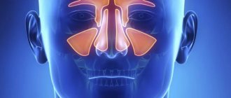
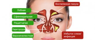
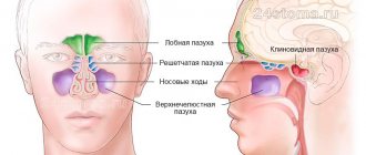
The human nose is a complex organ, which consists of four pairs of paranasal sinuses. Sinuses (sinuses) are air-filled cavities surrounded by skull bones and cartilage tissue. Anatomical features include narrow convoluted canals that connect the sinuses with the nasal turbinates and respiratory tract, which is shown in full by an MRI image. The facial part of the skull contains bilateral air cavities:
- The maxillary sinuses are adjacent to the root part of the teeth of the upper jaw, part of the nasopharynx and are located on both sides of the nose. Very voluminous areas that can hold up to 18 cubic centimeters of air, in the form of an inverted tetrahedral pyramid.
- The frontal sinuses are located in the scales of the frontal bone in the supraorbital region.
- The ethmoid sinuses are located in a porous area between the bones of the upper jaw and the frontal bone, have access to the nasal passages, and are connected to the brain and orbits.
- The sphenoid cavity has cavernous sinuses. The anatomically complex structure and deep location cause difficulties in diagnosis.
The tasks of the sinuses include:
- maintaining a constant temperature and optimal humidity level for the sensitive receptors of the nasal mucosa;
- ensuring mobility and maintaining the shape of the facial bones;
- participation in correct articulation of speech;
- regulation of pressure inside the nasal area;
- head weight relief.
The mucous membrane of the sinuses has the ability to self-cleanse. Inflammation of the sinuses and accumulation of purulent contents lead to the development of infection, blockage, and neoplasms in the cavities. MRI of the sinuses completely visualizes changes in inflammatory diseases. According to the localization of the process, sinusitis is divided into:
- sinusitis - infection of the maxillary sinuses;
- frontal sinusitis - infection in the frontal sinuses;
- ethmoiditis - an inflammatory process in the ethmoid sinuses;
- sphenoiditis is an infection of the sinuses of the sphenoid bone.
The similarity of symptoms of diseases of the nose and air sinuses are very similar. These include: congestion, headache, fever, runny nose. It is important to understand that the process occurring in cavities can transform into the nasopharynx, dental canals, and brain. There has been a noticeable increase in the transformation of acute forms into chronic and difficult-to-treat forms of the disease; sinusitis is especially susceptible to this. An MRI examination allows you to determine the source of infection and begin treatment in a timely manner.
Radiation doses during research
During an MRI examination, a person does not receive any radiation, since the device uses the properties of a magnetic field. The principle of operation of the equipment is that hydrogen protons line up in one line, forming a force field. After this, the particles are “activated” due to a radio pulse, which causes the protons to return to their original positions in a state of rest.
At the same time, a certain amount of energy is released, which is read by equipment sensors. Based on the information received, layer-by-layer images of organs are created.
Regardless of the information content and what MRI of the sinuses shows, in medical practice such diagnostic methods as computed tomography (CT) and multislice computed tomography (MSCT) of the nasal sinuses and nasopharynx are also used. These methods of studying the human body provide a certain radiation dose.
Due to the procedures, the average radiation dose is considered safe at 4 mSv (this is a radiation dose safe for the health of the body that biological tissue can absorb). So, when taking an X-ray of the head, a person receives a dose of radiation that is equal to 0.02 mSv.
For comparison, when conducting an MSCT and CT examination of the nasal sinuses, this radiation dose indicator can be 1 mSv due to the large number of images.
Sinus cyst
In terms of the prevalence of ENT abnormalities, sinus cysts rank second after inflammatory diseases of the air sinuses. This is due to the structure of the mucous membrane that forms the sinuses. A cyst is a fluid formation in the mucous membrane.
The location of formation affects the size, shape and internal contents. In the nasal cavities, a cyst can be congenital or acquired, arising from sinus tissue, as well as when the gland duct is blocked. From the microscopic formation, over time, a voluminous cavity with a soft, elastic shell is formed. The contents of the cyst are formed by fluid.
The clinical picture of a paranasal sinus cyst is related to its size and location. For a long time, a person does not even suspect the presence of a neoplasm. But over time, the cyst grows, and specific symptoms appear. When visiting an ENT doctor, patients complain of:
- episodic or regular headaches;
- feeling of constant discomfort in the upper jaw;
- when diving to depth, an existing headache appears or intensifies;
- frequent cases of sinusitis;
- the flow of mucous, serous-purulent contents along the back wall of the pharynx, in some patients it is permanent;
- difficulty breathing - in case of rapid growth of the cyst or formation of a polyp.
The clinical picture of the cyst has similar symptoms to many other diseases. That is why an examination in the form of an MRI of the nose is prescribed, in order to differentially exclude paranasal sinus cysts from other pathological conditions. An MRI for a cyst shows the structure of the nasal bones, the exact location of the formation in case of planned surgical treatment.
After treatment, MRI makes it possible to evaluate its effectiveness. The ability to repeat the examination after a short time is also an advantage of performing MRI of the paranasal sinuses.
MRI for cysts, as well as for sinusitis and other diseases of the nasal air cavities, is possible in open and closed type tomographs and angiographs.
Features of the procedure
The MRI technique itself is based on resonance stimulation.
It allows you to get a clear image of bones and tissues, take a photo or record a video on a medium.
If you have signs of sinusitis, you will need to undergo an MRI of your sinuses. They can be seen very clearly in the image, not only to make a diagnosis, but also to develop a direction for further treatment.
Since the procedure costs quite a lot of money, many people think, will an MRI show sinusitis? The answer is yes. In this case, the picture turns out to be much clearer and more readable than when using X-rays, CT scans and other diagnostic methods.
The essence of the MRI method
Diagnostic examination using magnetic resonance imaging technology relies on the action of electromagnetic waves on body tissue under conditions of an intense magnetic field. The magnetic field affects hydrogen atoms, which are present in all structures of the human body. They, in turn, form a response in the form of a radio wave. Atomic responses are converted into images using a computer.
The result of this interaction between the body and the apparatus is layer-by-layer images. A three-dimensional pattern appears on the tomograph screen. The radiologist can turn it to the sides and enlarge it for a detailed study of a particular area being examined.
What are the advantages and disadvantages of CT?
The advantages of this diagnostic method include:
- high information content of the study and accuracy of the results;
- short duration of the procedure;
- relative safety due to rapid CT scanning;
- detection of pathologies at early stages;
- detailed study of bone structures.
The main disadvantage of the method is the use of x-ray radiation. The human body tends to accumulate a dose of radiation. This means that CT scans cannot be done frequently. In addition, the use of CT with contrast often causes allergies. Therefore, before carrying out allergy tests, allergy tests are performed. This procedure (even without the introduction of contrast) is prescribed to pregnant women very rarely, when there is no other research option.
The video explains the difference between CT and MRI:
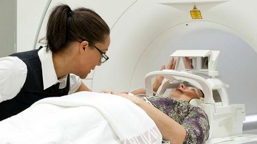
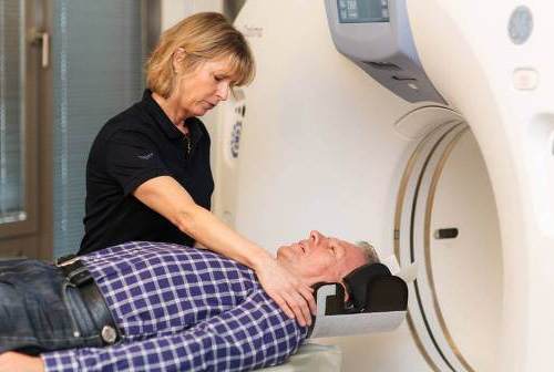
How is an MRI of the sinuses performed?
The patient is required to arrive at the clinic in advance. If you plan to administer a contrast solution, it is better to arrive an hour before the start of the examination. Before the examination, it is important to remove all metal objects - watches, jewelry, removable dentures, remove glasses, remove keys, phone, key fob from your pocket. Some clinics recommend changing clothes to a loose cotton hospital gown before an MRI examination to avoid the presence of forgotten metal products.
As a rule, MRI for cysts is performed without contrast agent. Its administration is necessary for neoplasms, which shows their location, exact size and shape. Before injection of a contrast agent, an allergy test is performed to prevent unwanted reactions.
The contrast is a gadolinium solution that can concentrate in tissues. Depending on the saturation of contrast, the doctor can judge the density and structure of a particular internal organ. A contrast solution is injected intravenously, about an hour before an MRI for a cyst, so that there is enough time for the solution to spread through the bloodstream.
The patient is helped to take a horizontal position on a special couch. For convenience, a special cushion can be placed under the head. Thanks to good air ventilation, the tomograph can be quite cool. The patient may be offered a blanket. After the preparatory stage, the couch with the patient is placed inside the apparatus tunnel.
MRI of the sinuses is accompanied by a specific noise and crackling sound. The patient must be prepared for this. It is imperative to remain still to obtain high-quality, most accurate images. If a person suffers from claustrophobia and cannot remain still, general anesthesia can be performed.
After an MRI of the paranasal sinuses, the patient returns to normal life. It is recommended to drink a lot of clean water to quickly remove the contrast agent.
Which method of studying the sinuses is better - MRI, CT or MSCT?
It is quite difficult to answer this question unambiguously, since each of the presented methods has its own characteristics and subtleties. Therefore, a certain type of diagnostics is used to diagnose specific diseases.
Types of tomographs and their distinctive characteristics:
- A good display of the condition of soft tissues when scanning in an MRI machine contributes to the fact that this type of diagnosis is used mainly to identify tumors. However, it is contraindicated for use in patients who have metal objects in their bodies (pacemakers, endoprostheses, etc.).
- Not every person is able to remain motionless throughout the entire study period. In this regard, it is advisable for a separate group of patients to be prescribed CT or MSCT - examinations that are less sensitive to movements.
- MSCT is considered the most innovative method for examining the nasal sinuses. However, compared to MRI, which is absolutely safe, with MSCT a person receives a certain dose of radiation, which can cause the development of cancer.
Based on the above, we can draw the following conclusion: a specific diagnostic method should be chosen taking into account the complexity of the clinical picture, the presence of contraindications, as well as the individual characteristics of the human body. Therefore, the choice of a specific diagnostic technique should be made by the attending physician.
Indications for MRI of the sinuses
Symptoms that bother the patient for a long time are the basis for prescribing an MRI of the paranasal sinuses. The attending physician bases the diagnosis decision on the presence of the following signs:
- regular headaches in the forehead, eyes, upper jaw;
- memory disorder, sleep disorder;
- recent injuries and bruises of the nose;
- prolonged runny nose that does not disappear after long-term treatment;
- difficulty breathing, feeling of congestion on one or both sides;
- nasal voice;
- assumption of the formation of a cyst in the maxillary sinuses;
- frequent sinusitis, frontal sinusitis, ethmoiditis;
- anatomical pathologies of the air sinuses;
- Allowing the formation of a brain tumor;
- accumulation of blood in the paranasal sinuses;
- migraine of unknown etiology.
The presence of even one sign is grounds for an MRI of the nose.
In what cases are they prescribed?
Such studies are prescribed in the presence of certain clinical symptoms, including:
- frequent pain in the head area;
- chronic inflammation;
- prolonged, persistent runny nose;
- allergic rhinitis;
- injuries of the nose and facial bone;

If the patient has a prolonged runny nose, an MRI or CT scan is indicated.
- toothache;
- labored breathing;
- nosebleeds;
- pain when pressing on the forehead or nose.
MRI is not recommended for diagnosing fresh injuries. In these cases, computed tomography is performed.
Contraindications to MRI of the paranasal sinuses
Regardless of what MRI diagnostics show, you cannot decide on its implementation on your own. The attending physician does not prescribe an MRI of the nose for the following conditions:
- the patient’s body contains metal pins, plates, clips, staples that cannot be removed for examination, except for titanium structures;
- the presence of electronic devices embedded in the body - a pacemaker, a neurostimulator, a defibrillator, an insulin pump, a hearing implant;
- Ilizarov apparatus is a conditional contraindication; MRI of the nose can be performed after its elimination;
- excessive weight of the patient - the tomograph device does not make it possible to perform diagnostics for people weighing more than 150 kg; sometimes the study is possible in an open-type MRI tomograph;
- first three months of pregnancy;
- breastfeeding is a relative contraindication to MRI of the sinuses; if this is a necessary diagnostic measure, milk is expressed and not given to the child for 2 days after the procedure;
- acute period of an infectious disease (sinusitis, sore throat, etc.);
- renal failure requires refusal of contrast MRI of the nose;
- unconsciousness of the patient, drug or alcohol poisoning;
- psychiatric disorders in which the patient experiences phobias and cannot remain in a stationary position; in this case, for example, for MRI of a cyst, it is advisable to use general anesthesia.
Some of the contraindications are not a strict ban on MRI of the nose. After assessing the risk and feasibility of conducting an MRI study, only the doctor makes a decision on its implementation.
What are the pros and cons of MRI?
Currently, MRI examination is very popular. However, there are not only advantages, but also disadvantages of such a diagnostic procedure, which is reflected in the table.
| Advantages | Flaws |
| The relative harmlessness of the method due to the absence of x-rays and radiation | High cost of the procedure |
| 3D images | Duration of the study |
| Image clarity | Unreliability in identifying pathologies and anomalies of bone structures |
| Few contraindications | The need to comply with certain requirements when conducting diagnostics, for example, patient immobility |
Another advantage of magnetic tomography with the introduction of contrast should be noted. The substance that is administered to the patient intravenously does not contain iodine. Thanks to this, you don't have to worry about developing allergies.
There are practically no contraindications for performing MRI
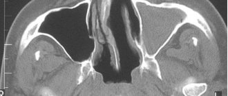
What does MRI of the paranasal sinuses show?
MRI of the sinuses is prescribed when other methods of examining the patient do not provide a complete diagnostic picture. This is necessary when data is needed on the condition of the difficult to access sphenoid or ethmoid sinuses located behind the osteochondral structures of the skull.
Images obtained during magnetic resonance imaging provide detailed information about:
- pathologies of the formation of parts of the nasopharynx;
- defects in the structure of osteochondral structures;
- injuries to the facial part of the skull - cracks, fractures, displacement of bone fragments;
- diseases of the nose of a regular nature;
- thickening of bone walls
- inflammatory foci;
- single- or multi-chamber cystic and tumor formations;
- malignant neoplasms even at the very initial stages;
- infections of the skull bones;
- degree of symmetry of the paranasal cavities;
- accumulation of blood or other fluids in the paranasal sinuses.
A radiologist decodes and describes in detail the images obtained during MRI. He also draws up a conclusion on proving or challenging the preliminary diagnosis. The description time can take from 30 minutes to 2 days, depending on the clinic’s workload. The images are sent to the patient in printed and electronic form along with a full diagnostic report. MRI results must be provided to the attending physician to determine treatment tactics and the quality of therapy.
MR imaging (MRI) and X-ray computed tomography (CT)
Magnetic resonance imaging and x-ray computed tomography are methods for studying various organs through computer reconstruction of images in the form of two- (slices) or three-dimensional images. Currently, these are the most accurate methods for diagnosing structural disorders of bones, joints, brain, blood vessels, internal organs, and soft tissues.
We perform tomography in the MRI and CT department of the Central Clinical Hospital of the Academy of Sciences on the street. Fotieva, 12, building 3. The department is equipped with powerful, latest equipment. You can sign up for the study by phone. Call for travel details.
Tomograph
In neurology, MRI is mainly used. This is the first (and currently the only widely available) way to see with your own eyes the foci of demyelination of the brain and spinal cord. A great help in the differential diagnosis of multiple sclerosis is gadolinium contrast. After intravenous administration of contrast, active foci of demyelination begin to “glow” on MRI scans. But difficulties in the differential diagnosis of inactive foci of demyelination still remain. We solve them with the help of laboratory examinations, studies of evoked potentials, reflexes and sensitivity, etc.
Foci of demyelination of the spinal cord Foci of demyelination of the brain
Modern MRI scanners are equipped with special programs for identifying a particular disease (epileptology program, research for diagnosing multiple sclerosis, examination of cerebral vessels in encephalopathy, etc.). MRI does not clearly see the bones of the skull, so it is advisable to evaluate them using X-ray CT.
Brain research. To study the brain, we most often use MRI, using special programs aimed at identifying a particular disease (epileptology program, contrast study for diagnosing multiple sclerosis, study of cerebral vessels for encephalopathy, etc.). MRI does not clearly see the bones of the skull, so it is advisable to evaluate them using X-ray CT.
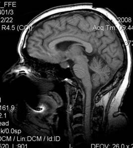
MRI brain scan
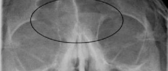
Examination of the paranasal sinuses and hearing organs. Here, computed x-ray tomography (CT) will provide more information. In general terms, the condition of the paranasal sinuses can also be assessed using MRI scans, which we do in almost all cases of headaches (the sinuses are visible on MRI scans of the brain).
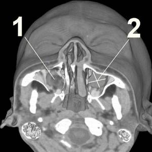
Computer tomogram 1 – normal maxillary sinus 2 – maxillary sinus cyst
the joints and spine using MRI scans. On MRI scans, cartilage, intervertebral discs, spinal cord, ligaments and tendons, joint fluid, swelling and inflammation are clearly visible.
MRI scan of the spine 1 – normal intervertebral discs 2 – spinal cord canal 3 – disc herniation protrudes into the spinal canal
The lungs and lymph nodes of the chest are clearly visible on X-ray computed tomography (CT) scans. Even small foci of inflammation, tumors, scars after illnesses, signs of pleurisy, tuberculosis, etc. are visible. CT is much more informative than a regular x-ray or fluorogram.
CT lungs
The abdominal and pelvic organs can be examined using CT or MRI in various cases. Contrasting techniques are often used.
The pancreas and adrenal glands are usually subjected to MRI to exclude hormonally active tumors (insulinoma, adrenal adenoma, pheochromocytoma, etc.). For more accurate diagnosis, special contrast techniques are used.
the teeth and jaw bones using X-ray CT when looking for the cause of trigeminal neuralgia. Various sources of irritation of the trigeminal nerve are clearly visible on tomograms: the exit of filling material beyond the apex of the tooth, cysts, destruction of tooth roots, tumors.
CT jaw
MR angiography and CT angiography are a method of examining blood vessels followed by three-dimensional reconstruction. In many cases, CT angiography can replace complex and risky routine X-ray angiography.
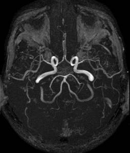
Cerebral vessels - MR angiography
Where can I get an MRI of the sinuses?
It is difficult to independently understand the huge amount of information and find the right diagnostic clinic. That is why the website msk-mrt.ru was created, the work of which is aimed at providing information assistance and support to people. Here you can choose a convenient clinic and plan your route.
The price of diagnostics depends on the type of tomograph, the doctor’s workload and the level of the clinic. All prices can be found on the clinic’s page. We interact with a variety of medical institutions, so we can provide discounts on magnetic resonance imaging.
Our website will offer information about promotional offers, compulsory medical insurance cooperation programs, benefits for children and certain categories of patients. Contact the operators by phone and get the necessary advice, answers to your questions, and information on prices. In addition, here you can read reviews from patients who used the services of the clinics and were diagnosed according to our recommendations.