One day hospital 3rd KO
Kravchenko
Olga
10 years of experience
Oncologist (CHI)
Make an appointment
Uterine cancer is a serious pathology that manifests itself in the form of a malignant tumor formed from modified endometrial cells - the inner mucous membrane lining the uterine cavity. This is one of the most common cancers developing in women. The most dangerous period for the development of a tumor is the interval between 65 and 70 years; in general, the incidence increases during menopause, i.e. after 50 years.
Kinds
In accordance with histological characteristics, uterine cancer is divided into the following forms:
- adenocarcinoma, which is the most common type;
- squamous cell form - the least aggressive, successfully treatable;
- glandular-squamous, formed from glandular cells of the endometrium;
- leiomyosarcoma, developing from cells of the muscle layer;
- clear cell, a rather rare form, accounting for 1-5% of all cases;
- mucinous, which is characterized by increased mucus formation;
- serous, in which a tumor with a multi-chamber structure and the secretion of serous fluid is formed.
Can endometrial cancer be seen on ultrasound?
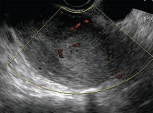
The most important part of diagnosing endometrial cancer is performing an intravaginal ultrasound. Using an ultrasound machine, the doctor can detect echographic signs of endometrial hyperplasia, endometrial polyps, an increase in endometrial thickness of more than 4-5 mm in menopausal women and other signs that may indirectly indicate the presence of a malignant neoplasm in the uterus. If such changes are detected, it is necessary to perform hysteroscopy with taking an endometrial biopsy and histological examination of the resulting sample.
At the moment, hysteroscopy with endometrial biopsy is the gold standard for diagnosing hyperplastic processes and detecting endometrial cancer.
To plan the scope and preliminary staging, a full examination is carried out before the operation in accordance with international oncological standards for the treatment of oncological diseases:
- a complete set of general clinical tests
- MRI of the pelvic organs;
- computed tomography of the pelvis, abdomen and chest with contrast.
Symptoms
It is important not to miss the first signs of uterine cancer and visit a gynecologist on time. A cause for concern should be copious watery discharge, which is periodically mixed with a small amount of blood. Over time, the discharge intensifies and develops into bleeding. In general, any vaginal discharge during menopause may be a symptom of uterine cancer. Signs of endometrial cancer in women of reproductive age are bleeding between periods and heavier than usual menstruation, often accompanied by pain. In addition, general symptoms of cancer appear:
- feeling of constant fatigue, weakness;
- lack of appetite, sudden weight loss;
- anemia, pale skin;
- nausea, malaise;
- with tumor growth – pain in the pelvis or back.
The appearance of uncharacteristic discharge, especially bloody ones, should be a good reason for a visit to the gynecologist.
Causes and risk factors
It is still not known exactly how the malignant process starts, however, the factors that increase the likelihood of uterine cancer have been studied quite well. The most significant of them:
- disturbances in the production of hormones, primarily an increase in estrogen levels;
- obesity, diabetes, hypertension;
- benign neoplasms of the uterus – endometrial hyperplasia, polyps, fibroids;
- too early or late start and end of menstruation;
- no history of childbirth;
- long-term hormonal therapy;
- hereditary predisposition;
- chemoradiation treatment of tumors in other organs;
- lack of regular sexual intercourse for a long time.
The presence of one or more of the listed signs does not mean that a woman will necessarily develop symptoms of a uterine tumor. However, those who are at risk should visit a gynecologist at least once a year to monitor the condition of the reproductive organs.
What test should you take for cervical cancer?
The main test for cervical cancer is a cytological examination of a stained smear taken from the cervical canal. It has many advantages.
This study:
- Simple
- Inexpensive
- Fast - Once doctors test for cervical cancer, results can be obtained the same day or the next
- Painless - does not require either general or local anesthesia
- Does not harm health, and therefore can be performed with any frequency
- Has high sensitivity and specificity for a screening method
Tests for cervical cancer should be taken regularly. If they are all negative, it is enough to do research once every 3 years, starting from the age of 21. But if background and precancerous processes are detected, more frequent monitoring is required. The study will have to be completed annually. However, for most women this is not a problem. Considering how much a cervical cancer test costs, screening will not be financially burdensome.
The importance of a smear for oncocytology can hardly be overestimated. In developed countries, this test detects stage 0 or stage 1 cervical cancer in 70-80% of cases. That is, only 20-30% of women have the disease at stage 2 and above. In this case, the prognosis of the pathology is much worse. The likelihood of a complete cure is low.
Women often ask what blood test shows cervical cancer. There are several tests that help in diagnosis. For early detection of pathology, a blood test using PCR for oncogenic types of papillomavirus is used. If they are detected, the woman is monitored, since she is at risk for cervical cancer.
For patients who are already being treated for cancer, it is possible to take a test for cervical cancer to determine tumor markers in the blood. An increase in their concentration indicates the ineffectiveness of treatment or relapse of the tumor. A decrease in the amount of squamous cell carcinoma antigen indicates the success of therapy or ongoing remission of the cancer.
Stages
The development of a tumor is preceded by the so-called zero stage, when malignant cells are located on a microscopic area of the mucous membrane. According to the degree of growth of the malignant neoplasm, four stages of endometrial cancer are distinguished.
- The process has spread to the entire endometrium, but has not yet affected other layers of the uterine wall. The lymph nodes are not affected, there are no metastases.
- Malignant cells invade the muscle layer and can spread to the cervix.
- All layers of the uterine wall are affected, the malignant process spreads to the vaginal vault and affects regional lymph nodes.
- The tumor has spread to neighboring organs - intestines, bladder, etc. Lymph nodes are affected, metastases have spread to distant organs - lungs, liver, bone structures.
Basic diagnostic measures for RTM:
- Physical examination. A gynecological examination is performed to visually assess the condition of the vaginal mucosa and cervix. Bimanual diagnostics determines the condition of the parametriums.
- Laboratory research. Detailed biochemical and clinical blood tests to assess kidney and liver functions also require a blood test for the presence/absence of CA-125 tumor markers, a general urine test and a blood coagulation test.
- Instrumental diagnostics. Endometrial aspiration biopsy is recommended. Separate diagnostic curettage of the uterine cavity is also performed. An ultrasound of the abdominal cavity, pelvis and retroperitoneal space is mandatory.
As an additional measure, an MRI is prescribed to assess the depth of invasion and the transition of the malignant tumor to adjacent organs. To determine the extent of surgical intervention, a clarifying diagnosis of damage to the lymph nodes is carried out.
Diagnostics
For uterine endometrial cancer, the symptoms and signs of the disease are not specific. Therefore, instrumental and laboratory studies are of utmost importance for diagnosing a tumor.
- Transvaginal ultrasound. Allows you to describe the structure of the endometrium and control its thickness.
- Hysteroscopy. The procedure for examining the internal cavity of the uterus using a special optical system. During the examination, a biopsy of pathologically altered tissues may be performed.
- Biopsy. An aspiration biopsy is a procedure to remove a sample of the endometrium using a thin, hollow needle.
- Histological analysis of the biopsy specimen. Microscopic examination of the prepared sample is necessary to confirm the diagnosis and to determine the type of malignant cells.
- MRI of the uterus. As a rule, it is carried out at the third or fourth stage of tumor growth to identify the extent of spread, affected lymph nodes and neighboring organs.
Attention!
You can receive free medical care at JSC “Medicine” (clinic of Academician Roitberg) under the program of State guarantees of compulsory medical insurance (Compulsory health insurance) and high-tech medical care.
To find out more, please call +7, or you can read more details here...
Can endometrial cancer be cured?
Today, endometrial cancer of the uterus is not a death sentence. With regular preventive examinations by a gynecologist and detection of malignant neoplasms in the uterus in the early stages, the disease is curable. Correct and timely treatment allows you to get rid of the tumor, prevent its recurrence and metastasis. In terms of mortality, this pathology ranks last in the list of diseases of the female genital organs. When selecting a treatment regimen for uterine adenocarcinoma, the patient’s age, general condition, and stage of the disease are taken into account.
Stage I usually requires surgery to remove the uterus and its appendages. At the second stage, along with extirpation of the uterus and appendages, adnexectomy of the surrounding lymph nodes is also assumed (possible penetration of tumor cells into them).
Please note that a favorable prognosis for endometrial cancer depends on the quality of the surgical stage, which in turn largely depends on the technical equipment of the operating room and the qualifications of the operating team.
Endometrial cancer in the final stages requires the use of radiation and chemotherapy - exposure to ionizing radiation on diseased cells or therapy with drugs.
Treatment
To successfully treat endometrial cancer, different methods are used, which are selected based on the prevalence of the cancer process, the type of tumor, its location and structure.
- Extirpation of the uterus. During the operation, as a rule, the uterus and fallopian tubes are completely removed, especially if we are talking about a woman who already has at least one child. If the tumor size is small, the cervix is preserved to reduce the risk of complications during the recovery period. At the fourth stage, surgical treatment is not performed.
- Radiation therapy. Treatment is carried out before surgery to reduce the size of the malignant tumor, and also after surgery to prevent relapse due to remaining tumor cells. At the inoperable stage, radiation is used to prolong the patient's life.
- Chemotherapy. Often, cytostatic drugs are prescribed in combination with radiation treatment before or after surgery, as a palliative in inoperable cases and in case of relapse of the disease.
- Hormonal method. Effective for the treatment of hormone-dependent tumors, it is used mainly for the treatment of young patients with a highly differentiated type of cancer.

The use of nanotechnology in the treatment of cancer
For MEDICAL PLAZA specialists, each patient is an individual case that requires a personalized, comprehensive approach. We treat endometrial cancer at all stages, and also work to prevent relapse by diagnosing abnormal abnormalities.
The main step in the treatment of endometrial cancer is surgery.
It includes complete removal of the uterus and, in patients with high-risk uterine cancer, removal of the pelvic and para-aortic lymph nodes. Removing the entire block of lymph nodes, especially near large vessels and nerves, is always associated with great risk for the patient. However, thanks to the use of ICG technology in our surgical center, it has become possible to minimize this risk and avoid unnecessary tissue trauma by identifying and removing only a few lymph nodes responsible for communication with the uterus, without damaging the remaining lymph nodes and surrounding structures.
Chemotherapy and radiotherapy, which are prescribed in later stages, are also an important part of the treatment, but success largely depends on the surgical stage, and therefore on the qualifications of the surgeon and the quality of surgical equipment.
At Medical Plaza, surgical treatment is carried out in an operating room equipped with the most modern equipment from leading global manufacturers, as well as using the DaVinci robotic surgical system. Robotic surgery allows for the most precise removal of a tumor with minimal trauma to the patient.
The advantages of this technology can be briefly described as follows:
- the volume of blood loss decreases by more than 4 times;
- the time spent in hospital is reduced by almost 3 times;
- the need for blood transfusion, intraoperative and long-term postoperative complications is minimized.
Diagnosis and treatment of uterine cancer in Moscow
If you have discovered signs of uterine cancer, contact the Medicina clinic to be examined using modern high-precision diagnostic equipment. Consultations regarding diagnosis, and if the diagnosis is confirmed, qualified treatment is carried out by oncologists-gynecologists of the highest category according to protocols used by leading clinics in the world. Our patients have our own laboratory, which performs all types of tests, and a comfortable hospital with attentive, professionally trained staff.
Forecast
The prognosis for survival from uterine cancer, as well as complete recovery and relapses, depends on many factors. First of all, this is the time of cancer detection; the earlier the tumor is diagnosed, the more favorable the prognosis. The type of malignancy, the patient’s age, the presence of concomitant diagnoses, the chosen treatment tactics and the body’s response to therapy also influence.
Average prognosis for five-year survival if cancer is detected at the appropriate stage:
- in situ – 90%;
- I – 75%;
- II – 69%;
- III (A, B, C) – 58%, 50%, 47%;
- IV (A, B) – 17%; 15%.
Recurrence of uterine cancer is possible, most often it occurs within 2 years after treatment. The relapse rate is on average about 10%, it depends on the type of tumor and the degree of differentiation.
Questions and answers
How long do they live with uterine cancer?
The lifespan of a woman after detection of a uterine tumor directly depends on what stage of development the disease is at. After surgery performed at an early stage, the patient can live safely for decades. Patients with stage 4 without medical care can die within a few months, and with quality treatment they can live for more than five years.
What does uterine cancer look like?
It is impossible to independently detect a cancerous tumor of the uterus. A full diagnosis is required in a specialized medical institution to confirm or refute the suspicion of uterine cancer.
How does uterine cancer manifest?
The main symptom of a uterine tumor is bleeding from the vagina, which appears between periods and even during menopause. If any bleeding occurs during these periods, you should immediately consult a gynecologist. The earlier a tumor is detected, the higher the chances of a complete cure.
Attention! You can cure this disease for free and receive medical care at JSC "Medicine" (clinic of Academician Roitberg) under the State Guarantees program of Compulsory Medical Insurance (Compulsory Medical Insurance) and High-Tech Medical Care. To find out more details, please call or visit the VMP page for compulsory medical insurance
How long does it take for endometrial cancer to develop?
The endometrium is the mucous membrane of the uterus, creates favorable conditions in the cavity for blastocyst implantation, but is also susceptible to any fluctuations in hormonal levels. The membrane undergoes cyclic changes during the menstrual cycle. This is also where the emergence of various types of neoplasms begins.
Malignant tumors progress over several years. Often the disease is initially asymptomatic. The neoplasm does not affect the woman’s well-being until the later stages. This is precisely the reason why patients come to us even when uterine cancer poses a real danger to a woman’s life. Of course, this is bad, but in no case should you give up. When selecting a scheme to combat the disease, the degree of differentiation is used.
| Type of tumor | Characteristic |
| I | Endometrial adenocarcinoma G1 with a high degree of differentiation. Occurs with endometrial hyperplasia. Solid areas account for up to 5%, the remaining 95% are glandular structures. |
| II | Endometrial adenocarcinoma G2 is a moderately differentiated tumor (50% glandular structures, 6-50% solid). |
| III | Poorly differentiated or solid (G3) adenocarcinoma of the uterus. Solid structures exceed 50%, although glandular ones are also diagnosed. |