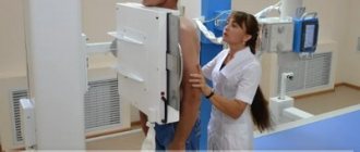
The anatomical structure of the lungs, their ability to be filled with air that freely transmits x-ray radiation, makes it possible to obtain, during fluoroscopy, an image that reflects in detail all the structural elements of the lungs. However, darkening in the lungs on an X-ray does not always reflect changes in the tissues of the lung itself, since other organs of the chest are located at the level of the lungs and, therefore, the radiation beam, passing through the body, projects on the film a superimposed image of all organs and tissues , falling within its range.
In this regard, if any darkened formation is detected in the image, before answering the question of what it could be, it is necessary to clearly differentiate the localization of the pathological focus (in the tissues of the chest, diaphragm, pleural cavity or, directly, in the lungs).
Main syndromes on radiographs
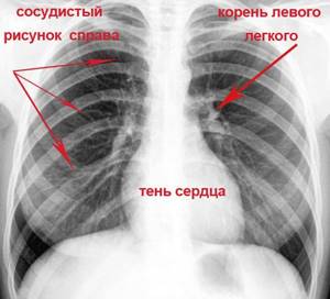
On an x-ray taken in an anterior projection, the contours of the lungs form pulmonary fields over the entire area, intersected by symmetrical shadows of the ribs. A large shadow between the pulmonary fields is formed by the combined overlap of the projection of the heart and great arteries. Within the contour of the lung fields, one can see the roots of the lungs located at the same level with the anterior ends of the 2nd and 4th ribs and a slight darkening of the area caused by the rich vascular network located in the lung tissue.
All pathological changes reflected on x-rays can be divided into three groups.
Dimming
Appear on the image in cases where the healthy part of the lung is replaced by a pathological formation or substance, causing the displacement of the air part by denser masses. As a rule, it is observed in the following diseases:
- bronchial obstruction (atelectasis);
- accumulation of inflammatory fluid (pneumonia);
- benign or malignant tissue degeneration (tumor process).
Change in pulmonary pattern
This group of changes can be considered the most common. Despite the huge list of lung pathologies, all possible changes in the X-ray picture can be attributed to one of 5 syndromes:
- total (complete) or subtotal (almost complete) blackout;
- limited dimming;
- round (spherical) shadow;
- ring shadow;
- focal darkening.
Enlightenment
The clearing in the image reflects a decrease in the density and volume of soft tissues. As a rule, a similar phenomenon occurs when an air cavity forms in the lung (pneumothorax). Due to the specific reflection of x-ray results on photographic paper, areas that easily transmit radiation are reflected in a darker color due to the more intense effect of x-rays on the silver ions contained in photographic paper; areas of a denser structure have a light color. The wording “darkening” in the image is actually reflected in the form of a light area or focus.
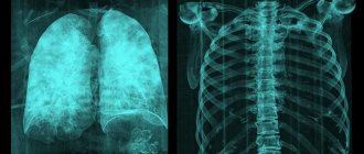
X-ray showing a pulmonary pattern of healthy lungs
Smoker's lungs
A person who smokes, by definition, is at risk for pulmonary diseases, and quite serious ones at that. Such citizens should undergo fluorography as often as possible, but not violate safety standards. Once every six months is the most appropriate time.
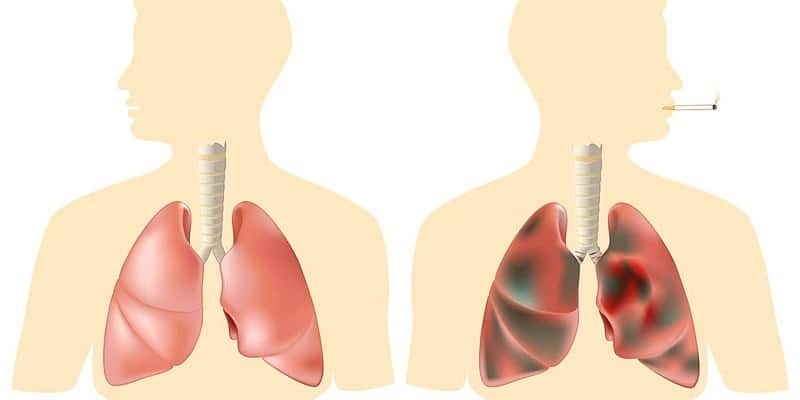
Smoking itself can cause darkening in the lungs - a large amount of foreign substances in the form of tar affects it (the average annual “clogging” of the bronchi is about a glass). In addition, the natural lung tissue is replaced with scar tissue. This circumstance greatly complicates a full diagnosis, since the spots caused by the disease can be covered by the darkening of the “smoker”.
Total blackout syndrome
Total darkening of the lung on an x-ray is a complete or partial darkening (at least 2/3 of the lung field). In this case, gaps are possible in the upper or lower part of the lung. The main physiological reasons for the manifestation of this syndrome are the lack of air in the lung cavity, an increase in the density of the tissue of the entire surface of the lung, the content of fluid or any pathological content in the pleural cavity.
Diseases that can cause such a syndrome include:
What is bronchoscopy?
- atelectasis;
- cirrhosis;
- exudative pleurisy;
- pneumonia.
To carry out differential diagnosis of diseases, it is necessary to rely on two main signs. The first sign is to assess the location of the mediastinal organs. It can be regular or offset, usually in the direction opposite to the darkening focus. The main landmark in identifying the displacement axis is the shadow of the heart, located mostly to the left of the midline of the chest, and less to the right, and the stomach, the most informative part of which is the air bubble, always clearly visible on the images.
The second sign that makes it possible to identify a pathological condition is an assessment of the uniformity of darkening. Thus, with uniform darkening, atelectasis can be diagnosed with a high degree of probability, and with heterogeneous darkening, cirrhosis can be diagnosed. Interpretation of the results obtained using the radiographic method consists of a comprehensive assessment of all visually detected pathological elements in comparison with the anatomical features of each individual patient.
Follow-up examination
To conduct further examination, the doctor may send the patient to a pulmonologist or oncologist, where he will be shown to undergo certain specific procedures. Here are the most common ones:
- This may be a diaskintest, which can determine the presence of tuberculosis. If we compare this procedure with Mantoux, which quite often gives a false and inaccurate result, such an examination does not respond to BCG, which often shows a complete absence of a problem in the child. This is an ideal opportunity to diagnose tuberculosis as accurately as possible.
- The study of sputum of children and adults is another mandatory analysis that is carried out in the laboratory. Based on the results obtained, it is possible to detect tuberculosis bacilli, the presence of malignant cells, as well as various impurities that may be characteristic of certain pathological conditions.
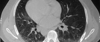
- Quite often, doctors prescribe a more modern examination method - computed tomography of the lungs. This is an additional, informative method for diagnosing diseases of the lung tissue, pleura and mediastinum. It turned out that this is the most reliable method.
- Lung bronchoscopy or tracheobronchoscopy may also be used. This procedure is performed using medical endoscopes that are quite flexible in design and are inserted through the nose. Through this form of examination, you can see the lungs and also take material, completely painlessly. The collected material, as a rule, is subjected to subsequent diagnostics - bacterial, histological and cytological.
If a doctor suspects lung cancer based on an x-ray, a tumor marker test may be prescribed. The analysis makes it possible to detect specific proteins that are usually produced by emerging malignant tumors.
Limited dimming syndrome
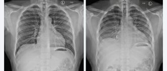
To identify the causes of limited darkening of the pulmonary field, it is necessary to take an image in two directions - in direct projection and lateral. Based on the results of the obtained images, it is important to assess the localization of the darkening focus. If the shadow in all photographs is located inside the pulmonary field and is similar in size to its contours or has a smaller volume, it is logical to assume a lung lesion.
If there is darkening adjacent to the diaphragm or mediastinal organs with a wide base, extrapulmonary pathologies (fluid inclusions in the pleural cavity) can be diagnosed. Another criterion for evaluating limited shades is size. In this case, two possible options should be considered:
- The size of the darkening clearly follows the contours of the affected part of the lung, which may indicate an inflammatory process;
- The size of the darkening is smaller than the normal size of the affected segment of the lung, which indicates cirrhosis of the lung tissue or blockage of the bronchus.
Particular attention should be paid to cases in which there is a darkening of normal dimensions, in the structure of which light foci (cavities) can be traced. First of all, in this case, it is necessary to clarify whether the cavity contains liquid. To do this, a series of photographs are taken in different positions of the patient (standing, lying down or bending over) and changes in the level of the estimated upper limit of the liquid contents are assessed. If fluid is present, a lung abscess is diagnosed, and if it is not present, then the likely diagnosis is tuberculosis.
Important! The detection of several cavities with limited darkening of the lung is characteristic of pneumonia caused by staphylococcus. Such a lesion has an unfavorable prognosis, and often treatment is only possible through surgery.
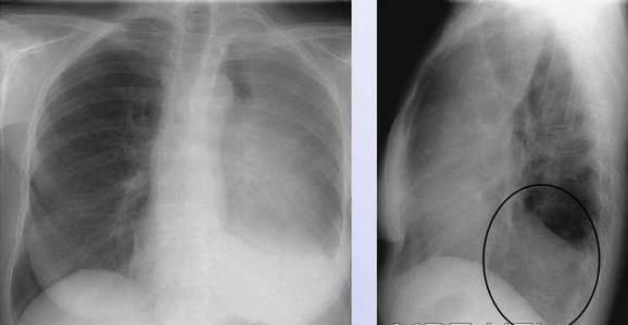
X-ray shows limited darkening of the lungs in two projections
What is "frosted glass"?
“Frost glass” are areas of compacted lung tissue that are less transmittable to X-Ray rays. In another way, they are called foci of compaction of lung tissue. On tomograms, such areas resemble light spots or a cloudy coating. Ground glass lesions indicate that the alveoli of the lungs are filled with fluid (transudate, blood), and not air. They may also indicate a decrease in pulmonary pneumatization due to fibrotic changes. Alveoli participate in gas exchange, supply cells with oxygen and remove carbon dioxide, so filling their cavity with a liquid substance or connective tissue cells is unacceptable.
If the total area of the “frosted glass” increases, this indicates a progressive infectious-inflammatory disease. The patient begins to worry about lack of air, shortness of breath, and cough.

Round shadow syndrome
I identify round shadow syndrome when the spot on the lungs has a round or oval shape on two photographs taken perpendicular to each other, that is, from the front and the side. To decipher the results of radiography when a round shadow is detected, they rely on 4 signs:
- form of shading;
- localization of darkening relative to nearby organs;
- clarity and thickness of its contours;
- structure of the internal shadow field.
Since the shadow reflected on the image within the lung field may actually be located outside it, assessing the shape of the darkening can greatly facilitate diagnosis. Thus, a round shape is characteristic of intrapulmonary formations (tumor, cyst, infiltrate filled with inflammatory contents). An oval shadow in most cases is the result of compression of a round formation by the walls of the lung.
The structure of the internal shadow field is also highly informative. If, when analyzing the results, the heterogeneity of the shadow is obvious, for example, lighter foci, then with a high degree of probability, it is possible to diagnose the disintegration of necrotic tissue (with disintegrating cancer or disintegration of tuberculous infiltrate) or the formation of a cavity. Darker areas may indicate partial calcification of tuberculoma.
A clear and dense contour indicates the presence of a fibrous capsule, characteristic of an echinococcal cyst. Round shadow syndrome includes only those shadows that are more than 1 cm in diameter; shadows with a smaller diameter are considered lesions.
3.Types of tumors
Here are some of the most common types of benign lung tumors:
- Hamartomas
. Hamartomas are the most common type of benign lung tumor and one of the common causes of the formation of solitary pulmonary nodules. This type of lung tumor is formed from the tissues of the lining of the lungs, as well as fatty and cartilage tissue. As a rule, hamartoma is located on the periphery of the lungs. - Bronchial adenoma
. Bronchial adenoma accounts for about half of all benign lung tumors. It is a heterogeneous group of tumors that arise from the mucous glands and ducts of the trachea or large airways of the lungs. Mucous adenoma is one example of a true benign bronchial adenoma. - Rare lung tumors
can appear in the form of
chondromas, fibromas, lipomas
- benign lung tumors consisting of connective or adipose tissue.
About our clinic Chistye Prudy metro station Medintercom page!
Ring shadow syndrome
A ring-shaped spot on the lung on an x-ray is the easiest syndrome to analyze. As a rule, a ring-shaped shadow appears on an x-ray as a result of the formation of a cavity filled with air. A mandatory condition under which the detected darkening is classified as ring-shaped shadow syndrome is the preservation of a closed ring when taking pictures in all projections and in various positions of the patient’s body. If in at least one of the series of photographs the ring does not have a closed structure, the shadow can be considered an optical illusion.
If a cavity is detected in the lung, the uniformity and thickness of its walls should be assessed. Thus, with a large and uniform thickness of the contour, one can assume the inflammatory origin of the cavity, for example, a tuberculous cavity. A similar picture is observed with an abscess, when purulent melting of tissue occurs and the contents are removed through the bronchi. However, with an abscess, the remains of pus most often remain in the cavity and their complete removal is quite rare, so usually such a cavity is a tuberculous cavity.
The unevenly wide walls of the ring indicate the process of decay of lung cancer. Necrotic processes in tumor tissue can cause the formation of a cavity, but since necrosis develops unevenly, tumor masses remain on the inner walls of the cavity, creating the effect of an “uneven” ring.
Important! The main difficulty in assessing the ring-shaped shadow is determining the localization of the formation, since in most cases a similar syndrome is observed in extrapulmonary processes (deformation of the ribs, gases in the intestines, gases in the pleural cavity).
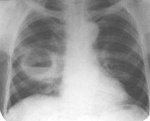
The image shows a ring-shaped shadow in the lower lobe of the right lung
2. Causes of benign tumors
The reasons why benign lung tumors appear are poorly understood. But in general, they often appear after health problems such as:
Inflammatory processes caused by infection:
- Fungal infections – histoplasmosis, coccidioidomycosis, cryptococcosis, aspergillosis;
- Tuberculosis
- Lung abscess
- Pneumonia
Inflammation not associated with infection:
- Rheumatoid arthritis;
- Wegener's granulomatosis;
- Sarcoidosis.
- Congenital pathologies such as lung cyst and others.
Visit our Oncology page
Symptoms of hilar pneumonia
Hilar pneumonia is manifested by the following symptoms: high body temperature (up to 400C), cough, shortness of breath. With prolonged pneumonia, body temperature can remain at 37-38 0C for weeks. The cough is initially dry, but over time it becomes wet and painful. With the bacterial nature of hilar pneumonia, pronounced symptoms of intoxication occur:
- severe general weakness;
- increased sweating;
- headache;
- insomnia.
Hilar pneumonia in adults often occurs without the pain in the chest, side or under the shoulder blade typical of pneumonia. In children, the disease develops rapidly, and the symptoms of intoxication are more pronounced than in adults.