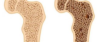
Magnetic resonance imaging of the brain is an imaging diagnostic procedure based on the effect of interaction between the electromagnetic field and hydrogen atoms, which make up about 50% of all atoms in the human body.
MRI is performed on a special machine, which is a giant magnet.
Brain examination using MRI scans is used to diagnose diseases, injuries, and monitor progress after surgery.
Benefits of MRI
Magnetic tomography allows you to obtain a three-dimensional image of the areas under study in three projections. During the procedure, the device takes many slices, the thickness of which can be set individually and is usually 2-4 mm.
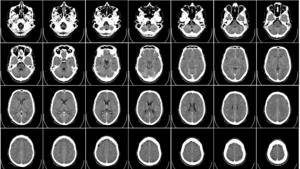
Images taken using a tomograph
Obtaining a large number of sections allows you to examine the entire organ and detect even the slightest abnormalities and pathologies.
What types of tomographs are there?
Modern magnetic tomographs are available in various variations with a wide variety of characteristics.
All tomographic devices are divided into:
- open;
- closed.
Despite the fact that conducting a study in an open tomograph is usually considered more comfortable for the patient, closed devices have greater power and detail. If the patient does not have a strong fear of closed spaces and does not have weight restrictions, it is recommended to conduct the study in a closed-type apparatus.
Tomographs are also divided according to the strength of the magnetic field radiation, the unit of measurement of which is called Tesla. Magnetic tomographs can be:
- low-floor – power up to 1.0 T;
- high-field – radiation intensity above 1.0 T.
Low-field tomographs do not provide a clear and detailed picture. A study using a high-field tomograph will allow you to examine the diagnosed area with the highest accuracy.
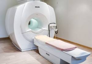
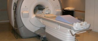
Modern high-field tomograph
The DiMagnit clinic has a closed-type tomograph from Philips, whose power is 1.5 Tesla. Using the device, it is possible to obtain images of the highest quality and detail.
Should you be afraid of the procedure?
Some patients are worried before the test. But their fears are in vain - magnetic resonance imaging is absolutely painless, and the effect of magnetic radiation on the body is safe.
Unlike other types of radiation diagnostics, MRI does not use ionizing radiation. The magnetic field does not have a carcinogenic or mutagenic effect on the cells of the body. Magnetic resonance scanning can be performed as often as needed.
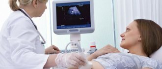
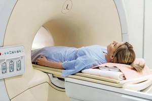
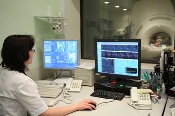
Carrying out an MRI procedure
The patient being examined lies on his back on a couch in a separate room where there is a tomograph. He is asked to wear headphones against the noise of the tomograph to protect his hearing in order to prevent panic from being in a closed room.
If the study is done with contrast, then a catheter is installed nearby to supply the drug. During the MRI, the staff is in an adjacent room, but the patient can talk to them.
When conducting the study, you must lie still for 25-30 minutes ; in special cases, when conducting a study of all parts of the spine, the time is increased to an hour.
It takes up to 40-50 minutes to decrypt data, diagnose and write to a magnetic disk. The results are delivered to the client.
The difference between MRI and CT and ultrasound
Magnetic resonance diagnostics has a number of advantages compared to ultrasound and computed tomography.
Ultrasound examination allows you to obtain a two-dimensional image of the area under study, but does not allow you to see a three-dimensional image of soft structures.
Computed tomography can be compared to MRI in terms of image clarity, but has a number of serious contraindications. CT is more often used to visualize hollow organs and bone structures, while MRI is much more effective at visualizing soft tissue.
What does an MRI show?
Magnetic resonance imaging is successfully used to diagnose diseases:
- thyroid gland;
- liver;
- gallbladder and ducts;
- pancreas;
- kidney;
- spleen;
- joints;
- spinal cord;
- vessels of the head, neck, abdominal region;
- pelvic organs;
- soft tissues;
- etc.
All of the above anatomical structures are perfectly visualized on MRI images. The diagnostic results make it possible to accurately identify abnormalities in the functioning of the organs being examined.
Who is prohibited from the procedure?
Diagnostics is contraindicated:
- with pacemakers;
- endoprostheses with ferromagnetic properties;
- metal brackets on the veins or arteries of the brain;
- implants with electronics in the middle ear;
- metal particles in the organs of vision.
These things affect the results of the study, and the magnetic fluxes created by the equipment provoke malfunctions in the operation of the stimulators. In cases of high levels of obesity, diagnosis is impossible due to the small size of the device.
Relative prohibitions on diagnosis include the first quarter of gestation, fears of tight spaces, foreign structures on the teeth, tattoos with metallic dye, foreign particles with metal found in other parts of the body. Diagnostic testing is not advisable for patients with a reusable medical device (pump) for insulin administration.
In what cases is MRI prescribed?
The wide capabilities of magnetic resonance diagnostics make its use indispensable in the following cases:
- the need for a primary diagnosis;
- conducting a comprehensive survey;
- preparation for surgery;
- monitoring the effectiveness of the therapy and treatment methods used.
In each individual case, the choice of diagnostic technique is made by the attending physician. Magnetic resonance imaging is more often used than other methods to detect diseases and injuries of soft tissues.
The MRI technique is indispensable for diagnosis:
- Neoplasms.
Magnetic resonance scanning can clearly identify the boundaries and size of the tumor and the extent of its invasion into soft tissue. No other radiation diagnostic technique is capable of providing such a clear and detailed picture of diseases.
MRI also makes it possible to determine the nature of the tumor with a high degree of probability. Malignant neoplasms have unclear boundaries and grow into surrounding tissues. Benign neoplasms, as a rule, are clearly differentiated from healthy tissues.
- Brain diseases.
The greater accuracy of magnetic resonance diagnostics makes it possible to visualize such small anatomical structures as the pituitary gland and the sella turcica. Also, MRI with contrast of the brain shows high efficiency for diagnosing demyelinating diseases (multiple sclerosis, Parkinson's disease, etc.), as it allows one to clearly see the structure of altered nerve tissues.
Brain images obtained with contrast-enhanced MRI of the brain are particularly clear because magnetic waves are difficult to image solid anatomical structures and there are no artifacts from the skull in the brain images.
- Diseases of the intervertebral discs.
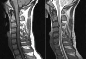
MRI images of the spine
Magnetic resonance imaging is the only diagnostic method that allows you to see intervertebral discs. Even modern diagnostic methods, such as computed tomography, allow you to see only the space between the vertebrae, while MRI gives a complete picture of the condition of the discs, the possible presence of hernias and protrusions.
The use of MRI is not limited only to the above diseases, but is used when it is necessary to identify and monitor a wide range of pathologies, congenital developmental anomalies, consequences of injuries and previous surgical interventions.
Who is prescribed MRI?
A neurologist writes a referral to patients:
- with frequent cephalgia, attacks of dizziness;
- suspicion of a formed neoplasm of any etiology;
- the need for examination before surgery;
- infectious pathologies, including meningitis, encephalitis;
- decreased hearing or visual acuity not associated with diseases of these organs;
- pathological changes in blood vessels, stenoses, aneurysms;
- pathologies of the auditory or optic nerve;
- MS, dementia;
- developmental defects of GM of congenital etiology;
- epilepsy, including post-alcohol forms;
- deterioration in concentration or memory;
- traumatic lesions;
- checking the results of cancer therapy
- impossibility of performing CT.
In childhood, diagnostic measures are recommended for children:
- with delays in motor and mental development;
- logoneurosis or other disorders of speech functionality;
- strange actions at home and in society;
- convulsions, fainting.
On a paid basis, you can undergo diagnostics without a referral from a local physician.
When to Apply Contrast
Magnetic resonance diagnostics can provide a very high degree of clarity of the resulting images. In most cases, the use of contrast is not required.
But when it comes to diagnosing tumors and small anatomical structures, a contrast agent can still be used.
The staining agents are made from the rare earth metal gadolinium and are injected intravenously into the patient during an MRI.
MRI contrast agents are much better tolerated than CT contrast agents. This makes the use of the dye safe even for patients with kidney pathology and does not require a preliminary test for creatinine, which is necessary for CT diagnostics with contrast.
MRI with contrast is used in the following cases:
- suspicion of a neoplasm;
- the need for differential diagnosis of a malignant tumor;
- study of the pituitary gland;
- the need to diagnose demyelinating diseases.
The use of contrast allows one to obtain a comprehensive picture of the disease, its course and the effectiveness of the therapy used.
MRI of the spine with contrast
Sometimes, in order to obtain a more informative result, an MRI with contrast is prescribed . A solution based on the element gadolinium is used for staining. Unlike iodine used in CT scans, it is non-toxic, there are no allergic reactions, this contrast is well tolerated and quickly excreted from the body. A contrast agent is introduced to improve the clarity of the images and the information content of the study.
High-definition images show all structures and foci of the disease. Most often, safe drugs are used for MRI with contrast: Omniscan and Prohans; Magnevist and Dotarem are also used. The drug is administered intravenously, very rarely through a catheter into the spine. Preparations with gadolinium are safe; during the entire period of their use, there are no known cases of harmful effects on the body. Their administration is well tolerated and no side effects are observed.
If side effects occur, they go away on their own after some time and no additional treatment is required. Nausea may occur, intensify to vomiting, a feeling of a rush of blood, and heat may be felt in the lower back. Such side effects usually develop in the patient within an hour after administration, and during this time it is necessary to remain in the facility and leave it after being fully convinced that there is no reaction to the study.
Contraindications to the use of a contrast agent during MRI of the spine are:
- early pregnancy;
- kidney disease;
- allergies in the development stage.
Contraindications for MRI
Despite the fact that magnetic resonance diagnostics is a safe technique, the study has a number of absolute contraindications, in the presence of which diagnostics are prohibited:
- presence of a pacemaker, neurostimulator, insulin pump;
- vascular clips on the arteries of the brain;
- the patient’s inability to maintain a stationary position for various reasons;
- early childhood up to 5 years;
- the patient’s weight is more than 130 kg and body girth is more than 150 cm;
- first trimester of pregnancy;
There are also a number of conditions in which MRI examination is carried out with caution:
- severe pain syndrome, in which it is difficult for patients to remain in a motionless position for a long time;
- fear of confined spaces;
- psychical deviations;
- second and third trimester of pregnancy.
The presence of various prostheses and implants in the patient's body may be a contraindication to MRI if they are made of metals sensitive to magnetic radiation. Modern medical devices are most often made of titanium and other materials that are inert to the effects of magnetic fields. Their presence in the body does not interfere with MRI.
Indications for MRI of the spine
It's no secret that timely studies guarantee an accurate diagnosis and complete cure. An MRI is prescribed by the attending physician, this can be a neurologist, surgeon, or traumatologist. The patient himself can also undergo examination and, after receiving the results, consult a doctor.
Patient complaints and indications for MRI:
- Cervical region. For loss of consciousness, dizziness, surges in blood pressure, noise in the ears and head, numbness of the neck and upper limbs, pain when turning the head, frequent migraine attacks.
- Thoracic department. For pain in the shoulder blades and behind the sternum, if you hear a crunching sound in the vertebrae when moving, if the pain intensifies with exercise.
- Lumbar and sacrum. Restricted turns of the body, pain increases with physical activity, numbness in the lower extremities, difficulty urinating, a feeling of fullness in the intestines and rectum, and urinary incontinence.
- Coccyx. Pain and fistulas, the shape changes, when pressing with fingers, swelling is felt.
Once the diagnosis is established, MRI is indicated for the following diseases:
- for acute or chronic osteochondrosis;
- disc herniation or protrusion;
- spondyloarthrosis or spondyloarthritis, deformation of the joints of the vertebrae;
- myelitis, inflammation of the spinal cord;
- multiple sclerosis;
- for cancer, stage identification;
- congenital or acquired vertebral displacements;
- to monitor the dynamics of disease development.
How is an MRI performed?
Before the procedure, the radiologist questions the patient about the presence of contraindications to the study. The patient is asked to remove any metal accessories, including clothing with metal fittings, and lie down on a couch, which is then placed in the CT scanner tube.
During the diagnosis, it is strictly forbidden to move, as this may affect the clarity of the resulting images.
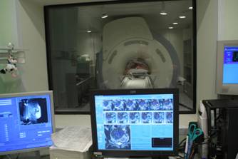
Scanning procedure
High-field tomographs produce a fairly high level of noise, which can cause some discomfort to patients. The medical department will provide you with headphones that will play pleasant music, drowning out the sounds of the operating device.
The tomograph scans the patient’s body in different projections and instantly transmits the images to the computer screen. The interpretation of the results by the research physician begins even before the procedure is completed.
Who will interpret the MRI of the head?
The results of the MRI diagnostics are deciphered by the radiologist conducting the examination. Carefully, without omissions, analyzes all the information received and draws up a conclusion. The person is given a tomographic examination protocol, where the specialist indicates the appearance, size, and structure of the areas being diagnosed. The protocol ends with a conclusion documenting the existing violations.
The patient receives a tomogram, an electronic version of the image recorded on an external storage medium. Certain clinics provide scans only on film, on disk/flash drive - upon the patient’s request. The doctor who prescribed MRI diagnostics of the brain (neurologist, urologist, endocrinologist) evaluates the data obtained.
The radiologist, when drawing up the descriptive part, the conclusion to the tomograms, should not assess the importance of the detected pathology, but clarifies the characteristics of the defective area.
The conclusion indicating the significance of the found disorders and the method of treatment is formed by the attending doctor, so the results of the examination must be given to him. The radiologist is able to refer the patient to a specific doctor of the required profile, determine when to do an MRI of the head, and choose the duration and type of examination.
Getting Results
Scan results are available immediately after the end of the study. Many of the images obtained are carefully examined by a radiologist, and a detailed report is drawn up describing both the normal anatomy of the area under study and possible abnormalities and pathologies.
15-30 minutes after the procedure, the patient is given a written report and a computer disk with the resulting images.
Magnetic resonance imaging is a modern, safe type of radiation diagnostics that allows you to obtain accurate and quick results and thoroughly examine the area under study. MRI helps to identify many diseases and abnormalities even at the initial stages of their development.
| MRI of the pancreas in Rostov-on-Don |
| MRI of the abdomen with contrast |
| MRI of the spine, what does it show? |
| MRI of ureters |
| MRI of the abdominal cavity, which organs are checked? |
| What does an MRI of the knee show? |
- Magnetic resonance imaging (MRI)
- Computed tomography (CT, MSCT)
- X-ray studies
- Mammography
- Department of Radionuclide Diagnostics
Magnetic resonance imaging is the most important modern diagnostic method based on the magnetic field. MRI has no radiation exposure and is considered harmless to the human body. MRI is often used to examine the brain and spinal cord, spine, joints, abdominal and pelvic organs, heart and blood vessels, and mammary glands. This method accurately identifies tumors in the early stages, allows one to predict the risk of stroke, and determine the cause of back pain. MRI has a high soft tissue contrast, so even different tissues within the same organ can be clearly distinguished on MR images. To increase the information content of MRI, special contrast agents are sometimes used. The drugs are well tolerated and safe for health. The need to administer a contrast agent is determined by the attending physician or specialist performing the MRI. Research does not require special preparation. An abdominal MRI should not be done on a full stomach, although a light breakfast will not interfere with the examination in any way. It is best to perform a pelvic examination with a full bladder, so it is recommended to drink several glasses of liquid 30-60 minutes before the MRI. The duration of an MRI examination is usually 20 minutes. If it is necessary to launch additional programs (decided by the doctor individually for each study), the study time can be increased. Pre-registration and inquiries by phone (495) 942-40-20. Registration hours: Monday-Friday from 8.00 to 20.00, Saturday - from 9.00. until 17.00.
Absolute contraindications to MRI are:
- Presence of a pacemaker
- Metal objects in the brain or eye area
Pregnant patients, overweight patients (more than 130 kg), with the presence of metal in the body, epilepsy, or fear of confined spaces should consult with a doctor - a specialist in radiation diagnostics before conducting the study.
MRI of the brain is performed to diagnose:
- Brain tumors
- Brain development abnormalities
- Ischemic strokes and hemorrhages
- Post-traumatic changes (brain contusion, cysts, hematomas)
- Vascular anomalies (aneurysms, arteriovenous malformations)
- Pathologies of the pituitary gland and pineal gland
- Pathologies of the eyeball and inner ear
- Focal changes in the brain in patients with inflammatory diseases of the brain (multiple sclerosis, encephalomyelitis, encephalitis)
- Focal changes in the brain in patients with high blood pressure or cervical atherosclerosis
And also to identify the causes of headaches and dizziness.
MRI of the spine is performed to diagnose:
- Hernias and protrusions of intervertebral discs
- Spinal cord diseases (spinal cord entrapment due to disc herniation, trauma, vertebral fractures, vascular, inflammatory and post-traumatic focal changes in the spinal cord)
- Tumors and metastases of the spinal cord, spinal nerves and vertebrae
- Post-traumatic changes (bruises, displacements, fractures of vertebral bodies, assessment of spinal cord compression)
- Inflammatory changes (discitis and spondylitis, abscesses and fistulas)
And also to identify the causes of back pain, pain and sensory disturbances in the legs and arms, and headaches.
MRI of joints is performed to diagnose:
- Traumatic ruptures of ligaments, tendons, menisci, labrum
- Degenerative changes in the menisci, labrum
- Deforming arthrosis (study of the condition of articular cartilage, severity of osteophyte development, assessment of subchondral changes)
- Inflammatory changes (arthritis, synovitis, bursitis, as well as bone abscesses and osteomyelitis)
- Traumatic bone changes (bruises and fractures)
- Avascular necrosis of bones
- Tumors and metastases of bones, muscles and soft tissues surrounding the joint
And also, to identify the causes of pain in the joints, impaired joint mobility, swelling of the soft tissues around the joints.
MRI of the abdominal cavity is performed to diagnose:
Focal changes in the liver (tumors, metastases, cysts, hemangiomas)- Other liver diseases (fatty hepatosis, cirrhosis)
- Cysts and tumors of the spleen.
- Tumors, cysts and pseudocysts of the pancreas.
- Cysts, tumors, angiomyolipomas of the kidneys.
- Tumors and nodules of the adrenal glands.
- Parasitic diseases of internal organs.
To assess the condition of the intra- and extrahepatic bile ducts and pancreatic ducts in case of cholelithiasis, obstructive jaundice, hepatitis, cholangitis, MR cholangiography is possible.
MRI of the pelvic organs is performed to diagnose:
- Tumors and polyps of the bladder.
- Anomalies of the development of the bladder and ureters.
- Prostate tumors, prostatitis, prostate adenoma.
- Tumors of seminal vesicles and vesiculitis.
- Tumors and developmental anomalies of the testicles.
- Ovarian cysts and tumors.
- Tumors and fibroids of the uterus, adenomyosis and ectopic endometriosis.
It can also be used as a diagnostic method for rectal tumors. Cardiac MRI is performed for:
- Assessment of the structure and function of the heart chambers, valves, large vessels, pericardium.
- Estimates of myocardial contractility of the left and right ventricles.
- Diagnosis of congenital and acquired heart defects, as well as for assessing the discharge of blood between the systemic and pulmonary circulation.
- Assessment of myocardial viability after infarction, as well as visualization of post-infarction scar
And also, MRI is used as a method of clarifying diagnosis in case of conflicting results of echocardiography. Cost of examination Pre-registration and inquiries by phone (495) 942-40-20 . Registration hours: Monday-Friday from 8.00 to 20.00, Saturday - from 9.00. until 17.00.