- Areas of medicine in which the procedure is used
- What does it show
- Types of ultrasound
- thyroid gland
- liver
- mammary glands
- gallbladder
- pancreas
- kidneys, adrenal glands and ureters
- joints
- hearts
- vessels
- during pregnancy
- uterus and appendages
- soft tissue
- eye
- Benefits of Ultrasound
- How is the diagnosis carried out?
- What is the result of the procedure
- When to do it
- Techniques
- Types of ultrasound in our clinic
Ultrasound examination is a procedure for examining the internal organs of a person, which is based on ultrasound.
An ultrasound machine has a sensor and transmitter in a small housing, which the doctor moves over the patient’s skin. The transmitter sends ultrasound into the patient's body, which is reflected differently from different tissues of the internal organs and changes its properties. The sensor, capturing the reflected ultrasonic waves, converts them into an electrical signal and sends them to the computer of the ultrasound machine, which converts the received signal into an image on the monitor screen. The ultrasound doctor analyzes the image on the screen and draws conclusions about the condition of the patient’s internal organs. The image on the ultrasound machine screen can be 2D, 3D or 4D in black and white or color. 2D is a classic version of ultrasound, and 3D and 4D are experimental image modes available only on new devices. 2D is a regular flat picture. 3D is a static three-dimensional image of an internal organ. 4D is a three-dimensional image of an organ in motion.
Ultrasound is a precise procedure that can detect the disease in its early stages. In addition, ultrasound is completely painless and safe for health.
Areas of medicine in which ultrasound is used
Ultrasound examination is used in almost all areas of medicine:
- Obstetrics and gynecology
. Ultrasound of the female genital organs, the course of pregnancy and fetal development. - Internal organs
. Search for pathology of all internal organs: liver, kidneys, pancreas, spleen, bladder, prostate gland and others. - Cardiology and Vascular Surgery
. Study of the heart, veins and blood vessels. Blood flow is examined using Doppler ultrasound. A 4D echocardiogram allows you to obtain a 3D image of the heart in dynamics. The heart is examined using both a sensor through the sternum and through the esophagus. - Ophthalmology
. Ultrasound is used to determine the position of the lens to measure the size of the eye. - Echoencephalography
. Studying the brain using ultrasound. - And others
What does an ultrasound show?
An ultrasound examination shows on the monitor screen of an ultrasound machine a two-dimensional, three-dimensional static or dynamic black-and-white or color image of internal organs, blood vessels and blood flow. The image depends on the model of the ultrasound machine itself.
The movement of blood flow and organs is determined using the Doppler effect, in which ultrasonic waves are reflected from moving objects with a changed frequency. Changing the ultrasound frequency gives an understanding of the nature of the movement of blood flow or the heart. In addition, Doppler ultrasound is used to construct a 3D image of organs when the transmitter and sensor move.
A 4D echocardiogram performed to study the heart allows you to obtain a three-dimensional image of the heart in dynamics on the monitor screen, allowing you to assess the volume, structure and functioning of this organ.
Depending on the patient’s complaints, the doctor selects the correct ultrasound operating modes and the location of the sensor in order to most accurately study the desired organ. The correctness of the diagnosis and medical conclusion largely depend on the professionalism of the ultrasound diagnostician. An ultrasound machine is simply a tool that can show pathology even at the earliest stage, but how the results of its work will be interpreted depends on the professionalism of the doctor.
Types of ultrasound
Ultrasound examination can be divided according to the types of organs that are diagnosed:
- thyroid;
- liver;
- mammary gland;
- gallbladder;
- pancreas;
- kidneys, adrenal glands and retroperitoneum;
- joints;
- heart;
- vessels;
- during pregnancy;
- uterus and appendages;
- soft tissues;
- mammary gland;
- eyes.
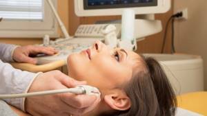
Ultrasound of the thyroid gland
The thyroid gland is part of the endocrine system and secretes hormones containing iodine. The thyroid gland controls cell growth and development of the entire body.
Ultrasound of the thyroid gland may be required if:
- excess weight or sudden changes in weight;
- high irritability;
- increased drowsiness;
- fatigue;
- sudden baldness;
- sore throat;
- the appearance of tumors to the touch;
- pregnancy planning;
- diabetes mellitus;
- age after 40 years;
- work in hazardous production;
- completion of a course of taking hormone-containing drugs.
During an ultrasound examination of the thyroid gland, the doctor moves an ultrasound probe along the patient's neck. The doctor evaluates the structure, size of the thyroid gland and the acoustic density of the organ, which can be:
- normal;
- reduced;
- increased;
- echo negative;
An ultrasound of the thyroid gland can show diseases such as:
- tumors;
- inflammation;
- cyst;
- hypothyroidism;
- hyperthyroidism.
To prepare for the procedure:
- Remove all jewelry from your neck and put on clothes that do not cover your neck;
- To avoid nausea, it is better not to eat before the procedure;
- For women, it is better to do the procedure on days 7-9 of the menstrual cycle.
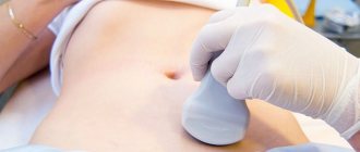
Ultrasound of the liver
The liver is responsible for removing toxins from the body, carries out hematopoiesis, synthesizes carbohydrates and proteins, and also performs other important functions.
An ultrasound of the liver may be required if:
- doctor's appointment;
- yellowness of the skin, mucous membranes and whites of the eyes;
- change in urine color;
- increased blood bilirubin;
- pain and discomfort in the right hypochondrium;
- vomit;
- nausea;
- long-term medication use;
- chronic liver diseases;
- alcohol addiction;
- suspicion of a tumor;
- abdominal trauma.
During an ultrasound examination of the liver, the doctor moves an ultrasound probe over the patient's chest and abdomen. The doctor evaluates the size and structure of the liver.
An ultrasound of the liver can show diseases such as:
- tumor;
- cyst;
- calcifications;
- cirrhosis;
- other disorders and diseases.
To prepare for the procedure:<.p contentScore=”12415″>
- Do not eat 8 hours before the procedure;
- If there is constipation, then do an enema 1 hour before the procedure;
- Before the procedure, do not drink more than 1.5 liters of water per day;
- For 5 days, eliminate foods that cause gas (bread, cabbage, soda, fruits and dairy products);
- Do it 2 days after examining organs with contrast.
Ultrasound of the mammary glands
The mammary gland is located in the female breast and is intended to produce milk for feeding the child.
Ultrasound of the mammary glands may be required if:
- Detection of a neoplasm upon palpation;
- chest pain or swelling;
- Enlarged lymph nodes;
- Discharge from the load;
- Changes in nipple shape;
- Chest injury;
- Pregnancy planning;
- Routine examination after 45 years;
- Diagnosis of lactation;
- Preparation for surgery;
- Preparation for IVF;
- Research of the condition of implants.
During an ultrasound examination of the mammary glands, the doctor moves an ultrasound probe over the patient's breast. The doctor evaluates the size and structure of the breast. In addition, the ultrasound diagnostician evaluates the skin, nipples, ducts, pectoral muscles, subcutaneous fat and regional lymph nodes.
Ultrasound of the mammary glands can show diseases such as:
- tumor;
- cyst;
- papillomas;
- size of lymph nodes;
- mastopathy.
To prepare for the procedure:
- Ultrasound should be done from 4 to 13 days of the menstrual cycle;
- Before the procedure, the woman takes off her clothes to the waist.
Ultrasound of the gallbladder
The gallbladder is an organ located just below the liver at the level of the last rib, responsible for the preparation and temporary storage of bile.
An ultrasound of the gallbladder may be required if:
- icteric syndrome;
- injuries in the area of the gallbladder and liver;
- pain in the liver area;
- suspicion of stone formation;
- suspicion of neoplasms;
- diagnosis during treatment.
During an ultrasound examination of the gallbladder, the doctor moves the ultrasound probe over the skin in the area of the liver and last rib. The doctor evaluates the size, structure and location of the gallbladder, looking for the presence of stones.
An ultrasound of the gallbladder can show diseases such as:
- tumor;
- dyskinesia;
- stones;
- cholecystitis;
- congenital pathologies.
To prepare for the procedure:
- Avoid foods that cause gas (bread, cabbage, soda, fruits, beans and dairy products);
- Do not eat 10 hours before the procedure.
Ultrasound of the pancreas
The pancreas is part of the group of organs of the digestive system and is responsible for the production of digestive enzymes and hormones that regulate the metabolism of proteins, fats and carbohydrates.
An ultrasound of the pancreas may be required if:
- pain in the left hypochondrium, surrounding and above the navel.
- yellow tint of the skin and mucous membranes;
- suspected neoplasm;
- nausea;
- vomiting;
- constipation;
- diarrhea;
- acute pancreatitis;
- increased body temperature;
- abdominal surgeries.
During an ultrasound examination of the pancreas, the doctor moves the ultrasound sensor along the skin in the area of the organ from the central part of the abdomen to the area of the left hypochondrium. In particularly difficult cases, an endoscopic ultrasound of the pancreas is performed, in which a sensor on an endoscope is inserted through the esophagus into the stomach and into the duodenum. The doctor evaluates the size, structure and location of the gallbladder, looking for the presence of stones.
An ultrasound of the pancreas can show diseases such as:
- congenital anomalies;
- pancreatitis;
- tumors;
- cysts;
- inflammation;
- diabetes.
To prepare for the procedure:
- Avoid foods that cause gas (bread, cabbage, soda, fruits, beans and dairy products);
- Light protein-free diet;
- Quitting alcohol, smoking and chewing gum;
- Using an enema before the procedure;
- Do not eat 10 hours before the procedure.
Ultrasound of the kidneys, adrenal glands and ureters
The kidneys are an organ that is part of the body’s urinary system and is responsible for purifying the blood. The adrenal glands are located on top of the kidneys and serve to produce vital hormones - adrenaline, norepinephrine and corticosteroids. The ureters connect the kidneys to the bladder.
Ultrasound of the kidneys, adrenal glands and ureters may be required if:
- headaches due to increased blood pressure;
- pain in the lumbar region;
- inflammatory processes;
- deviations in the results of urine and blood tests;
- renal failure;
- suspected neoplasms;
- accumulation of gases in the parenchyma;
- suspected presence of stones;
- kidney operations.
During an ultrasound examination of the kidneys, adrenal glands and ureters, the doctor moves an ultrasound probe over the skin in the area of the kidneys. An ultrasound diagnostician analyzes the size and structure of the kidneys, the presence of stones and other pathologies.
Ultrasound of the kidneys, adrenal glands and ureters can show diseases such as:
- tumors;
- cysts;
- inflammation;
- vascular diseases;
- pyelonephritis;
- hydronephrosis;
- formation of stones and sand.
To prepare for the procedure:
- For 3 days, eliminate foods that cause gas (bread, cabbage, soda, fruits, beans and dairy products);
- Do not eat 10 hours before the procedure.
Ultrasound of joints
Joints are movable joints of the bones of the skeleton that allow a person to move.
Ultrasound of the joints may be required if:
- painful sensation in the joint area;
- inflammatory process;
- injuries;
- the appearance of limited joint mobility;
- the appearance of a crunch;
- swelling.
During an ultrasound examination of joints, the doctor moves an ultrasound sensor over the skin in the joint area and analyzes the structure, functionality and presence of pathologies.
Ultrasound of the joints can show pathologies:
- muscles;
- cartilage;
- joint capsules;
- tendons.
Ultrasound examination of joints can detect rheumatic diseases in the early stages. Ultrasound is also performed during joint surgery.
No preparation is required for the procedure.
Ultrasound of the heart
The heart is a hollow muscular organ that provides blood circulation in the human body.
Heart ultrasound may be required if:
- ischemic disease;
- heart disease;
- heart rhythm disturbances;
- pericardial disease;
- thromboembolism;
- suspected neoplasm;
- infective endocarditis;
- arterial hypertension.
During an ultrasound examination of the heart, the doctor moves an ultrasound probe over the skin in the chest area. If there is not enough information when analyzing the heart with a sensor through the chest, then the ultrasound sensor is lowered through the esophagus, which allows you to get as close to the heart as possible. An ultrasound scanner analyzes the size and structure of the heart, the dynamics of heartbeat and blood flow. Modern ultrasound machines allow you to create a 2D and 3D image of the heart on the screen in black and white, color in static and motion. This allows you to study the heart in detail.
Ultrasound of the heart shows:
- organ anatomy;
- interaction of the heart with neighboring organs;
- the structure of chambers, walls and large vessels;
- heart valve function;
- pericardial condition;
- heartbeat dynamics.
No special preparations are required for an ultrasound examination of the heart. The patient comes to the doctor's office, undresses to the waist and lies down on the couch.
Ultrasound examination of the heart may be difficult if:
- overweight;
- some lung diseases;
- narrow intercostal spaces;
- wide ribs.
Vascular ultrasound
Blood vessels are tubular structures in the body through which blood moves.
Vascular ultrasound may be required if:
- stroke;
- atherosclerosis;
- thrombosis;
- thromboembolism;
- varicose veins;
- myocardial infarction;
- convexity of the vessel wall;
- suspicion of neoplasms;
- blood clotting disorders;
- dizziness;
- headaches;
- fainting;
- hypertension;
- memory problems;
- darkening or redness of the skin;
- impaired skin sensitivity;
- swelling;
- impaired blood supply to the fetus during pregnancy.
There are 3 methods of ultrasound examination of blood vessels:
- Doppler ultrasound (USDG)
- shows a two-dimensional image of blood flow and gives an idea of its presence and nature. - Duplex scanning (USD)
- shows not only a two-dimensional image of blood flow, but also allows you to evaluate the walls of blood vessels, their structure, the course of veins and arteries. - Triplex scanning
- demonstrates the direction of blood flow.
The integrated use of vascular ultrasound methods makes it possible to accurately diagnose diseases in the early stages and begin treatment in a timely manner.
During an ultrasound examination of blood vessels, the doctor moves an ultrasound sensor across the skin in the direction of the vessels, analyzing the blood flow and their condition.
Doppler ultrasound is done to examine blood vessels, arteries or veins:
- heads;
- neck;
- upper and lower extremities;
- eye.
Duplex scanning is done to examine blood vessels, arteries or veins:
- kidney;
- abdominal cavity;
- hearts;
- pelvis;
- fetus in the womb.
To prepare for the procedure:
- For 3 days, eliminate foods that cause gas (bread, cabbage, soda, fruits, beans and dairy products);
- In some cases, the procedure should be done on an empty stomach;
- Avoid alcohol and smoking.
Factors such as:
- serious condition of the patient;
- poor skin condition.
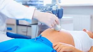
Ultrasound during pregnancy
During pregnancy, ultrasound examinations are performed scheduled and unscheduled.
Routine ultrasound during pregnancy is performed 3 times for:
- 11-14 weeks;
- 18-24 weeks;
- 30-34 weeks.
During the procedure, the condition of the uterus and embryo is assessed. Ultrasound also allows you to determine the sex of the child at 12-13 weeks of pregnancy.
An unscheduled ultrasound during pregnancy is performed if there is:
- suspicion of an ectopic or frozen pregnancy;
- suspicion of fibroids or other neoplasms;
- bloody issues;
- threat of miscarriage;
- previous infection during pregnancy;
- other complications and diseases during pregnancy.
Ultrasound during pregnancy is performed in 2 ways:
- transabdominal, in which the doctor moves the ultrasound machine sensor across the woman’s abdomen;
- transvaginally, in which a sensor is inserted into a woman’s vagina, is performed only in the first trimester of pregnancy.
To prepare for the procedure:
- For 3 days, eliminate foods that cause gas (bread, cabbage, soda, fruits, beans and dairy products).
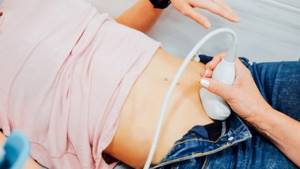
Ultrasound of the uterus and appendages
The uterus and appendages are the organs responsible for conception and gestation.
Ultrasound of the uterus and appendages may be required if:
- menstrual irregularities;
- pain and discomfort in the lower abdomen;
- therapy control;
- ECO;
- suspicion of infertility;
- preventive examination.
During an ultrasound examination of the uterus and appendages, the doctor moves an ultrasound probe over the skin in the lower abdomen and analyzes the condition of the organs. Ultrasound of the uterus and appendages is carried out in 2 ways: transabdominal, in which the doctor moves the ultrasound machine sensor along the woman’s abdomen; transvaginally, in which the sensor is inserted into the patient’s vagina.
Ultrasound of the uterus and appendages shows:
- abnormalities of the uterus;
- development of pregnancy;
- position and size of the organ;
- tissue structure;
- neoplasms;
- endometritis;
- other pathologies.
To prepare for the procedure:
- For 3 days, eliminate foods that cause gas (bread, cabbage, soda, fruits, beans and dairy products).
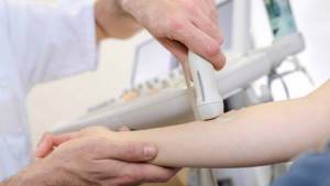
Ultrasound of soft tissues
Soft tissues include skin, fatty tissue, connective tissue, tendons, muscles, and lymph nodes.
Ultrasound of soft tissues may be required if:
- Pain in joints, muscles or ligaments;
- Regular occurrence of purulent formations;
- Suspicion of neoplasms in soft tissues;
- Rheumatoid diseases;
- Before soft tissue surgery;
- Enlarged lymph nodes;
- Therapy.
During an ultrasound examination of soft tissues, the doctor moves an ultrasound probe over the skin and analyzes the condition of the soft tissues.
Ultrasound of soft tissues shows:
- Various pathologies;
- Neoplasms;
- Tendon injuries;
- Complications after surgery.
No preparation is required for the procedure.
Ultrasound of the eyes
Eyes are the organs responsible for human vision.
An eye ultrasound may be required if:
- eye pathologies identified during examination;
- changing the size of the eye socket;
- hypertension;
- diabetes;
- suspected neoplasm;
- eye injury;
- retinal detachment;
- thyrotoxicosis;
- eye surgeries.
During an ultrasound examination of the eye, the doctor:
- in A-mode, instills an anesthetic drug into the eye and moves the sensor over the eyeball;
- in B-mode, lubricates the eyelid with gel and moves the sensor over the eyelid.
If necessary, for ultrasound examination, the doctor diagnoses the eye in two modes.
Ultrasound of the eyes shows:
- eye structure;
- eye size;
- lens injuries;
- neoplasms;
- foreign bodies;
- retinal detachment;
- rate of myopia progression;
- dynamics of processes inside the eye.
Norm and interpretation of abdominal ultrasound in children
Proper preparation for ultrasound facilitates clear visualization of all structures being examined. The doctor evaluates:
- sizes of organs and their individual parts;
- location features;
- quality of blood supply;
- wall thickness (for hollow organs);
- quality and edges (smooth, bumpy);
- homogeneity of the internal structure;
- the presence or absence of congenital anomalies;
- the presence or absence of pathological changes: inflammation, foreign bodies, stones, parasites, tumors, duct blockage, impaired blood supply, mechanical damage, etc.
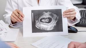
Normal and changed parameters, as well as identified pathologies, are recorded in the report, which, if necessary, is supplemented by a printout of problem areas or a video recording of the study. The attending physician interprets the results.
If the child’s parents have studied in advance the question of how to properly prepare for an ultrasound of the abdominal cavity, during the examination the doctor will receive a clear and complete picture of all internal structures, without distortions and artifacts. This will allow pathological changes or their absence to be identified in time in order to take action if necessary.
How is ultrasound diagnostics performed?
Ultrasound examination is carried out according to the following scheme:
- A patient walks into a doctor's office;
- The doctor asks the patient to expose an area of the body for diagnosis;
- The doctor asks the patient to lie on the couch, sit or stand for a successful examination;
- The doctor lubricates the ultrasound probe with gel, which allows it to glide easily over the skin. During an ultrasound, the doctor prepares the endoscope and probe for insertion into the body using a probe inserted into the body.
- The doctor begins to move the ultrasound sensor over the body, analyzing the internal organs. Or manipulates a sensor inside the patient's body to obtain the desired image.
- After the procedure, the doctor asks the patient to wipe the gel off the skin with a napkin and get dressed.
- The doctor provides an ultrasound report.
How to get to us?
Address: St. Petersburg, Kalininsky district, st. Rustaveli, 66 lit. G (metro station Grazhdansky Prospekt)
Phone, 8-967-342-13-11
From metro station Grazhdansky Prospekt
Free parking, plenty of parking spaces.
Art. m. Grazhdansky Ave. 7 minutes walk
Getting to us: Minibuses: 92, 121, 188, 199, 205, 208, 214, 284, 288,
Trams: 51, 100. Buses: 93, 121, 139, 177
Stop - Prosveshcheniya Ave. 106\ st. Rustaveli or Murino railway station
Metro Devyatkino
The MRI center and RIORIT clinic are a 30-minute walk away
or 10 minutes drive from Devyatkino.
By train it’s just one stop at the railway station. Murino.
Minibuses No. 205, 205a., 619 also go to us from Devyatkino
When to do an ultrasound examination
Ultrasound examination should be done:
- As prescribed by another doctor;
- On your own initiative, if something bothers you or as a preventive measure.
Ultrasound is usually prescribed for:
- Pain and discomfort;
- Swelling;
- Injuries;
- High blood pressure;
- Inflammatory processes;
- Deviations from the norm in tests;
- Before surgery;
- To control treatment;
- For preventive purposes.
Diet before abdominal ultrasound for a child
Dietary restrictions are an important stage in preparing a child for an ultrasound. Some foods and drinks can impair visualization by producing large amounts of intestinal gas. Accordingly, 2–3 days before ultrasound diagnostics it is necessary to completely exclude:
- brown bread, pastries;
- whole milk;
- legumes;
- fatty and fried foods;
- sweets;
- carbonated drinks, kvass;
- chewing gum;
- a number of vegetables and fruits: onions, mushrooms, all types of cabbage, grapes.
Priority should be given to light foods, steamed or baked dishes. The diet may include:
- porridges from various cereals cooked in water;
- boiled or stewed chicken, veal, fish;
- eggs (in limited quantities).
On the eve of the study, dinner should be as light as possible.
It is also important to ensure timely bowel movements. The diet must contain fiber to ensure regular bowel movements. Ideally, the child should go to the toilet the day before or on the day of the test. If your baby is prone to constipation, you should discuss with your pediatrician in advance the possibility of taking laxatives or a Microlax enema before the ultrasound.
Ultrasound techniques
Ultrasound examination is carried out in 3 modes:
- A-mode
. A research method that produces a one-dimensional image in which one coordinate is the reflected signal from the boundary of media with different acoustic resistance, and the other is the distance to this boundary. - B-mode
. A research method that provides a two-dimensional image in real time, with the ability to assess the morphological state. - M-mode
. A research method that provides a one-dimensional image, in which one is the distance from the sensor to the structure being located, and the other is time.
Dopplerography can be:
- Spectral, giving an idea of the movement of moving media;
- Continuous, allowing you to evaluate blood flows moving at high speed;
- Pulse, allowing you to study blood flow at any point;
- Tissue, allowing to evaluate the myocardium;
- Color Doppler mapping, which allows you to examine blood flows in the heart and in relatively large vessels;
- Power Dopplerography, which allows you to evaluate the vascularization of organs and pathological areas;
- Three-dimensional Doppler mapping, which conveys a three-dimensional picture of organs in real time;
- Combined options that allow you to obtain additional information.
Types of ultrasound in our clinic
We perform ultrasound:
- kidney;
- abdominal organs;
- during pregnancy;
- lymph nodes;
- thyroid gland;
- hip joint;
- soft tissues;
- Bladder;
- prostate gland;
- pelvic organs;
- ultrasound imaging with color Doppler mapping of blood flow;
- other organs.
You can make an appointment with an ultrasound diagnostician and find out the cost of the study by calling 8
,
+7 (4872) 49-57-57
.
You can also make an appointment for an ultrasound examination at our hospital through the online form on the website. All articles "
Ultrasound of the abdominal cavity for a child from 1 to 3 years old: what preparation is needed
As a child grows, his diet expands. Its menu includes more and more adult foods, including those that can cause constipation or excessive gas production. In order to adequately prepare a child aged 1–3 years for an abdominal ultrasound, it is necessary to both control the diet and maintain the required period of fasting. It is recommended to refrain from eating for at least 3 hours before the procedure, and not to drink liquids one hour before the test. If it is difficult for the baby to tolerate, 1-2 small sips of clean water are allowed.