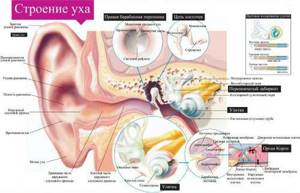
The external auditory canal and auricle are the initial sections of the hearing organ, the main part of which is located deeper in a special section of the temporal bone of the skull. Sound enters the middle ear, passes through the ossicles of the tympanum and enters the inner ear. Representing a labyrinth in the shape of a cochlea and semicircular canals, the inner ear contains receptors that transmit sound signals to the brain, as well as information about the position of the body - the vestibular apparatus is also located here.
The final target of the sound signal is the auditory center located in the temporal lobes of the brain. Cortical neurons process information from the vestibulocochlear nerve and form auditory sensations.
Only the outer ear is available for direct examination and study at a doctor’s appointment. The middle and inner ear can only be examined using instrumental research methods. The most informative and safest is magnetic resonance imaging. From the images you can evaluate the structure, anatomical features and any pathological processes.
The capabilities of MRI are expanded by the use of contrast agents during scanning. The preparations contain components with paramagnetic properties. After intravenous administration, the signal from pathologically altered tissues is enhanced. This way, tumors, vascular diseases, and inflammatory processes are better diagnosed.
Why is it important to promptly diagnose ear canal diseases?
- the middle and inner ear are located in close proximity to the brain and meninges, which increases the risk of their involvement in the event of an infectious lesion;
- many diseases have similar symptoms - it is important to make an accurate diagnosis as early as possible, when it is possible to prevent serious consequences;
- MRI is the most accurate and safe diagnostic method.
Which is better: MRI or CT
During MRI, the patient is not exposed to ionizing radiation, as with X-ray methods, and there are no restrictions on the number of procedures performed. Computed tomography gives a large radiation dose to the body and is not recommended for children and pregnant women.
In each clinical case, the choice of method remains with the doctor, so it is important to consult a specialist if symptoms appear. After examining the complaints and checking the ear using available methods, the doctor gives a referral for the most appropriate examination.
Computed tomography is more informative for injuries and damage to the skull, damage to bone structures, and accumulation of pathological fluid in the middle ear cavity. MRI will more accurately show the condition of soft tissues, tumors, inflammatory processes, and vascular diseases.
Leading specialists in the field of otolaryngology:
Volkov Alexander Grigorievich
Volkov Alexander Grigorievich, Professor, Doctor of Medical Sciences, Head of the Department of Otorhinolaryngology, Rostov State Medical University, Honored Doctor of the Russian Federation, I Full Member of the Russian Academy of Natural Sciences, Member of the European Society of Rhinologists.
Read more about the doctor
Book a consultation with a specialist

Boyko Natalya Vladimirovna
Boyko Natalya Vladimirovna , Professor, Doctor of Medical Sciences.
Read more about the doctor
Book a consultation with a specialist

Zolotova Tatyana Viktorovna
Tatyana Viktorovna Zolotova, Professor of the Department of Otorhinolaryngology, Rostov State Medical University, Doctor of Medical Sciences, Corresponding Member of the Russian Academy of Economics, Best Inventor of the Don (2003), Awarded: V. Vernadsky Medal (2006), A. Nobel Medal for Merit in the Development of Invention (2007) .).
Read more about the doctor...
Book a consultation with a specialist
Karyuk Yuri Alekseevich
Karyuk Yuri Alekseevich - otolaryngologist (ENT) of the highest qualification category, candidate of medical sciences
Read more about the doctor...
Book a consultation with a specialist

Page editor: Kryuchkova Oksana Aleksandrovna

Diseases of the ear, nose and throat. Preobrazhensky B.S., Temkin Ya.S., Likhachev A.G. Moscow. Medicine.
An ear examination consists of: 1) anamnesis, 2) external examination and palpation, 3) otoscopy - examination of the external auditory canal and tympanic membrane, and 4) determination of the state of the functions of the cochlear and vestibular apparatus.
When is an ear MRI performed?
The procedure may be prescribed by a neurologist or otolaryngologist if the doctor suspects the development of a certain disease and other research methods do not provide the necessary information to make a diagnosis. For already diagnosed diseases, MRI is used to monitor the effectiveness of treatment.
If the disease is detected at an early stage of its development, there is a chance of complete recovery. The following symptoms indicate the development of pathology:
- pain of various types inside the ear - sharp, shooting or irregular pressing;
- discharge from the ear canal – bloody, purulent, transparent;
- redness, crusting, itching, swelling;
- pain in the nasopharynx not associated with colds;
- periodic attacks of dizziness, headaches for no apparent reason;
- noise, unusual sound sensations in the ears;
- hearing loss – sudden or gradual;
- decreased tone of facial muscles;
- disturbances in gait and balance;
- suspicion of an oncological process.
Magnetic resonance imaging is used in surgical practice when planning an operation, to monitor postoperative changes and possible complications. In therapeutic practice, MRI of the ear is performed to determine the effectiveness of treatment and adjust it if necessary.
Particular attention should be paid to such a symptom as tinnitus, which occurs in one third of patients in one form or another. The cause of the condition may not only be a pathology of the middle or inner ear. Sound sensations arise as a result of irritation of the auditory nerve or have a vascular, muscular etiology.
Tinnitus may accompany the following diseases:
- spasm of the muscles of the middle ear, palate, temporomandibular joint;
- vascular pathologies – atherosclerosis, stenosis, developmental anomalies, heart defects;
- sulfur plug;
- otitis media of the external or middle ear, otosclerosis, labyrinthitis;
- tumors of the auditory canal, brain, vestibulocochlear nerve;
- osteochondrosis, instability of the cervical spine;
- long-term work under conditions of harmful production factors - noise, vibration;
- poisoning with benzene, methyl alcohol, ototoxic drugs (antibiotics from the group of aminoglycosides, erythromycins, the antitumor drug cisplatin, some diuretics, antidepressants, anti-inflammatory drugs).
The purpose of MRI is to determine the exact cause of tinnitus or to exclude pathology of the ear canal in order to begin treatment for the underlying disease.
Anamnesis.
When collecting anamnesis, in addition to general data (previous and concomitant diseases, profession, etc.), no less attention is paid to finding out the cause of the ear disease, its duration and the nature of the phenomena disturbing the patient. If there is subjective noise or dizziness, it is necessary to determine whether they are permanent or transient.
The patient should be allowed to independently describe the sensations he experiences without leading questions.
When there is suppuration from the ear, it is important to establish its duration, frequency, and the nature of the pus; whether it happened before, whether treatment was carried out, etc.
What diseases does tomography detect?
As a result of the study, the doctor receives a series of images that show in detail the structure of all parts of the hearing organ. Each area can be viewed in different projections and in the form of a three-dimensional model. MRI provides detailed information not only about the anatomical structure of the ear, but also about any pathology: inflammation, neoplasms, vascular disorders, deformations, sclerotic processes.
Diseases diagnosed using MRI of the ear:
- sensorineural hearing loss;
- neuritis of the auditory nerve;
- otitis – acute or chronic inflammation of the middle ear as a result of infection or injury;
- barotrauma, which occurs when there is a sharp change in external pressure on the eardrum in combination with insufficiently rapid equalization of pressure in the tympanic cavity (immersion in water or ascent, flying on an airplane);
- labyrinthitis – inflammation of the inner ear due to infectious diseases, transfer of inflammation from the middle ear or meninges, surgical interventions;
- mastoiditis - inflammation of the mastoid process of the temporal bone, most often a complication of otitis media or labyrinthitis;
- benign neoplasms - chemodectomas, hemangiomas, neuromas develop in the middle ear, the mastoid process is affected by osteoma, osteoblastoma, the inner ear is characterized by neuroma of the vestibulocochlear nerve;
- malignant tumors - squamous cell carcinoma, basal cell carcinoma, melanoma of the outer ear, cancer and sarcoma of the middle ear;
- metastases from tumors of another location;
- abscess - a limited accumulation of pus in any part of the ear, occurs as a result of inflammation, can move into the cranial cavity or mastoid process;
- cholesteatoma - a tumor-like formation of the middle ear, consisting of epithelial cells and cholesterol;
- injuries – rupture of the eardrum, traumatic brain injury, blow, burn, bite, foreign body;
- Meniere's disease is an increase in pressure in the inner ear, the causes are not fully understood.
Magnetic resonance imaging makes it possible to clarify the diagnosis, determine the stage of the pathological process, and the localization of the pathology. Detailed information allows you to select the most appropriate treatment strategy and monitor the dynamics of the disease.
The essence of the method
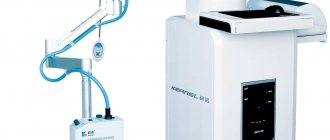
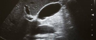
Ultrasound examination (ultrasound) of the ear is based on differences in the degree of absorption and reflection of ultrasonic waves by tissues with different densities. The ultrasound machine's sensor simultaneously sends and records an ultrasound signal. A black and white image of the outer ear is displayed on the monitor screen, each structure is coded with a certain shade of gray depending on its density.
Ultrasound examination can only reveal pathology of the outer ear. The middle and internal sections are hidden by the temporal bone, impenetrable to ultrasound waves.
Ultrasound allows you to see the external auditory canal and the auricle, surrounding tissues, the eardrum, and parotid lymph nodes. If you need to visualize blood flow in the ear structures themselves or in a tumor formation, the doctor may prescribe a Doppler study of local vessels. In this case, on the screen of the ultrasound machine you will see a red-blue image showing multidirectional flows. Blue color shows the blood flow from the sensor, red - in its direction.
Contraindications for magnetic resonance imaging
Before you sign up for the procedure, you should pay attention to the contraindications to its implementation. During scanning, the patient is exposed to a magnetic field, which should not be exposed to objects containing metals that react to the magnet with displacement or heating (iron, steel, cobalt, nickel), as well as magnetic devices.
Internal implants containing a magnet may fail under the influence of a tomograph and lead to irreversible consequences. Such devices include:
- pacemakers;
- stimulants of nervous and muscle activity;
- drug dispensers (pumps for administering insulin, analgesics, parenteral nutrition);
- cochlear implant.
Some cochlear implants have a removable magnet that allows for magnetic resonance imaging (MRI) scanning if necessary.
An obstacle to the procedure may be the presence of metal objects in the body:
- endoprostheses containing a high percentage of ferromagnetic materials;
- Ilizarov apparatus;
- clips on cerebral vessels;
- artificial heart valves;
- fragments, bullets, metal shavings or dust remaining in the body as a result of injury.
When visiting a radiologist, you must bring with you all the technical documentation describing the implant or prosthesis located inside the body. After studying the composition of the implanted material, the doctor decides on the possibility of carrying out the procedure.
Relative contraindications and restrictions include early pregnancy, claustrophobia, mental and severe somatic illnesses, severe pain, convulsions, epilepsy, age under 7 years, body weight more than 120 kg.
The contrast agent is not administered at any stage of pregnancy, as well as for people with renal failure, since the drug is excreted through the kidneys. For healthy people, an obstacle to contrast research is an allergic reaction to its components.
Price
Ultrasound diagnostics
| Name of service | Price |
| Duplex angioscanning of brachiocephalic vessels (great vessels of the head and neck) | 2 500 ₽ |
| Transcranial triplex scanning of the arteries of the circle of Willis | 2 500 ₽ |
| Digital duplex scanning of deep veins | 2 500 ₽ |
| Duplex scanning - Arteries and veins of the upper limb | 4 000 ₽ |
| Duplex scanning - Arteries of the upper limb | 2 000 ₽ |
| Duplex scanning - Arteries of the lower extremities | 2 500 ₽ |
| Duplex scanning - Abdominal aorta and renal arteries | 3 000 ₽ |
| Duplex scanning - Abdominal aorta and iliac arteries | 2 500 ₽ |
| Duplex scanning - Abdominal aorta with visceral branches | 2 500 ₽ |
| Duplex scanning - Veins of the upper limb | 2 000 ₽ |
| Duplex scanning - Veins of the lower limb | 2 500 ₽ |
| Duplex scanning of the renal arteries and kidneys | 2 500 ₽ |
| Duplex scanning - inferior vena cava and iliac veins | 2 500 ₽ |
Advantages
- The latest, constantly updated equipment
- Interest-free installments for all services
- Online consultations with an ENT doctor
- Visit of an ENT doctor to your home
- Friendly and qualified staff
- 24/7 ENT assistance
Vascular ultrasound allows you to get an objective picture of the state of the vascular system. Vascular diseases can cause life-threatening conditions: thromboembolism, strokes, heart attacks, so it is extremely important to detect their pathology in time. Ultrasound diagnostics of blood vessels establishes the structure of veins and arteries, the geometry of blood vessels, deviations in their anatomical structure, and identifies blood clots.
Ultrasound is often performed routinely before an upcoming operation in order to identify vascular pathologies that can cause serious complications or serve as an absolute contraindication to surgery.
How the procedure goes, preparation
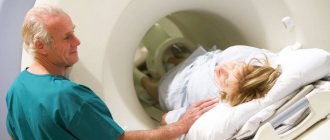
MRI of the ear in normal mode does not require special preparation. Magnetic resonance imaging with contrast involves stopping food intake 5-6 hours before the examination. You should conduct a kidney function test in advance and make sure there is no pregnancy. When you visit a radiologist, you need to take the results of previous studies and a doctor’s report on the current disease.
If the patient is agitated or suffers from mild claustrophobia, sedatives can be taken 30 minutes before the test. Children under 7 years of age find it difficult to remain still during scanning, so general anesthesia is indicated for them.
After a conversation with the radiologist, the patient removes all metal items of clothing, jewelry, accessories, removable metal dentures and leaves a mobile phone, plastic cards, money, and a bag. The procedure is carried out in comfortable disposable clothing, which is provided by the clinic.
The diagnosis continues for 15 minutes. During this time, the subject is exposed to a magnetic field in the tomograph body. Some centers provide examinations using open-type tomographs. In some cases, not only the ear is examined, but also adjacent structures - eyes, brain, blood vessels. The introduction of a contrast agent doubles the duration of the procedure.
During the procedure with contrast, you may experience a feeling of warmth and tingling in the injection area, headache, nausea, or an allergic skin reaction. Side effects are rare and do not last long.
Our address is near Zhulebino and Kotelniki metro stations
Contacts
Lyubertsy, microdistrict Gorodok B, st. 3rd Post Office, 102 5 minutes from the metro
Zhulebino
Kotelniki
+7(495)500-93-90
Make an appointment

New and modern application!
For your convenience, we have developed a mobile application, now you can sign up online in a matter of minutes and keep abreast of all the latest promotions and events of the medical center