Author
Kuibida Oleg Rostislavovich
Leading doctor
Candidate of Medical Sciences
Endoscopist
until September 30
We are giving 1000 rubles for all services for a visit in September More details All promotions
Gastroscopy
(full name -
esophagogastroduodenoscopy
, or
EGDS
) is an endoscopic examination of the upper parts of the gastrointestinal tract, which includes the esophagus, stomach, duodenum.
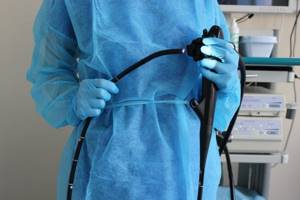
Gastroscopy is performed using special endoscopic equipment - a gastroscope. A gastroscope is a long, thin and flexible tube with a lens at the end. Since the gastroscope uses optical fiber, it is also called fibrogastroscope, and research with its help is called fibrogastroscopy
(
FGS
). A gastroscope is inserted through the mouth. The image transmitted from the gastroscope lens can be significantly enlarged, and the endoscopist conducting the examination has the opportunity to observe on the monitor a detailed picture of the condition of the patient’s mucosa.
What is EGD analysis?
First of all, let’s figure out what an endoscopy examination is. Esophagogastroduodenoscopy (EGD) is a diagnostic procedure aimed at examining the esophagus, stomach and duodenum, which is carried out by introducing a special device into the patient’s gastrointestinal tract - an endoscope. In fact, fibrogastroduodenoscopy allows you to evaluate the same parts of the digestive system, but usually takes place without examining the esophagus.
You can find out more about the service and its cost here >>
As for the recently widespread video gastroscopy , it differs from previous techniques by the location of a small video camera at the end of the endoscope tube. This addition allows the specialist to obtain a visual picture of the condition of the mucous membranes of the esophagus, stomach and duodenum on the computer screen by controlling a moving camera. In addition, it should be noted that the ability to save a video protocol of the study allows you to confirm the diagnosis and can save the patient from repeating the procedure.
To understand what EGDS is in medicine, you should realize that this technique, along with FGDS, allows you to identify many gastrointestinal diseases in the initial stages of development , when other diagnostic methods cannot give reliable results. These research methods are of particular importance in determining oncological tumors, even with small tumor sizes.
endoscopic used in the NEOMED clinic is equipped with the “i-scan” function (virtual chromoendoscopy), thanks to which it is possible to diagnose cancer at a very early stage . In addition, EGDS, unlike FGDS, makes it possible during the research process to take a small section of tissue for cytological and microscopic analyses, while minimizing discomfort for the patient.
You can find out more about the service and its cost here >>
Normal EGDS indicators
To decipher the research protocol, it is first necessary to determine what is the norm, after which a comparative analysis can be carried out. Each section of the gastrointestinal tract examined will have its own normal values.
- Esophagus. The first thing you need to pay attention to is the color and structure of the mucous membrane. Normally, the walls of the esophagus can have a color from pale pink to reddish, and the structure should be finely fibrous. In addition, the esophageal tube has 4 narrowings along its length and has longitudinal folding, which ends in a closed sphincter. This hole is called the cardiac hole. The length of the esophagus of a healthy person is 25-30 cm.
- Stomach. The mucous membranes of the stomach are usually more intensely colored than the esophagus and may even have a bright red color. The anterior wall of the stomach is smooth, shiny, and may be covered with a small amount of mucus. The posterior wall has a folded structure, which is why even the most flexible endoscope tube cannot examine the entire organ and allows for “silent zones.” The pylorus ends in an opening (a section of the stomach that passes into the duodenum). During EGD, gastric motility is also assessed.
- Duodenum. The organ looks like a small tube, with a diameter of 3 to 3.5 cm. The mucous membrane is usually pale pink. The fold is longitudinal, the only one, and has two duodenal papillae, which contain the bile and pancreatic ducts. These ducts connect to the gallbladder and pancreatic gland, respectively.
Research methods
There are the following digital diagnostic methods, which can be carried out by putting the patient into medicated sleep:
- video capsule endoscopy;
- video bronchoscopy;
- chromoendoscopy;
- narrow band endoscopy (NBI);
- video rectosigmoidoscopy;
- video colonoscopy;
- videogastroduodenoscopy.
Videogastroduodenoscopy (VGDS) is a visual examination of the duodenum, esophagus, and stomach using a videogastroduodenoscope. The procedure includes diagnostic steps regarding the upper gastrointestinal tract and additional steps: video recording, anesthesia, histological diagnosis and biopsy.
Indications for the procedure: pain in the upper abdomen; constant feeling of heartburn and nausea; complicated swallowing procedure, even during the period of eating carefully crushed food; altered taste sensations; lack of appetite.
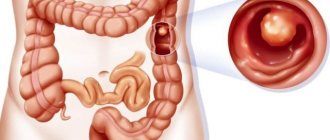
Videocolonoscopy is an endoscopic procedure for diagnosing the condition of the colon, which allows you to determine the presence of benign or malignant tumors, inflammatory processes and ulcerative formations. This type of study can be recommended by a doctor to confirm the diagnosis if indicated, and also as a screening for cancer after 35 years.
Indications for the procedure: presence of blood or mucus in the stool; severe pain in the lower abdomen; family predisposition to colon cancer and polyps; presence of suspicion of the development of cancer; the presence of previously identified polyps; during palpation, a lump in the abdomen is visualized; manifestation of diarrhea of unknown etiology; anemia of unknown origin; prolonged constipation.
Narrow-spectrum endoscopy - allows you to examine the condition and relief of the mucous membranes and select the most accurate place for a biopsy.
Chromoscopy - allows you to contrast lesions of the mucous membranes through the use of a dye. The most commonly used solution in medical institutions is methylene blue. Methylene blue is a dye that accumulates in the cells of intestinal metaplasia developing in the stomach or esophagus.
Video capsule endoscopy – allows you to examine the small intestine by swallowing a sterile, disposable capsule with a built-in camera and light. The signal from the capsule is automatically transmitted to a receiving device, which is attached to the patient’s belt, and then the information is transferred to a computer.
Indications for the procedure: failure to understand the etiology; Peutz-Jeghers polyp; celiac disease; suspected Crohn's disease of the small intestine; symptoms of intestinal bleeding without an established cause.
The capsule study is carried out for 8-12 hours, during which time the patient is in the hospital.
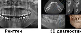
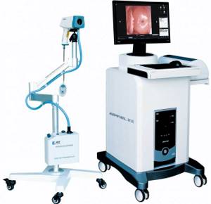
Digital endoscopy
Digital examination with an endoscope is a completely harmless and effective method for diagnosing various diseases; it allows the doctor to examine the patient’s internal organs without surgical intervention.
Digital endoscopy is carried out through the use of special digital electronic systems based on new technologies. The video systems used have a wide range of diagnostic capabilities. They are equipped with a high-definition format, which in turn allows the endoscopist to see minimal pathological changes. Digital endoscopy greatly simplifies making the correct diagnosis.
Preparation rules and diet
A week before the endoscopy, the patient will have to undergo a blood test for hemoglobin, biochemical and general clinical analysis, which will help identify hidden diseases related to absolute or relative contraindications. Their results should be ready a couple of days before diagnosis so that the doctor can assess all the risks.
Particular attention should be paid to the diet, the purpose of which is:
- reducing irritation of the mucous membranes of the digestive tract - exclude alcoholic drinks, canned food, pickles, hot spices and carbonated drinks from the diet;
- restoration of peristalsis - include cooked vegetables and fruits, buckwheat and rice groats, durum pasta in the diet;
- elimination and prevention of fermentation and putrefactive processes in the intestines, which can provoke gas formation - exclude sausages, legumes, cheeses, milk, fresh vegetables and fruits, bran from the menu.
It will not take long to follow special nutritional rules: 3-5 days will be enough to bring the gastrointestinal tract to relative normality. Otherwise, the patient can lead a normal lifestyle; there are no restrictions on physical activity.

Per day
On the eve of the examination, a light steamed meal is recommended. Last meal no later than 20:00. For dinner you can eat pureed soup, soft puree, poultry soufflé or casserole. After dinner, you can drink unlimited amounts of tea or water.
On the day of the event
There should not be a single meal before the start of endoscopy. Drinking is not limited, but the last portion of water should be drunk no later than 2 hours before the endoscopy. Smoking patients are advised to give up cigarettes and chewing gum. Those who are prescribed medication should not take pills in the morning. They can be accepted after the study.
Indications for endoscopy
- Dyspeptic disorders (heartburn, nausea, belching).
- Pain or feeling of pressure in the pit of the stomach or in the upper abdomen.
- Loss of appetite.
- Sudden weight loss for no apparent reason.
- Difficulty swallowing.
- Frequent vomiting.
- Anemia of unknown origin.
- Monitoring the course of bulbitis, gastritis, gastroduodenitis, peptic ulcer.
- Carrying out certain medical procedures (removing polyps or swallowed objects, installing stents, stopping gastric bleeding).
How is EGDS performed?
The procedure is performed under local anesthesia or under medicated sleep. The patient is placed on his side and an endoscope, a thin flexible probe equipped with a video camera, is inserted through the esophagus. The examination lasts 10-20 minutes, during which time the doctor determines the presence of a particular pathology and, if necessary, takes tissue samples for morphological examination and diagnosis of Helicobacter pylori infection.
Esophagogastroduadenoscopy at the Yusupov Hospital is performed by an experienced endoscopist Natalya Vidyayeva, who specializes in identifying pre-tumor pathology of the gastrointestinal tract, as well as tumors at an early stage. Our doctor completed an internship in Japan under the guidance of leading experts in the field of endoscopic diagnosis and treatment.
The Yusupov Hospital uses modern equipment that allows you to obtain high-definition endoscopic images with 80x magnification. Our doctors use additional technologies of narrow-spectrum endoscopy and chromoscopy, which allow us to visualize the pattern of blood vessels, evaluate the structure of the surface of the epithelium of internal organs and identify possible pathology at a very early stage.
Contraindications
Despite the information content and relative safety of the procedure, it is not permitted for all patients. In some cases, it can harm the patient's health. There are absolute and relative contraindications to gastroscopy.
Absolute contraindications:
- diseases of the esophagus in which it is impossible to pass an endoscope through it;
- acute ischemia of the heart muscle;
- strokes;
- shock of any origin;
- acute infections;
- severe insufficiency of respiratory and circulatory function;
- pathology of the thyroid gland, which interferes with the procedure;
- suspected organ perforation.
Relative contraindications:
- pregnancy;
- mental disorders;
- acute inflammatory processes in the upper respiratory tract;
- glaucoma.
If a person has conditions that act as relative contraindications to the study, it can be carried out, but only in extreme cases, if it cannot be avoided (if bleeding continues, when it cannot be stopped by other means).