mammary gland examination is one of the most modern, highly informative and effective diagnostic methods for various pathologies of the mammary glands. It is based on the principle of measuring and processing the difference in the attenuation of X-rays by tissues with different densities, which allows them to obtain a layer-by-layer image, which undergoes pre-processing and is displayed on the monitor.
It is important to understand that CT for breast cancer and other diseases is not the only research technique. Most often, it is prescribed to clarify certain results in cases where ultrasound or mammography does not allow them to be obtained. This is due to the fact that the information content and effectiveness of the technique may be higher or lower depending on the nature of the pathological changes to which the mammary glands have undergone. You can undergo a computed tomography scan of the mammary glands at the CELT multidisciplinary clinic. We have a modern tomograph, which allows our specialists to obtain the most accurate image and identify pathologies at an early stage of development.
Features of the CT technique of the mammary glands
The computed tomography technique involves creating a narrow beam of X-rays that pass through the patient's body, moving in a spiral. The sensor that records them also moves spirally, synchronously with their source. The data enters the computer, where special software is used to process it and display layer-by-layer sections of the part of the body being diagnosed on the monitor. The resulting image is presented in three-dimensional format.
It is necessary to understand that the thinness of the cut and the number of processed layers directly affect the quality of the resulting model. The thinner the section and the greater the number of layers that were processed, the smaller the pathological changes can be recorded. These indicators depend on the capabilities of the tomograph used in the clinic. Modern CT machines are very effective in diagnosing breast cancer. In accordance with studies by foreign specialists, they can detect the germination of a malignant neoplasm into the skin or muscles with almost 100% accuracy. At the same time, the effectiveness of the technique drops by almost a third when diagnosing carcinomas at the zero stage of development.
Contraindications to computed tomography

Computed tomography is a non-invasive, quick and painless diagnostic procedure that is an examination of internal organs using radiography.
There are not many contraindications to CT scanning. They are divided into absolute, which make the diagnostic procedure impossible, and relative - such contraindications can affect the diagnostic result, and before carrying out it, it is important to weigh all the factors.
Absolute contraindications:
- pregnancy or suspicion of it;
- age under 18 years without clinical indications;
- The patient's body weight is more than 130 kg.
Relative contraindications:
- severe claustrophobia;
- the presence of hyperkinesis - involuntary twitching of the body;
- a serious condition of the patient in which he will not be able to follow the doctor’s instructions.
When performing a CT scan, contrast is often used, especially when diagnosing pathologies of blood vessels, internal organs, and soft tissues, to clarify the results of the study. The contrast agent for computed tomography contains iodine, so there are also a number of contraindications for its use that must be taken into account before signing up for the diagnostic procedure.
Contraindications to CT with contrast:
- allergy to iodine;
- polyvalent, that is, multiple, or cross allergies in the anamnesis - among diseases that manifested themselves during life;
- any previous adverse reactions to an iodine-containing contrast agent during previous X-ray examinations;
- high levels of creatinine in the blood;
- diseases of the thyroid gland, liver and kidneys (decrease in glomerular filtration from 30 ml/min/1.73 m and below), accompanied by dysfunction of these organs;
- severe form of bronchial asthma;
- surgery on the thyroid gland (the study is possible after agreement with the endocrinologist, but temporary discontinuation of iodine-containing medications may be required);
- diabetes mellitus type II (the study is possible after agreement with the endocrinologist, but temporary discontinuation of glucose-lowering medications may be required).
The contrast agent is cleared from the body within one to three days. To speed up this process, you need to drink more water after the diagnosis. When performing a CT scan during breastfeeding, it is necessary to express and pour out milk within two days after the procedure.
Possible harm in the absence of contraindications is minimized, and the diagnostic benefit of computed tomography with contrast is difficult to overestimate.
Indications for CT scanning of the mammary glands
As already mentioned, computed tomography is prescribed to clarify the results of previous studies if it is not possible to determine the nature of the tumor and provide the following information:
- Precise localization of the tumor;
- Presence/absence of metastases;
- Tumor operability;
- Confirmation of a malignant breast neoplasm if it cannot be palpated;
- The condition of the lymph nodes that are located nearby.
CT is also used in cases where it is necessary to collect material for diagnostics, but ultrasound and mammography do not allow full control of the process.
Should I pump?
Computed tomography does not affect the taste and nutritional quality of breast milk. Therefore, the procedure is not contraindicated during lactation. There is no need to express before or after the procedure. A woman can breastfeed her baby immediately after the procedure. Of course, it is not forbidden to express a portion of milk if it calms an anxious mother. After all, stress and anxiety have a bad effect on lactation.
Computed tomography is a highly informative diagnostic procedure. Not contraindicated during breastfeeding, regardless of the child's age. You can do it immediately after childbirth, if there are indications.
Differences between CT, MRI, ultrasound and mammography. What's better?
Many patients ask the question of what is better: ultrasound, MRI or CT scan of the mammary glands? There is no clear answer to this question, since each technique has its own advantages and disadvantages, and when used correctly, can be as effective as possible. In the table below we will consider the information content of methods in determining certain diseases.
| Diagnostic technique | Value in different cases |
| Mammography | In 95% it allows to detect cancerous calcifications with low breast density, i.e. in the breasts of elderly patients in whom part of the glandular tissue is replaced by fatty tissue. The effectiveness of the technique drops to 45% when diagnosing benign neoplasms in young patients, since they are not visible in the glandular tissue. |
| Magnetic resonance imaging | It is carried out using a contrast agent and often leads to erroneous conclusions about the presence of a disease that does not exist. This is due to the fact that the dense tissue of the glands intensively captures the contrast agent, which can cause an incorrect diagnosis. This means that the effectiveness of the technique is lower with dense mammary glands. |
| Ultrasonography | The technique is effective in identifying tumors that cannot be palpated - 80%. However, the readings drop below 60% if the size of the tumor is less than 1 cm. Unlike mammography and MRI, breast ultrasound is less effective when there is a large amount of fatty tissue, but in combination with mammography it gives results in 98% of cases. |
| CT scan | The technique allows you to visualize glandular tissue, mammary ducts, lymph nodes, bones and cartilage of the breast. What does a breast CT scan show?
At the same time, the technique does not allow a good look at the vessels feeding the neoplasm and changes in the tissue structures around it. When trying to detect calcifications, the effectiveness of CT drops to 60%. |
Thus, each of the methods is effective in each individual case, therefore, when selecting them, in addition to the patient’s age, they take into account the condition of the mammary gland tissue, as well as the localization of the tumor and its structure.
How the MRI machine works
This type of research involves the use of special scanning equipment that allows you to take high-precision images of the examined organs and systems.
Based on the images taken, specialists create an informative 3D model of the internal organs.
In some cases, the study is performed using a contrast agent.
This technology makes it possible to identify the most minor pathologies in the early stages of development.
The operating principle of a magnetic resonance imaging scanner is based on the use of electromagnetic and magnetic waves, excluding radioactive radiation. Exploring one area takes no more than 30–50 minutes. At the time of scanning, the patient should be at rest, remaining motionless. This allows you to get correct and most accurate results.
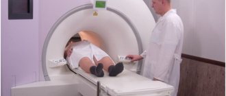
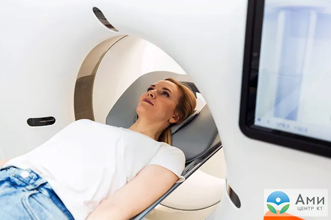
How is a CT scan of the mammary glands performed?
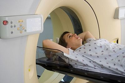
CT scans of the mammary glands are most often performed with contrast. This is an iodine-based substance that is safe for the human body and is injected through a catheter into the blood. This approach allows you to obtain a more accurate image. Before undergoing a CT scan, the patient will be asked to remove any metal objects, including piercing jewelry. If there are implants, shunts, pacemakers or other foreign objects in the body, you should inform your diagnostician.
The procedure is carried out after the patient has taken a lying position on the tomograph table, which smoothly moves into the scanner. The start of scanning is signaled by clicks and noise that appear during scanning. It is important that the patient lies still and relaxed. The procedure lasts 15 minutes or longer and is absolutely painless and cannot cause any discomfort.
Can I breastfeed immediately after the procedure?
A nursing mother, of course, worries. Is the study safe and will radiation affect breast milk? Many mothers, after the study, transfer the child to formula, and do not breastfeed for 1-2 days. There is an opinion that you need to express milk several times before offering it to your baby. These events are meaningless and unjustified.
After the CT scan, you can breastfeed your baby. By the end of the manipulation, there is no longer any radiation left in the woman’s body. The procedure does not spoil the quality of milk or reduce its production. After a chest tomography, there is no need to take a break, switch the baby to formula and delay the next feeding.
But there is an exception.
You should not breastfeed immediately after a CT scan with contrast agent.
This procedure is not contraindicated during lactation, but requires compliance with certain rules. During a CT scan with contrast, a solution is injected into the body. It improves tissue visualization and helps to clearly see the structure of blood vessels, kidneys, liver, and intestines.
The contrast remains in the body of a nursing woman for some time after the examination. You can breastfeed your baby once the contrast agent is cleared from the body. Depending on metabolism, this process takes from 6 to 24 hours. The doctor will give the nursing mother more detailed recommendations after a CT scan with contrast.
CT scan of the mammary glands at CELT - effective and convenient!
Our clinic has a powerful diagnostic base, which is represented by modern equipment. Multislice 64-slice tomograph "General Electric" allows you to conduct research in accordance with international safety and quality standards. The specialists of our radiology department have many years of clinical experience and actively collaborate with doctors of various specialties, which allows us to further increase the efficiency of diagnostics. You can make an appointment with doctors at our clinic online or by phone: +7.