directions
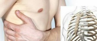
Every person knows what a coccyx is. These are vestigial small vertebrae in the shape of a triangle or pyramid, located in the lower part of the spine. But hardly anyone thinks or remembers that in fact the tailbone is not a developed tail, its rudiment, inherited from animals, the need for which has disappeared as a result of evolution. Today, the tailbone only reminds of the tail that once existed. The coccyx consists of 3-5 vertebrae, connected to the fifth sacral vertebra by a low-moving joint. Near the coccyx there are many nerve fibers, as well as a network of small blood vessels.
Our clinics in St. Petersburg
Structural subdivision of Polikarpov Alley Polikarpov 6k2 Primorsky district
- Pionerskaya
- Specific
- Commandant's
Structural subdivision of Zhukov Marshal Zhukov Ave. 28k2 Kirovsky district
- Avtovo
- Avenue of Veterans
- Leninsky Prospekt
Structural subdivision Devyatkino Okhtinskaya alley 18 Vsevolozhsk district
- Devyatkino
- Civil Prospect
- Academic
For detailed information and to make an appointment, you can call +7 (812) 640-55-25
Make an appointment
We draw your attention to the schedule of technological breaks in the CT and X-ray rooms.
With the help of the anterior section of the coccyx, ligaments and muscles involved in the functioning of the large intestine and genitourinary system are attached. The tailbone also distributes the load in the pelvic area, because serves as a support when bending, squatting, and moving.
What is the coccyx and its functions?
Before you learn how to do an X-ray of the coccyx
, you should become more familiar with the functions of this part of the spine. It is a rudimentary organ, which during the process of human evolution stopped developing into a tail, since there was no such need. The tailbone looks like the curved tip of the spine and consists of three to five vertebrae. It is attached to the sacrum via a joint with a cartilaginous disc, and the joint itself is so mobile that the coccyx can tilt back during childbirth.
Muscles are attached to the front of the coccyx, which are involved in ensuring the functioning of the genitourinary organs, as well as the intestines. The coccygeal plexus is also located here, thanks to which signals from the organs of the pelvic area are sent to the brain. The main load from sitting falls on the tailbone.
Diseases, injuries in the coccyx area
Pain in the coccyx area occurs not only in professional athletes, but also in any person at any age, regardless of his field of activity.
Such a small and at the same time such an important structure as the coccyx can cause a lot of inconvenience and pain leading to the inability to sit, stand up and walk normally.
The tailbone is a very vulnerable place that can be easily damaged. Most often, pain in the tailbone occurs as a result of injury, such as a fall, blow, or bruise. Sometimes the consequences of an injury do not immediately make themselves felt, a person forgets about the incident, and after some time pain appears, and not always in the tailbone itself. The patient may begin to suffer from chronic headaches and lower back pain, and diseases not directly related to the tailbone may develop.
Pain in the tailbone can also be felt due to the presence of a disease such as coccydynia. This disease is characterized by severe attacks of pain in the coccyx area, sometimes this disease becomes chronic.
To understand the causes of pain and why the patient experiences discomfort, the doctor prescribes an x-ray of the tailbone.
X-ray examination of the coccyx helps diagnose serious injuries, such as closed fractures, pathologies and other ailments.
If you are looking for where to get a quick and high-quality x-ray of the coccyx in St. Petersburg, then at the medical traumatology department you can take an x-ray of the coccyx using a modern high-tech digital device Clinomat. You are guaranteed safety, efficiency, reliability and sensitive attitude of qualified experienced doctors of the Medicenter. After receiving the image and deciphering it, you will be referred to an appointment with the doctors of our multidisciplinary center, if necessary, they will prescribe additional examinations and give a comprehensive consultation with the prescription of therapy.
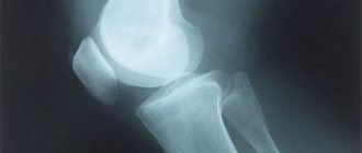
What can be seen on an x-ray of the coccyx
X-ray of the coccyx, as a rule, shows all changes in the bone structure of the coccyx, the presence of tumors, cracks, fractures, dislocations.
When is an x-ray of the coccyx prescribed?
An X-ray of the coccyx is a prerequisite for diagnosing the cause of pain and other symptoms noted by the patient.
It is impossible to make a diagnosis and prescribe treatment using palpation and questioning.
X-rays are mandatory in the following situations:
- a person cannot bend or fully straighten his back;
- there was a strong aching pain in the lower back;
- problems with urination appeared;
- the lower back muscles are constantly tense;
- the patient notes the spontaneous occurrence of “lumbago” in the spine;
- long sitting causes the patient severe discomfort or is impossible.
Preparation for an x-ray of the coccyx may also be necessary if pain in the pelvic organs appears after an injury. This is explained by the fact that the nerve endings of the coccyx are connected to these organs.
What can diagnostics show?
It is important to find out what an x-ray of the tailbone shows and what problems doctors can diagnose based on its results. The image will show the vertebrae and their spinous processes. By carefully examining the images, the doctor will be able to identify:
- fractures, bone integrity disorders, dislocations;
- discrepancy between the articular surfaces of the coccygeal joint, complicated by dislocation;
- presence of osteochondrosis;
- tumor processes and metastases in the coccyx;
- deformation of the connection of the coccyx with the sacral region;
- osteomyelitis;
- developmental defects.
Preparing for an x-ray of the coccyx
There are several rules for preparing for an x-ray of the coccyx.
First of all, a couple of days before the test, you should follow a diet that excludes the consumption of fatty, fried foods, foods that increase gas formation, and carbonated drinks. The night before the study, you must do a cleansing enema and take a laxative. No later than 2 hours before the x-ray of the tailbone, you also need to do an enema to empty the intestines and get a clear picture. To carry out an x-ray of the tailbone, it is not necessary to completely remove all clothing; it is important to simply remove all things that have metal elements. Areas not to be examined are covered with a special lead apron to protect against radiation.
How is an X-ray of the coccyx done?
It is important to remove all metal objects and decorations so that they do not affect the quality of the photo. To begin the diagnosis, the patient will be asked to lie on a table and assume a certain position. As a rule, several basic styling is used:
- direct projection - first you need to bend your legs at the knees and hip joint so that you can take a picture of the sacrum, then straighten your legs to take a picture of the tailbone;
- lateral projection - the patient lies on his side, hands behind his head, knees slightly bent;
- oblique projection – rarely needed, the doctor selects the position individually.
Having learned how to do the coccyx x-ray procedure, you don’t have to worry that it will cause discomfort and unpleasant sensations. After the diagnosis, the patient will be asked to wait outside the office while the doctor makes a transcript.
X-ray of the spine in the clinics of JSC "Family Doctor"
In most cases, an x-ray of a specific part of the spine is performed. At the Family Doctor clinics you can do:
- X-ray of the cervical spine
(including with functional tests); - X-ray of the thoracic spine
; - X-ray of the lumbosacral spine
(including functional tests); - X-ray of the coccyx
.
X-rays are performed in one or two projections (posterior and lateral). The essence of radiography of the spine with a functional test is that the picture is taken in the position of maximum possible flexion or extension. The position in which the photograph is taken is selected individually, depending on the mobility of which segment of the spine requires information.
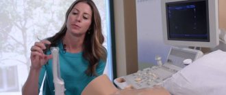
How is an MRI examination of the coccygeal area performed?
When a doctor recommends undergoing an MRI examination, a person immediately has thoughts about how the procedure will go and whether it will be painful. You can immediately calm down - this method is absolutely painless and safe.
Magnetic resonance imaging of the coccygeal area takes place in several stages:
The first step is to remove all metal objects from the body. All chains, rings, belts will need to be removed during the procedure, and all electronic devices - phones, tablets, watches - should be left in the preparation room.
Next, the patient takes a comfortable position on a special table, which slides into the MRI machine. You will have to lie on your back or stomach.
The tomographic scanner will begin its work. It is worth noting that the device sometimes makes specific noises. While the scanner is taking pictures, the patient will hear noises and tapping sounds. To rid yourself of these sounds, you can use earplugs or noise-canceling headphones.
The entire diagnosis usually lasts approximately 15-20 minutes. When the table with the subject leaves the MRI machine, the diagnostic procedure is considered complete.
You can find out the results of the study in about 1 hour. Occasionally this time can be increased to 2 days.
Questions about diagnostics
Dress code
You can enter the MRI room in any clothing that does not contain metal. When going to the clinic, it is best to wear loose, non-restrictive clothing without metal elements (zippers, rivets, hooks), in which you can lie comfortably. For women, we recommend bringing a T-shirt or not wearing a bra with metal wires or hooks.
Preparation
This tomography does not require any preparatory steps from the patient.
Is MRI harmful to health?
MRI is a completely harmless diagnostic method for the human body. This method of examination can be carried out at any age and for any disease an unlimited number of times, unless you have contraindications.
Contraindications
Some pacemakers and foreign objects in the body may pose serious limitations to tomography. In particular, cochlear implants, vascular clips, stents, heart valves and insulin pumps, pacemakers, neurostimulators, steel screws, staples, pins, plates, joint endoprostheses may be a contraindication to diagnosis. The patient must notify the radiologist about all implanted objects in the body. The diagnostician will be able, based on information about the composition and model of the implant, to assess the possibility of conducting diagnostics.
If you are having an MRI with contrast, be sure to report any allergies to medications or kidney problems. It is also necessary to inform the radiologist about a possible pregnancy.
Is it possible to do an MRI with braces and dental implants?
Dental implants and crowns are not a contraindication to magnetic resonance imaging. The magnetic field does not have any negative effect on them. Fixed brace systems can produce artifacts on tomograms during MRI of the head. If the light effect is too strong, the doctor will stop the study and offer the patient alternative diagnostic methods.
Is the device noisy?
Any MRI machine in working condition makes noises reminiscent of tapping. The open tomograph is one of the quietest installations. The noise from its operation is significantly lower compared to closed tomographs. If the sounds of the operating unit cause you anxiety, you will definitely be offered special noise-canceling headphones.
What should I do if I have claustrophobia?
An open tomograph is the optimal solution for patients suffering from panic attacks in a closed space. It is open on the sides on three sides and does not create a claustrophobic feeling.
Can I take sedatives before an MRI?
If you are a little nervous, before the tomography you can take mild sedatives, for example, valerian, motherwort infusion or afobazole. Taking sedatives does not have a negative impact on the quality of MRI.
Why is it important not to move during the test?
Any movement during the examination reduces the quality of the resulting images. Multiple motion artifacts may appear on the images, and the MRI results will be uninformative.
Can I do the research with an accompanying person?
Absolutely yes. You can invite any accompanying person from among your family and friends to the MRI room. It is important that your companion does not have metal implants or artificial pacemakers in his body.
Treatment
If a fracture of the coccyx was diagnosed during the examination, then first of all a special splint is applied . It is made in the form of a circle of cotton wool and gauze with fastening in the back area. Depending on the severity of the injury, it is worn from two weeks to one and a half months .
During this time, the patient must strictly adhere to bed rest, while lying on his side or stomach. Occasionally and very carefully, you can roll over onto your back for a short time. In this case, the patient can be treated at home with regular observation in the hospital.
Medications
Did you know that...
Next fact
Therapeutic treatment for a fractured coccyx includes the use of analgesics to relieve pain , as well as anti-inflammatory drugs to prevent the development of an inflammatory process at the site of impact. They can be used either as injections or as tablets.
Calcium supplements and vitamin D are also prescribed. If there is no damage to the skin or suppuration, pain-relieving ointments can be used.
Surgical treatment of a coccyx fracture
Surgical treatment of a coccyx fracture is used in the case of an open fracture, trauma with displacement or the presence of fragments, as well as severe damage to blood vessels and nerve endings.
In these situations, open reduction is performed. During this process, the fragments are connected and fixed. The operation is performed manually under local anesthesia. After the procedure is completed, the patient is given a plaster cast. Further treatment is carried out only in inpatient settings.
Rehabilitation after treatment and preventive measures
Usually, after four weeks of treatment for a coccyx fracture, a period of rehabilitation begins , which, as prescribed by a specialist, may include hirudotherapy, exercise therapy, physiotherapy, mud therapy, electrophoresis, and massage. After the injury has been eliminated, pain in the coccygeal region may remain for a long time in one situation or another (sitting, walking, defecation, etc.).
In this case, it is advisable to follow some recommendations that will help reduce them, as well as recover faster from injury:
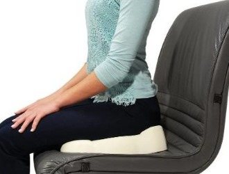
- When sitting on any hard surface, use a small pillow or other support;
- While sitting, lean forward and periodically move from buttock to buttock to remove excess load from the tailbone;
Read the rules for rehabilitation after a coccyx fracture. Do not sit on very soft surfaces so as not to put pressure on the coccyx area;- Drink at least two liters of water per day. It is also advisable to drink rosehip decoction, green tea and other drinks rich in vitamins and minerals;
- Include in your food as many foods as possible that contain calcium, silicon, and vitamin D. This can be milk and sour-milk products, eggs, seafood and sea fish, beans, lentils, corn, buckwheat and oatmeal, vegetable oils, various nuts. You should also eat vegetables and fruits daily, as they contain a large amount of fiber;
- Take baths several times a week in a sitting position;
- Sleep on relatively hard surfaces;
- If necessary, use painkillers and mild laxatives.
- Do not lift heavy objects, do not ride in vehicles with hard seats or ride a bicycle, and do not play sports.
Compliance with these recommendations will contribute to the rapid recovery of the coccygeal region and eliminate the consequences of injury.
Video: “Simple exercises for pain in the tailbone”
Treatment at home
In order to eliminate the consequences of a coccyx fracture in a short time, after eliminating the injury, you can carry out additional treatment at home.
To do this you can use the following methods that will help relieve pain and restore the tailbone:
- Take a small piece of magnet and lightly massage the coccygeal area with it. Carry out the procedure for twenty minutes three times a day;
- Peel a few raw potatoes and grind them into a paste. Apply the resulting mass evenly onto a thin cloth, fold it in half and apply it to the sore spot. Cover the compress with plastic wrap and cover the patient with a warm blanket. The duration of the procedure should be at least half an hour;
- Grind 50 grams of fresh comfrey leaves, pour in 1 glass of olive oil and simmer over low heat for about half an hour. After this, cool the resulting composition, filter, add 50 grams of beeswax and 20 drops of vitamin E. Mix the ointment well, transfer to a glass container and store in the refrigerator. It should be used in the morning and evening, applying to the coccygeal area with light massaging movements;
- Brew 2 tablespoons of fresh crushed geranium leaves with 1 liter of boiling water and simmer for 10 minutes. After this, cool the composition, strain and use for compresses several times a day;
- Mix 1 tablespoon of table vinegar and 2 tablespoons of honey. Rub this mixture into the coccygeal area three times a day. In between, lubricate the sore spot with olive oil;
- Combine 1 tablespoon each of cottage cheese, kefir, honey and vodka. Beat the resulting mixture well and use it as a compress in the morning and evening;
- Grind fresh wormwood leaves into a paste, wrap in a thin cloth and apply to the sore spot. The compress should be fixed, covered with a warm blanket and lie down for half an hour.
Indications
A patient may receive a referral for examination of the coccyx using a tomograph in the following cases:
- Anomalies in the coccygeal region;
- Possible displacement, fracture;
- Suspected tumor, metastases;
- Suspicion of cauda equina syndrome;
- Pain syndrome;
- To confirm previous studies;
- To identify the dynamics of treatment.

Previous Next
Recovery prognosis
It is quite difficult to give a prognosis for recovery after a fracture of the coccyx . Most likely, this can only be done by a specialist who observes the picture of the disease and treatment. The process depends on many factors.
These include the severity of the injury, the course of treatment, the rehabilitation period, etc. If the fracture is not too complex and the victim has completed a full course of treatment and rehabilitation, then, as a rule, the tailbone heals properly, the pain goes away, and the ability to work fully returns. But at the same time, for some time you can only do light work.
Interpretation of MRI results of the coccyx
An example of MRI decoding of the coccyx
Area of study: MRI of the coccygeal spine In a series of MR images weighted by T1 and T2 in the sagittal and axial planes, the structure of the coccygeal vertebral bodies is quite homogeneous, the intensity of the MR signal from the bone marrow of the coccygeal vertebral bodies is moderately increased on T2 and T1-weighted account of fatty degeneration.
Location of the coccygeal vertebrae according to type 1. There were no signs of bone marrow edema of the vertebral bodies. Direct MRI signs of post-traumatic changes are not determined. Presacral fatty tissue is not changed. Conclusion: Initial degenerative-dystrophic changes in the coccygeal spine. It is difficult for an ordinary person to independently understand and interpret the results that, after an MRI, will be given to him on a digital medium. Therefore, with the conclusion of the radiologist and the photographs, he should go for a consultation with the attending physician, who will make a final diagnosis based on the summary data of the examination, medical history and tomography data. In our clinic , after an MRI, you can have a free consultation with a neurologist or orthopedist . Doctor
- will answer all questions based on the results of the research and the conclusion received
- Helps explain tomography results without using complex radiological terminology
- will conduct an examination and, if necessary, offer treatment.
“Second independent opinion” service Medicine is an area where we want to be 100% sure . Therefore, at your request, we will be happy to offer you the service of a second independent opinion from the leading consultant of our clinic, Candidate of Medical Sciences , a doctor of the highest category with 18 years of experience in the field of tomography and radiology N.V. Marchenko.
Research result
X-ray of the coccyx allows you to clearly examine the condition of the lower part of the spine, establish the nature of the injury, and choose the optimal course of treatment. The doctor immediately examines the image and determines what kind of damage has been sustained. X-rays help identify problems and diseases such as:
- dislocations and fractures;
- tumor and metastases;
- osteochondrosis;
- deformation of bone tissue;
- osteomyelitis;
- pathological development of individual elements.
This effective research method has some limitations to its application. It is not recommended to do x-rays for pregnant women and children under 15 years of age; x-ray examinations for this category of people are used only in cases where the risk is truly justified. If possible, for such people radiography is replaced by MRI. Another problem is obesity, which reduces image clarity.