Ovarian hyperstimulation syndrome, what is it and how to avoid it?
Most women suffering from infertility have heard about ovarian hyperstimulation syndrome; fear of this complication of IVF often pushes women away from the program. Ovarian hyperstimulation syndrome (OHSS) is a condition based on the reaction of the ovaries in response to the administration of hormonal drugs (ovulation inducers), doses of which exceed physiological values. This condition was first described in 1930 using serum from pregnant mares.
Ovarian hyperstimulation syndrome is characterized by a fairly wide range of clinical manifestations: from minor changes in laboratory parameters to quite serious conditions requiring hospitalization.
The main reasons for its occurrence are high doses of hormonal drugs that are used to stimulate ovulation, as well as high levels of activity of the hormone estradiol, which is produced in growing follicles, and high levels of the hormone hCG. The main risk group for the formation of this syndrome are girls diagnosed with polycystic ovary syndrome, since this group has a high follicular reserve and a large number of follicles “come into growth” during induction.
Ovarian hyperstimulation syndrome is currently a well-studied syndrome; for this reason, reproductive specialists around the world are trying to perform ovarian stimulation using a minimal hormonal load, pursuing the main goal: to obtain the maximum number of mature and high-quality oocytes, to avoid ovarian hyperstimulation syndrome. All patients who are preparing for the IVF program undergo a thorough examination (in accordance with Order 107n of the Russian Federation), all possible risks are assessed by a reproductive specialist, and preventive measures are taken in the presence of risk factors for the occurrence of OHSS.
Women at high risk of developing the syndrome are recommended to undergo an IVF protocol followed by cryopreservation of embryos and embryo transfer in another cycle.
So, modern knowledge about the peculiarities of stimulation of patients with a high risk of hyperstimulation makes it possible to minimize cases of ovarian hyperstimulation through the use of minimal protocols and embryo transfer in an unstimulated cycle.
Advantages
The advantages of minimally invasive surgery include:
- Minimally invasive and low-traumatic, which contribute to rapid recovery.
- Less risk of developing adhesions. This is also facilitated by the use of carbon dioxide during laparoscopy instead of ordinary air.
- External invisibility of scars. They are small in size, 0.5–2 cm.
- Has high diagnostic information content.
- All manipulations are carried out under visual control; the risk of complications is low.
- Short rehabilitation period, which is very important for patients with infertility.
The cost of laparoscopy is higher than traditional laparotomy, since the intervention requires expensive equipment and a high level of physician training. However, if in our clinic the procedure is performed simultaneously for diagnostic and therapeutic purposes, it is cheaper.
Cancer risks and IVF: myth or reality?
A lot of fears among our patients are caused by rumors about an increase in the risk of cancer after the program. Indeed, IVF is an intervention in the human body - exposure to hormonal drugs.
Does the program really increase the risk of various cancers: breast, ovarian, uterine, colorectal cancer, melanoma, etc. or is this an unfounded fear? Scientists have been closely studying this issue for decades; as a result of their efforts, it was established that there is no pattern between ART and the occurrence of cancer.
Here are the results of international research in this area:
- According to one randomized study, including more than 100 thousand people and a follow-up period of 25 years, it was proven that the risk of developing ovarian and uterine cancer does not increase in patients after the program.
- According to the American Gastroenterological Association, a randomized study involving 20 thousand people with a follow-up period of 21 years also proved that there was no pattern in the occurrence of rectal cancer and the implementation of an IVF program.
- According to a large randomized study in Oxford (England), including more than 25 thousand patients, it was proven that IVF does not affect the risk of melanoma.
- According to many randomized studies (2010-2016), it was shown that there was no effect on the incidence of breast cancer in women after completing the program. The more “frequent” detection of breast cancer in this population group compared to other women is associated with careful examination and follow-up, early detection of the disease in women of the older age group who have a genetic predisposition to its occurrence. Moreover, one of the risk factors for breast cancer is infertility. This is also worth remembering.
- Moreover, a history of cancer is currently not a strict contraindication for ART, as it was before. Each case is discussed at a consultation of doctors, where the optimal tactics for managing the woman are determined.
Thus, hormonal drugs used in ART do not provoke the development of cancer. However, in underexamined women with the presence of precancerous diseases, their own increased background of endogenous hormones can activate an already existing process.
Causes of polycystic disease
The causes of PCOS include:
- frequent stress, depression;
- endocrine system disorders;
- improper metabolism;
- chronic infections;
- overweight;
- hereditary predisposition.
The disease can also develop in women who live for a long time in regions with poor ecology.
In the presence of one or more unfavorable factors, the ovarian capsules thicken and increase in size, under which numerous cystic formations begin to develop.
Will my child be “normal”?
One of the biggest concerns any pregnant woman faces is whether her baby will be “normal” or not? When it comes to IVF, concerns increase due to the introduction of an “unnatural” component into the equation, which can increase the likelihood of abnormal offspring.
Several studies have been published that demonstrate an increased risk of developing defects in children after IVF: Down syndrome (trisomy 21), Edward syndrome (trisomy 18) and Turner syndrome (monosomy X0). However, a careful analysis shows that most of the listed malformations are due to an increase in the average age of women entering the IVF program compared to comparable ages in fertile groups of the female population as a whole, and there is a connection between age and the occurrence of chromosomal abnormalities. For example, at age 30, a woman's chance of having a child with Down syndrome is approximately 1 in 1,000, while at age 40 the risk is approximately 1 in 150 and at age 45 it is 1 in 35. This occurs due to the “wear and tear” of the eggs over time.
Thus, the patient’s late reproductive age increases the risk of chromosomal abnormalities, both during spontaneous conception and after IVF.
What do world statistics say?
The disease is diagnosed in 4-12% of adult females. Representatives of the European and American Society of Reproductive Medicine claim that the disorder occurs more often, in 15-20% of women of fertile age. In cases of infertility in marriage, this figure reaches 22%.
Today, there is active debate regarding the diagnostic criteria for polycystic ovary syndrome and the in vitro fertilization (IVF) procedure. Experts around the world consider the use of hormonal drugs to be an integral stage of treatment, especially when planning IVF for polycystic syndrome (PCOS).
Many modern doctors say that this problem is related to excess weight. After IVF for polycystic ovary syndrome, overweight women become pregnant half as often. That is why in the USA treatment begins only after the patient’s weight has been reduced by 5-7%. This measure increases the chances of successful IVF for PCOS by an average of 15-20%.
Does freezing embryos affect the health of the unborn child?
The first “successful” pregnancy with the transfer of cryopreserved embryos was obtained in 1983. Consequently, this method is far from new, it is actively developing, and its technologies have been improved over the past 30 years.
The cryopreservation method allows embryos to be preserved for a long time, so if an IVF attempt is unsuccessful, the “saved” embryos can be used in the future. In cases where the IVF program was successful and pregnancy occurred, the remaining frozen embryos can be used in the future when the couple decides to have another child. An additional advantage of the method is the reduction in the number of repeated ovarian stimulations and follicle punctures, which significantly reduces the drug load on the female body.
It has been proven that the process of storing embryos frozen does not affect their further development, however, damage to the embryos can occur during the “freezing” and “thawing” stages. Currently, a more effective way of “freezing” is the vitrification method, i.e. rapid “freezing”, during which the survival rate of embryos is much higher (more than 98%) than after slow freezing (about 50%), which was popular before.
The number of progressive pregnancies after cryopreservation of embryos is comparable to the number of progressive pregnancies during the transfer of fresh embryos (on average, 41.6% and 44.3%, respectively) and depends on a number of factors, the main of which are: quality, thickness and preservation of the receptive properties of the endometrium, absence of cavity deformation uterus, embryo quality, woman’s somatic health.
Despite the arguments that doctors give in favor of embryo cryopreservation, most women continue to be extremely wary of this process. As a result of an extensive scientific study in Sweden, it was found that children born after a “cryotransfer” are less likely to have low birth weight and are more likely to be born at term compared to children born after a “fresh transfer”.
So, in modern conditions, “cryopreservation” is absolutely safe and effective in terms of the further development of pregnancy.
Hydrosalpinx is an obstacle to successful IVF
Quite often, the cause of non-pregnancy is hydrosalpinx, a pathology of the fallopian tube. Even more often, because of this diagnosis, patients hear annoying recommendations for tube removal. The fact itself is truly unpleasant. But today it has been proven that hydrosalpinx reduces the effectiveness of IVF. Let's figure out what to do in such a situation.
To understand this issue, first consider the role and function of the fallopian tubes.
So, normally the fallopian tubes are located on both sides of the uterine angles. They take the eggs that are released from the ovaries every month during ovulation, and fertilize the egg with the help of sperm. Thanks to the vibrations of the ciliated cells, the fertilized egg returns to the uterus, where implantation occurs.
Approximately 35% of female infertility is associated with tubal factor. Bilateral tubal factor is an indication for IVF. And there is a popular misconception that if a woman goes for IVF, it doesn’t matter whether the tubes are passable or not. After all, the tubes are not involved in the fertilization process, and the embryo is transferred by the doctor, patients believe.
Often, tubal damage leads to the formation of hydrosalpinx, and this is a serious factor that interferes with both independent pregnancy (if another tube is patent) and during the IVF procedure. Hydrosalpinx is diagnosed in 10-30% of women who embark on an IVF program.
How does hydrosalpinx interfere with IVF?
Many women are afraid to say goodbye to their tubes even when they understand that they are useless for pregnancy. And we will try to find arguments to protect women from senseless IVF attempts.
Hydrosalpinx (hydrosalpinxhydro-water and salpinx-tube) is a condition in which the fallopian tubes are welded together in the ampullary sections and fluid accumulates in them, which is very toxic to the embryo.
The cause of hydrosalpix is previous infections, such as chlamydia and gonorrhea, tubal inflammation, tubal surgery, endometriosis and other adhesions in the pelvis. During infections, the inner layer of the pipe is completely destroyed and they become clogged, which leads to the accumulation of toxic fluid.
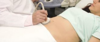
How can hydrosalpix be diagnosed?
Hydrosalpix is diagnosed using ultrasound, hysterosalpingography or laparoscopy. Ultrasound Hysterosalpingography Laparoscopy
How dangerous is this condition if you have been diagnosed with this?
There are several proven harmful effects of hydrosalpinx. a) The liquid that is in the tubes flows into the uterine cavity and at the same time mechanically washes away the embryo that is there. b) It has a very strong toxic effect and is harmful to the embryo. c) Such fluid disrupts the receptor apparatus of the endometrium, where the embryo should attach.
Due to the combination of all the reasons, the chances of implantation of fresh embryos and in cryoprotocols are very low and pregnancy does not occur, despite repeated transfers.
The likelihood of early miscarriages also increases in women with hydrosalpinx.
Such useless attempts are dangerous not only from the material side. Most importantly, a woman’s nervous system suffers due to long expectations and repeated disappointments.
There are many options, but only one is correct
What is often offered to women in medical institutions after they have been diagnosed with hydrosalpinx? And what way out of the situation will help you find parental happiness? The patient may be offered one of three treatment options:
- Removal of the affected fallopian tube
- Emptying fluid and restoring tubal patency
- Aspiration of fluid from the fallopian tube
In European medicine, there has long been a lot of evidence in favor of removing the source of infection. And in a sense, doctors and scientists have already lost interest in the issue of hydrosalpinx, because they have a clear and precise algorithm for what to do with such patients. Unfortunately, in our country, such patients can spend a long time visiting clinics, trying out options that do not increase the chances of pregnancy.
Studies have confirmed that after removal of the fallopian tubes, the chances of pregnancy in the IVF program increase. In women under 35 years of age, the likelihood of a successful protocol increases to 50 percent.
Restorative operations on the pipes did not show a very good effect - soon these pipes became fused again and hydrosalpinx occurred.
Aspiration of this fluid under an ultrasound monitor does not have any effectiveness, because the fluid in the pipes is quickly restored and the inflammatory focus continues its negative effect.
So, let's summarize. What should the patient do in such a situation? You will have to make the decision, because no one will remove the pipe without your consent. And this decision is not easy for many. If you do not want to become pregnant in the future, you can live with hydrosalpinx, in which case it does not interfere. And if you want to get pregnant, then you need to make a choice: a tube or a long-awaited baby. And after removing the source of infection, you can safely go through the protocol and be sure that there will definitely be a result. Good luck!
What is a “cryoprotocol”?
Cryoprotocol is a delayed transfer of embryos that are obtained as a result of IVF and frozen for use in one of the following cycles. The terms “cryotransfer” and “cryocycle” are from the same area. Initially, the scope of such protocols was limited to cases where patients froze their embryos during IVF for a pregnancy in the distant future. Nowadays cryotransfers are often performed for medical reasons.
Recommendations before the crypto protocol
This type of protocol is only possible after conventional IVF (with the exception of donor embryo transfer). If the cryoprotocol is prescribed for medical reasons, then the program preceding it is completed at the stage of obtaining embryos. Embryos are frozen using the vitrification method - only this method can ensure gentle freezing and thawing.
Read more about vitrification in the section about our cryobank.
When the cryoprotocol begins, the embryos are thawed and transferred to the expectant mother. Do you need any preparation for cryotransfer? The examination is the same as before a fresh IVF cycle. If the embryos were received recently, then most of the tests are usually still valid; only the expired ones need to be retaken. In addition, the attending fertility specialist can make individual prescriptions.
Preparing for embryo transfer
What can the patient do before the protocol to increase the chances of success? There is no need to prepare in any special way for the cryotransfer procedure. It is enough to follow a few simple rules:
- strictly follow your doctor's instructions;
- avoid heavy loads, but do not give up physical activity;
- beware of respiratory infections, hypothermia, and other unfavorable external factors;
- a few days before the procedure, abstain from sexual activity.
Make an appointment
Preparing for surgery
Before surgery, the patient needs to undergo examination:
- general blood and urine analysis;
- biochemical screening;
- examination for HIV, syphilis, viral hepatitis;
- blood group test, Rh factor;
- Ultrasound of the pelvic organs;
- smears for flora, cytology;
- ECG;
- FG;
- coagulogram.
Each patient is consulted by a therapist and an anesthesiologist. If necessary, a consultation with a specialist is prescribed. 5 days before the intervention, foods that contribute to the formation of gases should be excluded from the diet. You cannot drink alcoholic or carbonated drinks. The evening before the operation, a cleansing enema is given or osmotic laxatives are recommended.
Laparoscopy is performed on an empty stomach.
Features of the event
Before the operation begins, the anesthesiologist administers general anesthesia. The surgeon then makes 3-4 small punctures in the lower part of the anterior abdominal wall. Through the holes, micro-instruments and a video camera with a lighting element are introduced into the abdominal cavity.
For better visualization, the abdominal cavity is filled with gas, creating an artificial pneumoperitoneum. During diagnostic laparoscopy, the doctor examines the fallopian tubes, uterus, and ovaries one by one. Then a final diagnosis is made and a further surgical plan is drawn up. If endometrioid lesions or adhesions are detected, the surgeon removes them. Also, a gynecologist surgeon can perform manipulations of greater complexity (removal of cystic formations, fallopian tube plastic surgery, hysterectomy, etc.). Large tumors are crushed with a morcellator and removed. The resulting material is sent for pathomorphological examination. Then the doctor performs a second inspection of the abdominal cavity, sucks out the accumulated fluid, and checks the condition of the blood vessels. The operation is completed by removing the instruments and suturing the wounds.
The average duration of the procedure is about an hour, sometimes it can take up to two hours.