Does your abdominal area often swell? Does your weight seem to be increasing right before your eyes? Does your stomach hurt for no reason? Do an ultrasound of the abdominal cavity, you may have ascites!
COST OF SERVICES IN OUR CLINIC IN ST. PETERSBURG
| Abdominal ultrasound | 1200 rub. |
| Appointment with a urologist | 1000 rub. |
| Kidney ultrasound | 1000 rub. |
| Call free: 8-800-707-1560 *The clinic is licensed to provide these services | |
What is ascites and why is it dangerous?
The content of the article
The abdominal cavity is a separate anatomical region. It systematically produces moisture necessary to increase the efficiency of sliding of the abdominal visceral layers. During normal activities, the resulting effusion is absorbed fairly quickly.
However, it happens that free fluid begins to collect in the abdominal area. The cause of this anomaly may be a failure of blood circulation and lymphatic drainage or the presence of inflammatory processes. This disease is called “ascites,” which is better known as “dropsy.”
Dropsy of the abdomen is difficult to treat and leads to serious health problems. After all, the fluid accumulated in the peritoneal area creates ideal conditions for the formation of pathogenic microflora. It includes the causative agents of such dangerous diseases: hepatorenal syndrome, peritonitis, umbilical hernia, hepatic encephalopathy, etc.
Fortunately, today it is possible to determine the initial stage of development of ascites through the use of a non-invasive, very accurate and completely safe method - ultrasound of the abdominal cavity. Thanks to the procedure, it becomes possible to begin a timely fight against the disease with the help of medications.
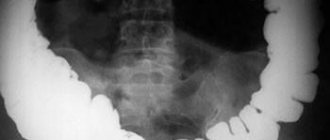
How are x-rays performed?
X-rays of the abdominal cavity are performed in both vertical and horizontal positions (standing and lying down). In some cases, the body is scanned in two projections, this allows a better assessment of the condition and structure of the internal organs. To get high-quality images, you must remain still.
You can take an abdominal x-ray at MedicCity every day from 9.00 to 21.00. Consult a doctor if you have problems with the gastrointestinal tract and do not self-medicate! Plain radiography allows you to quickly identify pathology. We successfully diagnose diseases of the stomach and other organs.
Before scheduling an x-ray, we recommend consulting with a gastroenterologist.
The material was prepared with the participation of a specialist:
What causes free fluid to accumulate?
The first and main cause of ascites is the appearance of any pathology in the pelvic organs. Initially, abdominal moisture collects in relatively small quantities (from 40 ml to 1 liter). Outwardly, it looks like swelling without inflammation.
The formation of the disease can also be influenced by a secretory disorder of proteins, the main task of which is to increase the degree of impermeability of the pathways and tissues that ensure the passage of circulating blood and lymph.
Such problems lead to the development of the following diseases:
- cirrhosis of the liver;
- chronic renal and heart failure;
- portal hypertension;
- lack of protein;
- lymphostasis;
- malignant or tuberculous lesions of the peritoneum;
- systemic lupus erythematosus;
- diabetes.
Often the problem appears in the presence of neoplasms in the area of the ovaries, mammary glands, on the serous walls of the abdominal cavity and pleura, as well as the digestive organs.
In addition, the accumulation of free fluid can be triggered by complications of amyloid dystrophy, hypothyroid coma, pseudomyxoma and the postoperative period.
Contraindications for carrying out
X-ray is a safe examination that has no absolute contraindications and allows you to quickly obtain clear images of internal organs. However, radiation diagnostics is contraindicated for pregnant women and children under 15 years of age.
Our studies are carried out using the latest generation Italray Clinodigit Compact digital X-ray machine. High-definition pictures are obtained in literally 10-15 minutes. Digital detectors are 5 times more sensitive than photographic film.
What signs indicate ascites?
At the beginning of its development, the pathology does not manifest itself in any way. The presence of a problem can only be determined by performing an ultrasound of the abdominal cavity.
Ascites begins to show its first negative signs after the quantitative indicator of abdominal moisture exceeds one and a half liters. In this case, the following changes occur in the patient:
- total body weight and abdominal area increases;
- unpleasant sensations of “bloating” in the peritoneum;
- health worsens;
- in men there is swelling of the scrotum;
- your legs begin to swell;
- systematic belching, nausea, heartburn appears;
- it becomes difficult to breathe;
- tachycardia and flatulence are observed;
- the umbilical node protrudes forward;
- suffer from painful and uncomfortable sensations in the abdominal area;
- urination and stool are disrupted.
When too much effusion collects in the peritoneum, the patient may even feel it moving and hear splashing.
After detecting a pathology on an ultrasound, the doctor diagnoses “dropsy,” but you need to understand that this is just a symptom of one of the true reasons for the formation of fluid in the abdomen. Therefore, a new examination will follow.
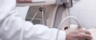
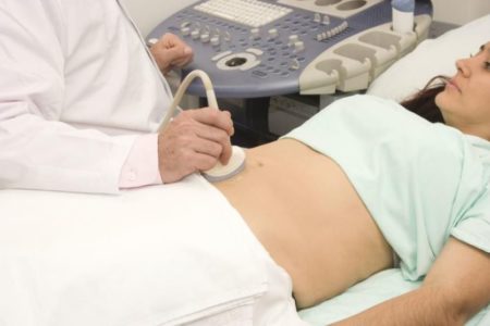
How to properly prepare for an abdominal ultrasound
One of the main advantages of ultrasound examination is that it does not have any restrictions or contraindications. In this case, ultrasound allows you to diagnose the presence or absence of many pathological processes in organs. But to get accurate results, you need to properly prepare for the examination.
The following rules must be adhered to:
- Three to four days before an abdominal ultrasound, it is prohibited to consume foods that can increase the formation of gases and include a large amount of fiber;
- A couple of hours before the procedure, you need to give a cleansing enema or use a laxative;
- To reduce gas formation the day before the ultrasound, you should take activated charcoal or Mezim.
Thanks to modern ultrasound methods, a specialist will detect the accumulation of free fluid in the abdominal cavity. Most often this is:
- the upper abdominal region, located directly below the diaphragm;
- areas under the liver;
- descending and ascending colon;
- lateral canals;
- the small pelvis, into which the effusion enters through the lateral canals.
Ultrasound today is one of the most informative and effective ways to detect a dangerous disease. The physical properties of the liquid do not allow it to reflect the ultrasonic wave, therefore a spot forms in the image in this place. Or rather, effusion, even in small quantities, looks on the monitor like a moving dark lesion. If the person is healthy, the procedure time does not exceed five minutes.
However, it also happens that for one reason or another it is not possible to find a transudate. Therefore, the doctor uses additional examination techniques:
- During the process of tapping the peritoneum, the sound changes. In the lower zone it becomes more deaf, and in the upper zone it becomes tympanic;
- there is a shift of the loops of the colon.
What does fluid behind the uterus mean on ultrasound?
It is worth noting that the mere presence of fluid in the pouch of Douglas is not a disease in itself, but may be a symptom.
The presence of a small amount of fluid behind the uterus in the middle of the menstrual cycle is considered normal. As a rule, this indicates pregnancy. In addition, blood can flow into the retrouterine space in small volumes during menstruation, which is also absolutely natural.
However, sometimes the causes of the appearance of fluid behind the uterus can be serious diseases (endometritis, peritonitis, endometriosis, purulent salpingitis, oophoritis, adnexitis and others), ectopic pregnancy, neoplasms on the ovaries, etc.
To accurately determine the nature of such liquid formations, additional examinations are carried out: puncture (sample of the component through laparoscopy), laboratory blood test, vaginal smear.
What types of ascites are there?
As mentioned above, dropsy is not an independent disease, but a sign of one of the pathologies, indicating that the process is in the last stages of development. Depending on the cause, ascites is divided into several forms, each of which differs in the individual severity of symptoms:
- initial - in the abdominal cavity the amount of fluid does not exceed one and a half liters;
- moderate – it is characterized by swelling in the area of the legs, shortness of breath, increased volume of the chest, constipation, heartburn, and a feeling of heaviness in the abdomen;
- massive - in this case, the quantitative indicator of the collected water reaches five liters.
The condition is dangerous: the patient’s respiratory and cardiac systems are disrupted, the abdominal walls become tense, and the free fluid becomes infected.
During bacteriological testing of transudate, which is mandatory, experts distinguish between:
- sterile dropsy – does not have the presence of pathogenic microorganisms;
- infected dropsy – pathogenic microbes are present.
Ascites detected in a timely manner can be treated with medications. However, there is also a so-called pathological persistent disease that cannot be cured.
What will the patient face after the diagnosis is confirmed?
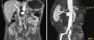
If an ultrasound confirmed ascites, the course of the treatment process will directly depend on the cause that caused the accumulation of excess fluid in the abdominal cavity. To make an accurate and reliable diagnosis, the doctor will prescribe additional diagnostics. The patient undergoes:
- general clinical and biochemical analysis of urine and blood;
- research on tumor markers and electrolyte metabolism;
- general x-ray of the abdominal and thoracic cavity;
- coagulogram showing the level of functionality of the blood clotting system;
- vascular angiography;
- CT and MRI of the peritoneum;
- hepatoscintigraphy, which will make it possible to view the liver with a special gamma camera;
- laparoscopy together with puncture of ascitic water may also be required.
If the patient has liver cirrhosis, he will additionally need to undergo portosystemic intrahepatic shunting. The manipulation is carried out using a mesh metal stent. The stent affects the space between the hepatic and collar veins. If an advanced form of the disease is observed, then organ transplantation is indispensable.
Determination of the volume of fluid behind the uterus by ultrasound
It is extremely difficult to determine the exact volume of such a formation using ultrasound, since the fluid spreads between the organs. To clarify the amount of liquid, the length of the vertical level of formation is analyzed. Thus, today the following criteria have been developed for assessing the amount of fluid in the retrouterine space:
- at a height of up to 10 mm, the formation size is insignificant;
- at a height of 10 to 50 mm - moderate;
- at a height of more than 50 mm – significant.
The data obtained must be compared with the patient’s menstrual cycle. Additional tests may be needed if your doctor has concerns about the cause of fluid behind the uterus.
What happens if you don't treat it?
The accumulation of transudate is not a disease, but a negative manifestation of complications associated with other pathologies. If treatment is not started on time, dropsy will cause dysfunction of the spleen and heart. Also, the problem often provokes the onset of internal bleeding, cerebral edema and the formation of peritonitis.
And the worst thing is the mortality statistics from dropsy - patients die in 50% of cases. Therefore, you should not be negligent about your health: you need to be examined on time and seek medical help. Inflammations and infections should be treated urgently and only in a clinic. It is also very important to be systematically examined by a doctor for preventive purposes and to strictly follow all his recommendations.
If you find an error, please select a piece of text and press Ctrl+Enter
Dangers
Pulmonary edema is life-threatening. Oxygen starvation develops in the lung tissues, and they can no longer cope with their function.
If the disease is not treated, serious complications can occur that threaten vital systems.
Possible complications include:
- Pressure jumps;
- Airway obstruction;
- Difficulty and depressed breathing;
- Extreme increase in heart rate;
- Fulminant swelling, which can be fatal.
Timely treatment can save you from such complications.