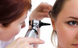
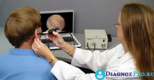
Otoscopy refers to a standard medical procedure for examining the ears in order to identify pathological processes that are characteristic of this part of the body. To carry out otoscopy, you need simple medical devices, such as a reflector, located in the doctor’s forehead and reflecting light rays from the source to the area of the organ being examined, and the light source itself. By directing the light beam, which is reflected in the reflector to the required area, the specialist examines the patient’s ear.
What is otoscopy: definition, essence and method of implementation
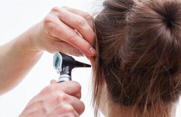
Ear otoscopy is a method for diagnosing diseases in otolaryngology. It is performed using an otoscope. This device is equipped with illumination, optics, and a video camera. Thanks to multiple magnification of the image on the monitor, the device allows you to identify defects, examine the organ in detail, and study the structure of the eardrum.
Otoscopy is used to diagnose and treat diseases of the ear canals and eardrum. Using this method, pathological changes in the ears (otitis, eczema) are detected and foreign bodies are detected.
Contraindications to otoscopy
Otoscopy is a completely safe procedure; there are no direct contraindications to it. There are only a few factors that make it difficult to carry out.
- Congenital ear abnormalities.
- Traumatic injuries causing obstruction of the ear canal.
- Severe swelling.
Otoscopy is a simple and safe procedure for the patient. This study is indispensable for diagnosing ear diseases. Its use helps the doctor resolve issues of differential diagnosis, timely identify the threat of perforation of the eardrum and prescribe the correct treatment.
Indications for the procedure
Otoscopy of the eardrum is performed by an otolaryngologist, less often by general practitioners, and in extreme conditions by ambulance workers using handheld devices.
Indications for otoscopy are pathological changes. They may be:
- sulfur plug;
- infectious and inflammatory damage to the eardrum;
- discharge of purulent exudate;
- injuries to the eardrum - from cotton swabs, other objects or as a result of blows to the head;
- suspicions of ear diseases: otitis media, eczema and others;
- hearing loss;
- bleeding;
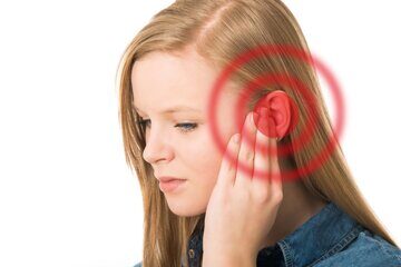
- inflammation of the ears, accompanied by pain and itching;
- feeling of a foreign body or suspicion of its presence;
- sensations of abnormal sounds - splashing, rustling, rustling.
Additional Information! For diagnostic purposes, otoscopy is used for preventive purposes or to assess the structure of the ear canals in the manufacture of hearing aids.
How to prepare
Preparation for otoscopy is carried out in the doctor's office. No preliminary measures are required from the patient. The only rule is not to use ear drops 2 to 3 hours before the test.
Preliminary procedures begin with an external examination. The otolaryngologist determines whether there are any contraindications or obstacles to otoscopy. They may be severe swelling, injury, or congenital anomalies that make it impossible to insert a funnel into the ear canal.
Further preparation includes:
- aseptic processing of instruments;
- removal of sulfur plugs;
- if necessary, clean the ears from pus and dead epidermal cells;
- selection of an ear funnel of the required diameter.
Cleaning the ear canals is carried out in 2 ways: dry or rinsing. In the first case, a piece of cotton wool is smeared with Vaseline, wrapped around a probe and used to remove wax or pus. For the second technique, warm water is drawn into a syringe for Zhanna, injected into the cavity, and after draining, the ear is dried with a swab.
Important! If the patient is diagnosed with a ruptured eardrum, the ear should not be irrigated. This risks fluid entering the middle ear and causing inflammation.
Technique for using otoscopy
- Before starting the study, the doctor selects a funnel whose diameter corresponds to the size of the ear canal.
- To examine, the specialist pulls the auricle slightly backwards and upwards.
- Then a funnel is carefully inserted into the membranous-cartilaginous part of the ear canal, the axis of which should coincide with its location.
In this case, the external auditory canal is straightened and most of it, along with the eardrum, becomes accessible for inspection. If the rules for inserting the funnel are violated, it will rest against the wall of the passage.
- In order to sequentially examine all their areas, the doctor can move the outer end of the funnel in the desired direction.
It should be noted that the funnel is not inserted into the bony part of the ear canal, as this may cause pain. In some patients, this procedure provokes a cough reflex. This is due to irritation of the endings of the vagus nerve located under the skin at the site of insertion of the funnel.
- Next, the examination is carried out using an otoscope. This allows the otorhinolaryngologist to obtain more detailed information about the condition of the object being studied and, if necessary, carry out a diagnostic or therapeutic puncture of the eardrum, removal of foreign bodies or any microsurgical intervention.
Depending on the type and design of the otoscope, the light sources and its direction may differ, but the description of the essence and sequence of diagnostic stages is the same in all cases.
Otoscopy: technique
To examine the ear canals, the patient is seated opposite the doctor. The patient's head is turned in the opposite direction from the otolaryngologist. Then:
- select the optimal size funnel;
- pull the auricle back and up to straighten the ear canal;
- carefully place a funnel heated to body temperature in the membranous-cartilaginous part of the ear canal - the instrument cannot be inserted into the bony area, as this provokes painful sensations, and if inserted incorrectly, the funnel will rest against the wall of the canal;
- inspect the cavity, moving the otoscope in the desired direction;
- if necessary, puncture of the eardrum is performed during otoscopy, microsurgical interventions, and removal of foreign bodies.
Note! Some patients cough during otoscopy. This occurs due to irritation of the vagus nerve by the device.
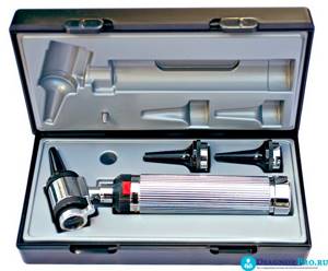
What is an otoscope
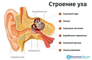
A medical device such as an otoscope, which is used in ENT practice, is a small device for examining the outer, middle and inner ears. This is a complex optical device that consists of:
- long handle for gripping;
- light source: halogen, xenon, in expensive models – fiber optic;
- a cone-shaped tip that is inserted into the ear canal.
Additional Information! Previously, the study was carried out using special funnels to expand the ear canals and a reflector placed on the doctor’s forehead. The latter is necessary to reflect the light beam from the lamp and direct it to the area being examined. In modern clinics, diagnosis is carried out using a special otoscope device.
There are several types of otoscopy instruments. They have a similar device. The devices differ in additional instruments.
The following types of otoscopes are distinguished:
| Diagnostic | Equipped with an insufflator - a device for massaging the eardrum |
| Operating | Equipped with open optics and the necessary instruments for surgical interventions |
| Pneumatic | Highly accurately evaluates the structure of the eardrum and tests it, with a sealed housing |
| Portable | Small in size, fits in a pocket, which allows it to be used on the go, equipped with a clip for fixation |
| With a camera | Contains a miniature camera; To display an image, the device is connected to a special monitor; data can be recorded on any device |
Features of the procedure in children
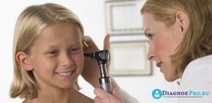
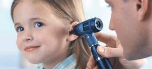
Otoscopy in children is not much different from the procedure in adults. However, there are a few key points:
- The essence of the manipulation is first explained to the child, they are reassured, and they are assured that the examination is painless.
- A parent or proxy is present during the examination. The guardian sits the baby on his lap, clamps his legs with his knees, presses his head to his chest and fixes his hands in his palms.
- Funnels of smaller diameter are used.
- The main feature of otoscopy in children is the direction in which the auricle is retracted. If in adults it is retracted upward and posteriorly, then in children it is retracted posteriorly and downward.
Important! Because children's eardrums are more sensitive, discomfort or pain may occur. It is especially pronounced in pathological changes - otitis media, perforation, eczema.
Is it painful to have an otoscopy?
Otoscopy is a quick, painless and easy diagnostic method. It does not require special preparation from the patient, and the manipulation itself takes from 5 to 15 minutes.
Usually the examination is painless. However, with pathological processes, especially inflammation, suppuration, damage to the eardrum, unpleasant sensations are possible.
Carrying out an otoscopy
Everyone knows a special round reflector with a hole in the middle, which is mounted on the head of an otolaryngologist. It is used to examine the cavities of the ears, nose, throat, nasopharynx and larynx. With otoscopy, it is possible to examine the ears in a completely painless way.
Content
- Carrying out an otoscopy
- Diagnosed diseases
- Normal otoscopic picture
At the beginning of the procedure, the doctor inserts a small tube made of metal or plastic into the ear canal. After insertion, the doctor pulls the auricle back and up, or in the case of children, back and down, and straightens the cartilage of the external auditory canal, bringing the eardrum into view. In order to better examine it, a reflector is used, with which a specialist regulates the illumination of the area in question. It reflects a dense beam of light reflected from the lamp opposite, transforming it into a beam. The beam through the tube is directed directly to the eardrum, as a result of which the specialist has the opportunity to examine its condition and identify pathologies.
General practitioners or family doctors use otoscopes in their work. The device looks like a light bulb; at its end there is a light source and a magnifying glass, as well as an ear tube. The otoscope is very easy to use - you just need to insert the tube into the ear, turn on the light on the device and, through a magnifying glass, examine all the interesting details of the ear canal and eardrum.
What does otoscopy show?
Diagnostic otoscopy reveals:
- causes of obstruction of the ear canal - wax plug or foreign body;
- pathological discharge - purulent, bloody, watery;
- diseases and neoplasms in the outer, inner or middle ear - otitis media, eczema, otomycosis, furunculosis, polyps, mastoiditis;
- deviations from the normal state of the eardrum - its rupture, redness, swelling, thickening, retraction;
- factors leading to hearing impairment.
Today, otoscopy is the only accurate method for diagnosing pathological changes in the ears. It is also carried out for preventive purposes or for the selection of hearing aids. In addition, an otoscope is used to evaluate the condition of the nose and throat.
Diagnosed diseases
Using an otoscope, it is possible to establish a disease of the eardrum and external auditory canal - redness, swelling, which is caused by an acute purulent inflammatory process of the middle ear. When this disease passes into the chronic stage and a rupture of the eardrum occurs, the specialist can see through an otoscope a hole in it and the fact that secretory fluid enters the area of the external auditory canal from the cavity behind the eardrum. This phenomenon is quite common for newborn children with purulent inflammation in the ears, when relief in the child occurs due to the rupture of the eardrum and the cessation of pressure of pus on it. At such a moment, you can easily see fluid flowing out of the ear.
In general, doctors call the following pathological conditions symptoms and indications for ear otoscopy:
- painful sensations in the ears, accompanied by itching, or the presence of noise;
- hearing impairment;
- diagnosed otosclerosis;
- various damages and injuries to the eardrum, ear canal, foreign bodies entering the ears;
- eczema of the outer ear;
- exostoses in the ear, furunculosis in the ear canals;
- otomycosis;
- the occurrence of sulfur plugs;
- malignant neoplasms.
All these signs can serve as a source of various pathological processes not only in the ears, but also in the brain.
With another disease - eustachitis - the mucous membrane of the Eustachian tube swells, becomes inflamed, the eardrum, visible through an otoscope, appears to be pulled inward and painted a matte shade of ice. The causes of eustachitis are enlarged tonsils in the nasopharynx. Also, when using an otoscope, you can easily identify a disease such as otomycosis or fungal infection of the external auditory canal. Due to the presence of this disease, a sharp deterioration in hearing may occur as the ear canal swells and the eardrum is damaged.
If the outward appearance of the ears is swollen and red, experts diagnose inflammation of the outer ear, while the otoscope helps to determine injury to the eardrum, its rupture, inflammation and other changes, and often inflammation simultaneously with this process in the middle ear.
If it is necessary to determine the mobility of the eardrum, otolaryngologists or family doctors use a Siegle funnel. In this case, for a detailed examination of the eardrum it is necessary to use biconvex magnifying glasses.