Author
Feshchenko Svetlana Yurievna
Candidate of Medical Sciences
Endocrinologist
until September 30
Prevention of fractures: diagnosis and treatment of osteoporosis with a 20% discount More details All promotions
Densitometry
(another name
for osteodensitometry
) is an x-ray examination that allows you to determine the mineral density of bone tissue.
As osteoporosis develops, bone density decreases, bones become less elastic and strong, and the risk of fractures increases. Densitometry can detect bone degradation in the early stages, whereas conventional radiography cannot detect bone loss of less than 25-30%.
An important advantage of densitometry is the low level of radiation - 10 times less than with radiography.
What is densitometry?
The name "densitometry" is derived from the Latin word densitas (density) and the ancient Greek metreo (to measure).
Devices for densitometry - densitometers - allow you to determine the density of materials by checking their resistance to the passage of light, ultrasound, and x-rays. They are widely used in various industries, including medicine. Based on their operating principle, medical bone densitometers are divided into ultrasonic and x-ray. Knowing what a densitometer is, it is easy to understand what a densitometry examination is. This is nothing more than a diagnostic study of bone tissue using a densitometer. Unlike histomorphometric studies, when a bone tissue sample is taken for analysis, densitometry is a non-invasive examination, it is painless and non-traumatic.
The result of densitometry allows you to determine how much bone density, their structure and the thickness of the outer layer differ from standard values. Based on the data obtained, a diagnosis is made and treatment is prescribed to improve metabolism in bone tissue and reduce the risk of fractures.
Densitometry for osteoporosis is used not only for primary diagnosis, but also to monitor the effectiveness of prescribed treatment. Control studies are carried out every 12–24 months and are stopped after stable treatment results are achieved.
Indications for densitometry
- Women, in the first few years after menopause (especially after removal of the ovaries).
- People with two or more risk factors for osteoporosis.
- All people who have had one or more non-serious fractures over the age of 40 years.
- After an injury (car accident, fall from a great height, sports injuries).
- People taking glucocorticoid hormones (prednisolone) and thyroid hormones for a long time.
- People who are suspected of having osteoporosis during an X-ray examination of bones.
- People receiving drug therapy for osteoporosis to monitor the effectiveness of treatment.
Densitometry is performed at the Belyaevo Medical Center
When is it necessary to undergo a densitometer examination?
Osteoporosis is an insidious disease; it is important to identify it in the early stages, when degenerative changes in bone tissue have not yet led to serious consequences. Although the disease most often occurs latently, there are a number of symptoms that can be used to suspect osteoporosis. It is also advisable to undergo a densitometry procedure if you have certain risk factors. Examination with a densitometer is recommended for:
- pain in the spine, a feeling of heaviness in the shoulder blades, fatigue;
- causeless appearance of lameness, changes in gait;
- decreased height by more than 3 cm, frequent fractures;
- the onset of menopause due to natural or surgical reasons;
- presence of cases of osteoporosis in close relatives;
- rheumatoid arthritis;
- underweight (body mass index lower than 18.5);
- long-term use of drugs that negatively affect bone density;
- lactose intolerance and other reasons for refusing to consume dairy products;
- diseases that cause problems with calcium absorption
A densitometric study is necessary if at least one of the above factors is present, especially if we are talking about age over 60 years. Another reason for decreased bone density is a low level of physical activity, so skeletal bone densitometry would be useful for people aged 50 and older who lead a sedentary lifestyle. This study is also carried out during pregnancy to determine the level of calcium in the pregnant woman’s body to prevent its deficiency and ensure the proper development of the fetal musculoskeletal system.
Risk factors for osteoporosis
- Age. The maximum density and strength of bone tissue is achieved, as a rule, by the age of 30, after which a gradual decrease in its mass begins.
- Gender factor. Women over the age of 50 have the greatest risk of developing osteoporosis. Women are four times more likely than men to develop this disease. Initially, women are lighter, their bones are thinner and their life expectancy is longer.
- Bone structure and body weight. Small and thin women are more prone to developing osteoporosis than large women. The same picture applies to asthenic men.
- Family history. Heredity is one of the most important risk factors for developing osteoporosis. If close relatives (parents, grandparents) have had signs of osteoporosis, such as a hip fracture after a minor fall, the risk of developing osteoporosis is quite high.
- A history of bone fracture.
- Taking certain medications. For example, long-term use of steroids (prednisolone) also increases the risk of developing osteoporosis.
- Woman after menopause. Women who had early menopause (before 45 years of age) and were not receiving hormone replacement therapy.
- After surgery to remove the ovaries.
- The presence of a somatic disease accompanied by excessive excretion of calcium from bone tissue (for example, hyperthyroidism).
- Initially thin bones.
- Excessive alcohol consumption.
- Low physical activity.
- Diet low in foods containing calcium and vitamin D.
Types of densitometric studies
Most often, densitometry of the pelvis, hip joints and spine is performed, but according to indications, examination of the limbs, jaws, and teeth may be prescribed. Depending on the devices used, the following types of densitometry are distinguished:
- X-ray;
- ultrasonic;
- computer
The latter is divided into MRI and CT (magnetic resonance and computed tomography). For each patient, the doctor selects the optimal research method, taking into account contraindications.
In our clinic you can undergo diagnostics and treatment of the spine and joints. We use the latest equipment, which allows us to accurately determine densitometric parameters in one procedure and obtain a three-dimensional image of the vertebrae and hip joints to make the correct diagnosis and prescribe the most effective therapy.
Features of densitometry
The examination has several varieties, differing in technique, and the most accurate among them is dual-energy X-ray absorptiometry (can be found under the simplified name “X-ray densitometry”). This is exactly the procedure that is carried out in our clinic. Its effectiveness is so high that it allows you to identify the slightest deviations from the norm.
In any case, this procedure is relatively quick (5-10 to 30 minutes) and painless. But regardless of what kind of densitometry will be performed, where you decide to do it, before the examination you need to follow a few simple rules.
Ultrasonography
Patients who are prescribed ultrasound densitometry are interested in what it is. This examination is performed using a special ultrasound machine that generates waves with a frequency of 20–1000 kHz. The device analyzes the propagation speed and broadband attenuation of ultrasound as it passes through bone tissue.
Ultrasonic waves cause microvibrations of bone tissue, which depend on its mechanical and structural properties. By measuring the parameters of these vibrations, it is possible to assess the condition of the surface and internal structures of bones and joints. The device uses a special program that compares the data obtained with standard indicators for different age categories. Ultrasound examinations are harmless, they can be used without restrictions, including for examination at any stage of pregnancy.
X-ray examination
It’s easy to guess from the name what X-ray densitometry is. This is a study of bone tissue, which is carried out on a special apparatus using x-rays. Passing through bones, X-ray radiation is attenuated, and the density of bone structures can be assessed by the degree of this attenuation. The latest devices make it possible to determine bone mineral density in different parts of the skeleton with an accuracy of 2–6% and identify osteoporosis even in the early stages.
X-ray bone densitometry is a more accurate method compared to ultrasound. Although the radiation dose from its use is low, it is not completely harmless, and therefore has limitations in its use. In particular, it cannot be used in diagnosing pregnant women and should not be used too often.
Computer diagnostic methods
Computer densitometry methods allow not only to measure mineral density, but also to obtain a three-dimensional image of the structures of the outer and cancellous layers of bone. This helps to establish an accurate diagnosis and also provides data necessary for surgical interventions.
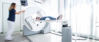
The computed tomography (CT) method is based on layer-by-layer scanning of bone tissue using thin beams of X-ray radiation. The radiation dose for CT is slightly higher than for X-ray bone densitometry. This examination is not applicable for pregnant women, but for others it can be repeated once every six months. Magnetic resonance imaging (MRI) is based on the effect of electromagnetic waves on atomic nuclei. This method allows you to visualize the structure of bone tissue. Unlike CT, the MRI method is harmless and has no restrictions on its use, except in cases where the patient has metal implants in the body. MRI can be used to diagnose pregnant women.
Where to do densitometry
Before choosing a clinic, it is important to pay attention not only to the qualifications of doctors, but also to the equipment of the medical institution. The Lancet Clinic is equipped with the most modern equipment: STRATOS X-ray bone densitometer, built on the basis of DXA technology with the Russian-language dR program and accessories, Kontron Medical, made in France. This model has several advantages: low radiation dose, which minimizes the harmful effects of x-ray radiation on the patient; high sensitivity of the device, which provides the most reliable results; the device reproduces the three-dimensional structure of the bone, which makes it possible to better establish the localization of the pathological focus.
Contraindications to the procedure
There are no absolute contraindications to densitometry. There are only some restrictions. At any stage of pregnancy, it is prohibited to use methods using x-ray radiation - x-ray densitometry and CT. During this period, only the use of ultrasound densitometry and MRI is allowed.
Densitometry is also impossible if the patient, due to pain or other reasons, is unable to take the position required for the examination and maintain it during the entire procedure.
Indications and contraindications
The densitometry procedure is necessary for patients who have:
- female age after 40 years, menopause, male age after 60 years;
- diseases of the parathyroid glands;
- in case of undergoing adnexectomy (removal of the ovaries);
- even a one-time fracture of any bone due to minor injuries;
- age over 30 years if the patient has a history of close relatives diagnosed with osteoporosis;
- taking medications that promote calcium leaching (glucocorticoids, anticoagulants, oral contraceptives, diuretics or psychotropic drugs, anticonvulsants, tranquilizers, etc.), as well as alcohol abuse and smoking;
- sedentary lifestyle;
- low body weight and height;
- periodic outbreaks of unbalanced nutrition - therapeutic fasting, diets, lack of a normal, complete diet;
- regular physical overexertion - heavy physical labor, active sports activities, etc.
There are no contraindications to the ultrasound method for diagnosing osteoporosis. X-ray densitometry, as mentioned earlier, is contraindicated for frequent use, as well as in cases of pregnancy or breastfeeding.
Preparing for the examination
To obtain reliable densitometry tests for osteoporosis, you must stop taking medications and supplements containing minerals such as calcium and phosphorus for several days prior to the procedure. You should also exclude cheese, cottage cheese and other foods high in calcium from your diet.
It is also necessary to notify the doctor in advance of the following facts:
- Pregnancy. Even the slightest possibility of pregnancy is a contraindication to examination using x-rays. In order not to harm the embryo or fetus, pregnant women are examined using harmless ultrasound and MRI methods.
- Presence of metal implants. Metal in the body is a contraindication to MRI, as it can negatively affect the reliability of the results and even lead to injury to the patient. In such cases, the doctor selects another research method.
- Recent undergone procedures such as CT with contrast, radioisotope scanning, radiography with barium. After these studies, a certain time must pass so that they do not affect the accuracy of the densitometry results, and the permissible radiation dose is not exceeded.
These recommendations must be followed when examining any location, including densitometry of the jaws and teeth. You must come to the procedure without jewelry, in loose clothing that will not interfere with the examination. Clothing should not have metal fittings - buttons, zippers, buckles, as they can interfere with the procedure and distort the results.
Don’t put off your visit until tomorrow, when a breakthrough can happen today!
The sooner you do this, the greater your chances of maintaining the health of your skeleton, the more active your life will be even at an older age, and most likely it will be without pain and suffering.
What is needed to prevent osteoporosis?
- Systematically consume more foods containing calcium.
- Include in your daily diet foods containing essential amino acids (meat, fish, soy, legumes)
- Lead an active lifestyle: exercise daily, walk at least 10 km a week or at least 60 minutes a day
- Stay in the sun for at least 30 minutes a day (if there are no contraindications)
- Quitting bad habits (smoking, alcohol abuse, coffee)
Thus, in order to fight osteoporosis, you need to eat right, actively engage in physical exercise, often be in the sun, and provide your body with sufficient amounts of calcium and vitamin D.
How is the densitometry procedure performed?
Those who are undergoing a densitometric examination need to know how bone densitometry is performed and prepare for the fact that they will have to remain completely still for some time, otherwise the pictures may be blurry. Depending on the chosen diagnostic method, the procedure can be carried out differently, but in general it goes like this:
- The patient is placed on a flat surface, upholstered with soft material, and asked to take a certain position. The spine examination is usually performed in the supine position, with the lower legs placed on an elevation so that the lumbar region is in contact with the table.
- The radiation source is usually located under the table, and the sensor is located above the patient's body. They move synchronously along the area under study and transmit the received data to the computer.
- During densitometry of the hip joint, the foot is fixed in a holder, which rotates the femur into the desired position.
- If an examination of the bones of an arm or leg is performed, the limb is placed inside the cavity of a special apparatus.
The procedure does not cause pain or discomfort, its duration ranges from half an hour (examination of the spine and hip joints) to 5–10 minutes (examination of the limb). At the end of the procedure, the orthopedist determines the bone density and issues a printout with the results.
What is densitometry, how is it carried out, x-ray examination, preparation for the procedure -MEDSI
Table of contents
- Principle and types of research
- Ultrasound examination
- X-ray examination
- Other types
- When is densitometry necessary?
- What contraindications exist?
- Preparation for the procedure
- How is densitometry performed?
- Decoding the analysis results
- Advantages of densitometry of teeth and other organs at MEDSI
Densitometry is a method for diagnosing the density and likelihood of bone fractures.
This test measures calcium levels, overall bone density and structure, and the thickness of the surface layer of bones. Thanks to this study, it is possible to identify osteoporosis at an early stage and begin its treatment in a timely manner. It also helps prevent calcium deficiency in the body of a pregnant woman, which is important for the proper development of the fetus.
Principle and types of research
Depending on what part of the body and under what conditions needs to be examined, different examination options can be used. It is possible not only to examine the bones of the pelvis, spine or limbs, but also to densitometry the teeth.
Ultrasound examination
The inspection device emits ultrasonic waves with a frequency greater than 3 MHz. In the process, the speed at which the wave travels through the bone tissue varies depending on its density.
All data on changes in this speed are recorded and then used for diagnostics and comparison with normal values.
This procedure is completely painless and harmless and is suitable during pregnancy or other restrictions related to the ban on radiation.
X-ray examination
This method is based on the use of x-ray radiation. It is retained in tissues with a dense mineralized structure.
The analysis can be used to examine the vertebral, lumbar and femoral spine, as well as the extremities: hands and feet.
This study is highly accurate and makes it possible to visualize the bone structure. It allows you to detect bone loss at an early stage.
The radiation dose that the body receives in this case is low, but is dangerous for pregnant women, since the rays can affect the normal development of the fetus.
Other types
Magnetic resonance imaging and computed tomography can also be used to diagnose osteoporosis.
A separate area is densitometry of teeth using radiovisiography (computer x-ray). Studies have shown that this type of analysis makes it possible to study the degree of damage to the hard tissues of the tooth (dentin) by caries.
When is densitometry necessary?
The doctor prescribes this type of study for the following symptoms:
- Frequent bone fractures, especially the femurs
- Fractures, dislocations associated with the vertebral region
- Regular back pain, the causes of which are unknown
- Diseases of the gastrointestinal tract, kidneys, liver
- Diabetes
There are also a number of diseases for which densitometry is recommended:
- Metabolic disorders associated with the removal of calcium and phosphorus from the body
- Neurological diseases
- Long-term hormonal treatment
- Previously diagnosed osteoporosis
- Endocrine system disorders
For preventive purposes, different types (including dental densitometry) of this study are used in the following cases:
- Monitoring the bone density level of the female body during pregnancy
- Monitoring the presence of calcium and phosphorus in the body at the stage of formation of tubular bones in the fetus
- Monitoring the progress of osteoporosis treatment
- Preventive examination of the condition of bone tissue in people over forty years of age
What contraindications exist?
There are not many of them, and they are related to the nature of the type of densitometry used:
- If the patient is pregnant, X-ray examination should not be used.
- Pain or disturbances in the lumbar region, due to which the patient will not be able to lie still for at least half an hour
Preparation for the procedure
There are a number of simple steps that will help you prepare for densitometry:
- A few days before the test, you need to remove foods that contain a lot of calcium (such as cottage cheese) from your diet.
- It is also necessary to temporarily stop taking medications containing phosphorus and calcium - you should consult your doctor about this.
In case of existing limitations of the study, it is necessary to take into account their presence:
- The patient should notify the doctor about pregnancy. In this case, she will be prescribed an examination using ultrasound waves, since x-rays are harmful to the body that is still developing
- If you plan to conduct a magnetic resonance examination, you need to warn the doctor about the presence of metal implants, as they may distort the results. And if some of them are present, it is absolutely impossible to perform an MRI. In this case, the doctor will decide what type of test is best to prescribe.
- The doctor must also be notified if shortly before this study the patient underwent:
- Radioisotope scanning
- Computed tomography with contrast agent
- X-ray examination with barium
In all these cases, the physician must determine exactly when it is best to undergo the examination, so that due to previous tests there is no distortion of the results or excessive stress on the patient’s body.
All these rules apply to the procedure for dental densitometry.
How is densitometry performed?
For the examination, a special device is used - a densitometer. The process itself goes as follows:
- The subject's clothing should be loose so as not to interfere with the examination.
- The patient sits on a couch or table in a position that depends on the organ being examined (when scanning the spine - lying on his back, etc.)
- A sensor and detector are placed above the analyzed area
- The device moves along the studied area, during which data on reflected and absorbed waves is transmitted
- The procedure lasts up to half an hour, during which the patient must remain motionless
- All results are saved on the computer and can be used in the future
If one of the limbs (hands, feet) is being examined, then for this purpose it is placed in a special cavity of the device. In this case, the procedure lasts much less - only a few minutes.
Decoding the analysis results
Based on the results of the examination, the doctor receives two main types of data:
- T-score. Its value is derived from comparisons of bone density with a normal value of 1 point or more. If it is less than specified, then:
- Number from -1 to -2.5 – decrease in mineral density
- Below -2.5 – osteoporosis with a high probability of fractures
Advantages of densitometry of teeth and other organs at MEDSI
- Anyone can sign up for a consultation easily and simply: by calling 8 (495) 7-800-500
- More than 20 clinics in different parts of Moscow are always ready to receive patients
- Radiologists and diagnosticians of highly qualified categories provide a full range of services: prepare clients for examination, accompany the process and decipher data
- The use of modern high-quality equipment allows you to get the most accurate results and start treatment on time
Decoding the results
The data obtained are assessed according to two criteria - T and Z. The T-criterion shows how the patient’s bone density indicators differ from the norms for young people of the same sex. A value less than -2.5 indicates osteoporosis with a high risk of fractures. The Z-score characterizes bone density in comparison with norms for people of suitable age. If this indicator is negative, then bone density is below age norms.
Now that we know what densitometry is and how it is performed, all that remains is to choose the right clinic for diagnosis and treatment of osteoporosis. The accuracy of diagnosis and the effectiveness of treatment depend on the level of equipment used, as well as on the qualifications and experience of doctors.
Our clinic is equipped with the latest densitometry equipment and employs the best specialists in Moscow. We use the most accurate diagnostic programs and effective treatment protocols, which allow us to detect osteoporosis at the initial stage and successfully treat it.
Who needs densitometry?
- all women over 45 years of age;
- athletes;
- patients with diseases accompanied by a decrease in bone mineral density, such as rheumatoid arthritis, chronic kidney disease (nephritis) or liver disease (hepatitis);
- patients with type 1 diabetes mellitus (adolescent or insulin-dependent);
- patients with thyroid diseases accompanied by a long-term increase in hormone levels;
- patients with diseases of the parathyroid glands;
- patients with indirect clinical or radiological signs suspicious for osteoporosis;
- patients taking long-term medications that cause loss of bone mineralization.
- patients with frequent fractures.
