Histological examination, in fact, is a method for diagnosing a disease at the tissue level - examination by a pathologist under a microscope of a specially prepared section of pathological tissue allows one to get an idea of the essence and severity of the pathological process, identify its features and, thanks to this, accurately make a diagnosis and select the required treatment. In oncology, histological examination plays the role of an “arbiter” - only the pathologist’s conclusion about the presence of pathological cells gives the clinician the right to make a diagnosis of cancer. Histological examination is widely used in almost all medical specialties and is always performed after any operation, but in oncology it is of particular importance.
How is material obtained for histological examination?
The process of obtaining a piece of tissue for histological examination is called a biopsy. The following types of biopsy are distinguished:
- Core biopsy. It is performed with a special needle and an automatic biopsy gun.
- Aspiration biopsy. It is used to collect the liquid contents of formations, for example, cysts or pleural cavity.
- Fine needle aspiration biopsy. A subtype of aspiration biopsy, material is removed using a syringe and a special needle with a pointed or scalloped edge. With this method, it is possible to obtain for research not only liquid contents, but also a piece of tissue.
- Aspiration-cutting biopsy. With this method, both tissue fragments and cellular material are taken with one needle for research.
- Trephine biopsy. The collection is made with a trephine, an instrument that is a hollow tube with a pointed end. Used to take a biopsy from bone tissue.
- Excisional biopsy. It involves removing the entire pathological formation (for example, a tumor node) entirely. A good example of an excisional biopsy is the removal of an enlarged lymph node when lymphoma is suspected.
- Scarification biopsy. The sampling is performed by cutting off a thin layer of tissue and is widely used to study skin tumors.
- Punch biopsy. Performed with special biopsy forceps, it is often used for diseases of the gastrointestinal tract.
- Brush biopsy. The material for research is taken using a special brush by scraping it - for example, from the wall of the bronchus.
- Loop biopsy. The histological material is collected using a loop simultaneously with the operation of the coagulator. Used in otolaryngology and gynecology.
- Imprint. It is used when it is necessary to study discharge from an erosion or ulcer - a glass slide is applied to the ulcerated surface.
- Smear-imprint. The material for research is removed with a scalpel, spatula or a special brush and transferred to a glass slide.

Small intestine
Biopsy is important in diagnosing diseases of the small intestine. Oral biopsy material is traditionally taken from the trigeminal ligament. Endoscopic biopsy is currently used most often, as it allows several targeted biopsies to be performed in a short time in more comfortable conditions for the patient. The volume of pieces obtained from a punch biopsy is usually sufficient to make a diagnosis in diffuse lesions of the small intestinal mucosa, if at least 3 biopsies are taken from areas distal to the duodenal bulb (to avoid misinterpretation of the morphological picture associated with Brunner's glands). In diseases with a segmental nature of the lesion, several biopsies are taken from more distant parts of the small intestine, which requires a longer endoscope of smaller diameter. Histological examination can be useful for making a diagnosis even with a normal macroscopic picture.
A small bowel biopsy is the standard test to confirm the diagnosis of malabsorption syndrome. When diagnosing celiac disease, histological examination of biopsies of the small intestine is extremely necessary, even with a positive test for the presence of endomysial antibodies or tissue transglutaminase in the blood. A biopsy should be performed before treatment is given as the above tests may be false positive
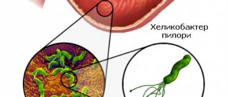
Infectious lesions of the small intestine can also be determined by histological examination. Giardia and a number of other protozoal pathogens can cause inflammatory changes in the mucous membrane of the small intestine. Typing of mature adult pathogens, their trophozoids, or intermediate life cycle forms within the epithelium or on its surface can help establish an accurate diagnosis. In some patients, morphological manifestations are similar to eosinophilic gastroenteritis; the diagnosis of the latter can only be established after parasitic infestation has been excluded.
In patients with immunodeficiency conditions (post-transplant disease, HIV infection), pathogens such as Isospora belli, Cryptosporidia, Cyclospora and Microsporidia can be detected in biopsies of the small intestine. Other pathogens detected in the small intestine in immunodeficiency states are cytomegalovirus, Candida fungi, histoplasma and Mycobacterium avium-intracellulare complex. When collecting material from such patients, it may be useful to use large biopsy forceps. It is also recommended to use forceps without a fixing needle to avoid mechanical damage to the mucous membrane and loss of exudate on its surface.
Tumors of the 12th colon are detected by endoscopic examination with biopsy. The technique for collecting material depends on the location and size of the tumor. Duodenal and jejunal polyps can occur in 33-100% of patients with a history of familial adenomatous polyposis (FAP). Gastric polyps in patients with FAP most often look like fundic gland polyps. They do not undergo malignant transformation, but require biopsy verification to exclude adenoma. Duodenal polyps are usually adenomatous and are found mainly in the ampulla or periampullary zone. Upper gastrointestinal polyps can develop synchronously or metachronously with respect to detected polyps in the colon. Adenocarcinoma arising from periampullary adenoma is an easily recognized condition and is the most common cause of death in patients with FAP after colon cancer. Patients with FAP should be included in a surveillance program, although the effectiveness of this strategy remains to be confirmed.
Several literature sources describe the occurrence of pancreatitis after biopsy of the major duodenal papilla, but complications after endoscopic examinations with biopsy of the small intestinal mucosa or removal of duodenal adenomas from areas adjacent to the major duodenal papilla are rare.
How is a biopsy performed?
A biopsy is usually performed under local anesthesia, if the formation is located deep enough - under ultrasound or x-ray control. The patient does not require any specific preparation (except, perhaps, a psychological attitude). The procedure time is not limited - if a scarification biopsy or a smear is performed, it takes literally a few minutes, and if we are talking about a biopsy of a deep-lying formation, especially near large vessels or nerves, then it takes much longer.
In the process of performing a biopsy, the needle is passed to the formation and, depending on the chosen method, either aspiration is performed simultaneously with the needle moving towards the center of the formation (if an aspiration biopsy is performed), or a needle is inserted into the formation and a column of tissue is fixed inside it (if a core biopsy is performed ).
The process of obtaining tissue using a biopsy game is currently either semi-automatic or automatic. In a semi-automatic process, the groove with the needle is advanced manually into the pathological focus, and the activation of the cannula in the needle, which captures the tissue, occurs automatically using a spring-loaded trigger mechanism. The automatic version uses a special biopsy gun, and the doctor only needs to bring it to the area of interest.
It is also possible to perform a biopsy during a diagnostic endoscopic examination, such as endoscopy or bronchoscopy. In this case, the collection of histological material occurs almost painlessly for the patient, although it causes some discomfort.
In addition, tissue removed during surgery will be sent for histological examination. It should be noted that in some cases, an operation may be prescribed just to obtain histological material (for example, the appointment of separate diagnostic curettage of the uterus and cervical canal in order to establish the cause of a gynecological disease).
Book a consultation 24 hours a day
+7+7+78
Features of the procedure
In our medical center in Moscow, the analysis of the histology of the cervix or other organs is carried out according to a repeatedly tested algorithm:
- sampling material using one of the biopsy methods - pinching, puncture, excisional, smear taking, curettage;
- fixing the sample in a special solution, treating it with paraffin and obtaining the thinnest sections;
- staining the section with a dye and studying it using an electron or light microscope.
How many days it takes to obtain histology results depends on the situation. The rapid analysis required to determine the extent of urgent surgical intervention can be performed within one or several hours. The best results are obtained by standard histology, which takes 7-10 days. Histology is a highly accurate diagnostic method, and if all the rules are followed, the possibility of error is practically eliminated.
By calling the MedBioSpectrum clinic, you can find out how much it costs to send material for histology and what the procedure is, as well as make an appointment with a doctor to receive a substantive consultation.
Types of histological examination
First of all, the histological examination itself is carried out, that is, the study of specially prepared tissue taken during a biopsy under a microscope. The process of preparing tissue for research includes several stages.
- A piece of fabric is dehydrated and impregnated with paraffin, thereby forming a small cube in which the fabric is enclosed.
- Next, sections are made from the paraffin cube using a microtome, and the thickness of the sections can reach 3 micrometers.
- The sections are transferred to glass, prepared for staining and stained using one of the known methods in order to facilitate the identification and differentiation of individual tissue structures during the study.
Thus, the process of preparing tissue for histological examination is quite lengthy, which explains the receipt of a doctor’s report several days after the biopsy. However, in cases where urgent research is required (for example, assessment of resection margins during surgery or examination of a sentinel lymph node in breast cancer), the tissue preparation process can be reduced to half an hour, but in this case the quality of the resulting histological specimen is significantly lower, so after During the operation, a thorough study of the biomaterial is carried out as planned.
It is no coincidence that the description of the process of preparing tissue for histological examination is described in such detail, since there are often situations when the patient wants to get a second opinion or continue treatment in another institution. In this situation, it is advisable to take ready-made histological preparations (“slides”) and, if possible, paraffin blocks, since the pathologist conducting the study can, if necessary, cut several sections from the block, use a different type of histological dyes and conduct additional studies on a larger amount of biopsy material. material.
What studies can complement the study of sections under a microscope? First of all, this is an immunohistochemical study, which makes it possible to determine the histological identity of the tumor, which is extremely important, for example, when studying metastasis from an undetected primary lesion. In this case, the expression of various genes and their proteins in the tissue is assessed. Also, in breast cancer, an immunohistochemical study makes it possible to determine whether the tumor has receptors for estrogen and progesterone, and, accordingly, to prescribe the chemotherapy and hormonal therapy required for the identified molecular subtype. Finally, determination of the expression of the Her2/neu protein allows us to conclude whether a patient with breast cancer or gastric cancer is indicated for targeted therapy.
Separately, I would like to dwell on the cytological examination. Unlike histological examination, in which the subject of study is tissue, cytological analysis examines individual cells for pathological abnormalities. Cytological examination is less accurate than histological examination, but allows us to give a conclusion about the nature of the disease in cases where it is impossible to obtain a piece of tissue (for example, when analyzing pleural effusion - whether it is associated with a tumor lesion of the pleura or not - or peritoneal washings in the case of suspected metastatic screenings of ovarian cancer).
Esophagus
Esophageal malignancies can be diagnosed by biopsy in 95% of cases, except in situations where obstruction prevents adequate imaging and biopsy from the lesion. 8 to 10 biopsies should be taken. Additional brush cytology can improve diagnostic capabilities.
The most common inflammatory changes in the esophagus occur with reflux esophagitis, which develops with gastroesophageal reflux disease (GERD). Endoscopic examination with biopsy is indicated in the diagnosis of Barrett's esophagus, or to exclude infectious or malignant lesions of the esophagus masquerading as gastroesophageal reflux disease. Erosive changes detected during endoscopic examination correlate well with the histological picture, but single erythema is an unreliable criterion for diagnosing esophagitis. On the contrary, histological abnormalities (inflammatory cell infiltration, including neutrophilic and eosinophilic leukocytes) can be detected in biopsies from patients with GERD with a normal endoscopic appearance of the mucous membrane. A biopsy and collection of material for cytological examination from an abnormal-looking mucous membrane are necessary to exclude malignant, infectious processes, some autoimmune diseases and Barrett's esophagus.
Barrett's esophagus is a condition in which the normal lining squamous epithelium is replaced by metaplastic, specialized intestinal epithelium. Its diagnosis requires a biopsy during endoscopic examination. Based on the detection of metaplasia of the esophageal mucosa, patients are included in national cancer programs (registered and periodically examined). Histological examination reveals the lining of the mucous membrane with columnar epithelium devoid of a brush border. The latter is distinguished from the gastric epithelium by the presence of goblet cells, which can be recognized by additional Alcian blue staining.
Destruction of the mucous membrane in the area of Barrett's esophagus is often accompanied by the formation of large ulcerations in the esophagus, and inflammation-induced atypia of epithelial cells at the edges of the defect can be mistakenly regarded as dysplasia. In such cases, intensive drug therapy leads to healing of the mucous membrane and correct subsequent histopathological interpretation of biopsy specimens.
A biopsy is also performed to detect dysplasia or adenocarcinoma. If dysplasia is established or its presence is suspected, a 4-quadrant biopsy should be performed at 1-2 centimeter intervals, as well as additional pieces should be taken from any pathologically changed areas of the mucosa. A 2-cm biopsy misses 50% of cancers in patients with severe dysplasia compared with a 1-cm biopsy. Although large forecept biopsies have previously been recommended, a retrospective analysis found that 4-quadrant biopsies missed the same number of cancers at 2 cm. crayfish when collected with both large forceps (4/12, 33%) and standard size forceps (6/16, 38%). The collection of bipsy material from the mucous membrane of the esophagus should be carried out using the method of rotational aspiration, in which the open forecept is brought close to the end of the endoscope, the endoscope is turned to the wall, aspiration is performed, the forecept is extended, closed and the piece is removed. It is reported that in patients with severe dysplasia who refused surgery, this technique (carried out at 3-6 month intervals) made it possible to effectively diagnose cancer, which at the time of detection in 96% of cases was located within the mucous membrane.
High-resolution endoscopic examination and methylene blue chromoendoscopy increase the detection rate of short segment Barrett's esophagus through targeted biopsy. Chromoendoscopy with Lugol's solution and methylene blue increase the detection rate of squamous cell carcinoma and neoplastic changes in the Barrett's esophagus, respectively, although the value of using methylene blue in monitoring patients with Barrett's esophagus remains controversial.
The study of biopsy samples using flow cytometry with DNA analysis makes it possible to identify patients with aneuploidy, polyploidy, and, using p-53, loss of heterozygosity, which indicates an increased risk of developing cancer.
Local lesions in the esophagus can be removed by endoscopic mucosal resection. In this case, a physiological solution is injected into the submucosal layer in order to elevate the pathologically changed area, and then it is removed using loop electroexcision. This technique has been successfully used to remove neoplastic lesions in areas of Barrett's esophagus and to remove benign tumors of the esophagus.
Infectious esophagitis develops in patients with immunodeficiency conditions caused by systemic antiimmune therapy, inhalation of steroid drugs, malignant tumors (after chemotherapy), diabetes, and AIDS. The most common causative agents of infectious esophagitis are fungi of the genus Candida, herpes simplex virus, and cytomegalovirus. Fungal esophagitis is recognized by the presence of a white coating against the background of inflamed mucosa. Brush biopsy and sampling are performed, but cytological examination of smears after brush biopsy is a more sensitive method. Viral esophagitis is manifested by the formation of ulcerations. A biopsy should be taken from both the edge and the center of ulcerative defects. Histological examination is usually informative, but in case of AIDS, the collection of a large number of pieces (up to 10) is required. Isolation of the virus in culture facilitates diagnosis, but this method is less sensitive than histological examination aimed at diagnosing cytomegalovirus infection.
Indications for histological examination
Histological examination is the most accurate diagnostic method, which should be used when all other studies taken together cannot clearly indicate a specific diagnosis.
First of all, a histological examination is indicated for patients with suspected malignancy or with an already confirmed diagnosis of cancer. In the first case, the result of the study gives the doctor the right to make a final diagnosis. In the second case, a histological examination is indicated in order to:
- monitor dynamics, for example, confirm distant metastasis;
- change treatment tactics according to the results, for example, if a patient with breast cancer received chemotherapy before surgery, and the results of postoperative histological examination did not show a complete response of tumor cells to treatment, in some cases it is possible to prescribe tablet chemotherapy in the postoperative period.
Further, histological examination is indicated in the case of diseases that are essentially benign, but have the potential for malignancy. These are, for example, intestinal polyps; in themselves, they do not pose a threat to life, but they can potentially degenerate into a tumor, therefore, after removal, polyps must be examined to determine whether tumor structures have appeared in them.
Widespread use of histological examination is also observed in non-tumor diseases. In gastroenterology and gynecology, the study of tissues and cells, in addition to excluding the tumor process, is also intended to assess inflammatory processes and their dynamics, and identify endocrine disorders. Histological examination also plays an important role in the treatment of autoimmune diseases, allowing one to assess the degree of activity of the pathological process and the severity of the lesion, as well as the response to therapy.
Finally, histological examination, or rather its subtype—cytological examination—has found its place in screening. Examination of a smear-imprint from the cervix allows one to determine a precancerous condition or an already developing tumor, and the simplicity and effectiveness of this method has made it possible to include a smear in screening for cervical cancer.
It should be noted that there are no contraindications as such for conducting histological examination, since the collection of histological material is a minimally invasive and well-tolerated procedure. Possible complications may be associated with the patient's individual reaction to anesthetics or with blood clotting disorders. In any case, the patient is prescribed rest and cold at the biopsy site, and analgesics for pain.
What can the analysis show?
Histology analysis is not prescribed in all cases. Histology of neoplasms is necessary to determine the inflammatory process in the gastrointestinal tract, to diagnose pathological changes in the female genital organs, to determine the causes of infertility, and to diagnose cancer.
In rare cases, analysis shows false results; according to statistics, 98% of histology shows true results, which makes it possible to establish a diagnosis and determine further actions.
More fresh and relevant information about health on our Telegram channel. Subscribe: https://t.me/foodandhealthru
We will be grateful if you use the buttons:
Result of histological examination
The result of the histological examination will be ready a few days after the biopsy, which is associated with the step-by-step preparation of the tissue being examined and the examination of the biomaterial by a pathologist under a microscope. The result can be either a short conclusion indicating the established tissue structure of the pathological formation (for example, “invasive lobular cancer”), or a detailed description of all changes in the structure of the tissue and its constituent cells.
The attending physician should decipher the histology results, since the histological examination is prescribed by him and for a very specific purpose. The resulting conclusion from a cytological or histological examination will help the doctor in establishing a clinical diagnosis and in selecting the currently required treatment methods.
Histological examination is one of the methods for diagnosing malignant neoplasms and a number of benign diseases. Euroonco doctors are highly qualified to interpret study results and prescribe the required treatment.
Book a consultation 24 hours a day
+7+7+78
Histology analysis: interpretation
The conclusion (deciphering the histology analysis) indicates all the results of the histology analysis, but only specialists can correctly decipher the medical terms. By contacting a doctor, the patient can receive complete information about their health status, and if dangerous diseases are detected, receive qualified assistance.
How much does it cost to decipher a histology analysis?
Of course, the price of deciphering histological analysis depends on the clinic where you conduct it. If you want to get a high-quality second opinion from an oncologist, then you need to pick up the blocks with histological material and bring them or send them to another clinic. If a repeated conclusion and interpretation of the histology analysis is carried out without histological material, this means that this is not a “second opinion” or a “histology revision”, but a confirmation of the diagnosis of one doctor by another doctor.
When it comes to such an important area of medicine as oncology, deciphering the histology analysis can significantly improve the course of treatment, and sometimes even save the patient’s life.
Our company BK Medical Logistic offers a complete revision and interpretation of histology analysis after a special study in an Israeli clinic specialized in oncology and oncological diseases, with high-tech laboratories and competent doctors known all over the world.
The price of deciphering a histology analysis may vary, but on average it is 550-650 US dollars.
How is repeated histology analysis performed?
At the moment, only our company can deliver histological material from Ukraine or Russia to Israel for medical examination. We use special refrigerators and packaging to ensure that the results of the histology analysis reach the clinic in Israel safe and sound.
After the blocks are delivered to Israel, the histology tests are re-checked and studied in the laboratory, after which the doctor prepares a special report with the results and a transcript of the histology.
How to apply for a repeat histology analysis?
Even if you just want to ask questions and get information about the revision of histology tests, you can contact us by phone listed at the top of the site. CALLS AND CONSULTATION ON THIS NUMBER IS FREE!
Colon
Visualization of a pathological focus in the large intestine necessitates its pathohistological evaluation. If the number of polyps is too large for simultaneous removal, a representative number of pieces should be taken. The smallest polyps detected during screening sigmoidoscopy should be biopsied; larger polyps should be removed entirely during a subsequent colonoscopy. Morphological confirmation of the presence of an adenoma or adenocarcinoma should serve as a reason for examining the entire colon. Published data on the value of identifying hyperplastic polyps during sigmoidoscopy remain controversial. Many US gastroenterologists do not believe that these polyps carry an increased risk of severe proximal neoplasia.
For colitis, endoscopic examination with biopsy helps in establishing the extent of the process, differential diagnosis and treatment planning. An acute biopsy in a patient with bloody diarrhea can distinguish spontaneously resolving acute colitis from a primary or recurrent attack of chronic ulcerative colitis or ischemic colitis. Terminal ileal biopsy may be helpful in the diagnosis of Crohn's disease, infectious ileitis, and lymphoid nodular hyperplasia. Both Crohn's disease and ulcerative colitis are associated with an increased risk of colorectal cancer. For these diseases, in order to identify dysplasia, it is recommended to conduct an endoscopic examination 8 years after the initial diagnosis, if the process was right-sided. In patients with left-sided localization of pathological changes, the risk of developing cancer increases by the 15th year from the onset of the disease. In patients with pancolitis, the most commonly used approach is 4-quadrant sampling every 10 cm (every 5 cm over the distal 25 cm). In left-sided colitis, a biopsy from the proximal colon should also be performed to determine the extent of the process.
Tactical approaches to dysplasia lesions in patients with chronic colitis continue to evolve. If the area of the mucous membrane with dysplastic changes is large, has an uneven surface, or is combined with a stricture, surgical treatment is required. However, a typical type of adenoma that has developed in an area of the colon with signs of chronic colitis should be removed with the collection of biopsy material from adjacent areas of the mucous membrane. If the adenomatous polyp is removed entirely and there are no signs of dysplasia in the surrounding mucous membrane, we can assume that adequate treatment has been carried out for a benign sporadic adenoma and continue to monitor the patient with repeated endoscopic examinations.
It remains unclear how many pieces and in what location should be harvested in chronic diarrhea and endoscopically normal colon. The diagnosis of microscopic colitis is established after identifying the corresponding histological signs in patients with chronic watery diarrhea, in the presence of a normal endoscopic picture and the absence of dysbiosis. Biopsy fragments obtained by flexible fiber optic sigmoidoscopy may be adequate to diagnose this condition.