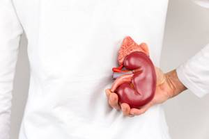
The kidney is a paired organ, shaped like a bean, which, through its urinary function, regulates the chemical constancy (homeostasis) of the human body. Located in the lumbar region. The length of one bud ranges from 11.5 cm to 12.5 cm, thickness from 3 cm to 4 cm, length from 5 cm to 6 cm.
The weight of the kidney ranges from 120 to 200 grams. The kidney is covered with a connective capsule and includes systems for excretion and storage of urine. Blood supply is provided by the renal arteries emerging directly from the aorta.
The main job of the kidneys is excretory. From 1700 to 2000 liters of blood passes through a normally functioning kidney per day, from 120 to 150 liters of primary urine and from one and a half to two liters of secondary urine are formed.
The kidneys perform essential work to regulate the acid-base balance of the blood plasma and maintain water-salt balance. They remove the final products of nitrogen metabolism and toxic substances from the body, take part in the metabolic processes of proteins and carbohydrates, and remove drug residues.
Unhealthy lifestyle, nutrition, and congenital hidden pathologies have led to the fact that the number of people with kidney diseases has recently increased.
The most common diseases include:
Nephrolithiasis (urolithiasis) – formation of dense stones (calculi) in the kidneys;
Pyelonephritis is an inflammatory process that affects the tubular system of the kidney, predominantly of a bacterial nature. May have an acute, chronic or chronic course with exacerbation;
Nephroptosis (wandering, mobile kidney) – the disease is expressed in abnormal mobility of the kidney. As a rule, the right kidney is displaced more often than the left. The main symptom is pain in the hypochondrium or iliac region.
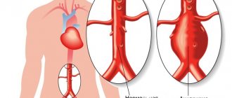
Hydronephrosis is an enlargement of the calyces and renal pelvis due to problems with urine outflow.
Renal failure is a syndrome in which all kidney functions are impaired. Water, nitrogen, electrolyte and other metabolisms are disrupted. It can be in both acute and chronic stages.
Glomerulonephritis is damage to the glomeruli of the kidneys (glomeruli).
Since the kidneys and bladder are part of the same excretory system, very often, for a more accurate diagnosis, the ultrasound procedure of the kidneys and bladder is combined.
What is USDG of renal vessels and arteries?
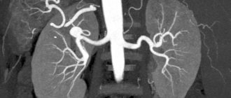
Ultrasound scanning of renal vessels is used as an additional diagnosis of the condition of the urinary system. It is resorted to when it is necessary to understand not only the structural features, structure and functions of the kidneys, but also the vessels that supply them with blood.
Doppler ultrasound provides information about the diameter of veins and arteries, their location inside the kidneys, and also allows you to measure the speed of blood flow.
Such diagnostics do not pose a danger to the human body and do not cause pain. In this regard, there are no restrictions on age and frequency.
Indications
Ultrasound Dopplerography of the blood flow of the renal vessels will be prescribed for:
- Complaints of pain (including acute pain due to renal colic) and discomfort in the lumbar region.
- Swelling of the legs, face.
- Suspicion of vesicoureteral reflux - reverse flow of urine into the kidney.
- Injuries to the abdomen and lower back - they can lead to damage to blood vessels.
- The appearance of protein in the urine, an increase in the number of leukocytes according to the results of urine tests.
- Suspicion of neoplasms in the kidneys.
- Hypertension with unknown cause.
- A number of endocrine or cardiovascular diseases.
- Preparation for kidney surgery and organ transplantation.
Dopplerography of the renal vessels can be performed repeatedly as many times as necessary. This allows us to evaluate the dynamics of changes and the effectiveness of therapeutic measures.
Indications for research
The following indications are distinguished for performing ultrasound examination:
Lower back pain
The lower back is the area where pain is localized in kidney diseases. Pain can occur either on one side or on both sides at the same time. To make an accurate diagnosis, ultrasound with Dopplerography of the renal vessels is recommended.
Swelling of the legs, face
Swelling of the legs and face may be a consequence of kidney dysfunction. In turn, the kidneys may suffer due to impaired blood flow in their vessels and insufficient blood supply.
Difficulty urinating
As the main element of the urinary system, the kidneys have a direct influence on the process of urination. If it is difficult and accompanied by pain, it makes sense to conduct a full examination - ultrasound plus Doppler.
Increased blood pressure
An increase in blood pressure is a clear sign of hemodynamic disturbances in the vessels. If at the same time the patient experiences other alarming symptoms related directly to the functioning of the kidneys (lower back pain, swelling of the extremities), he needs to undergo an ultrasound with Doppler to identify pathologies of blood flow in the vessels of the kidneys.
Attacks of renal colic
Renal colic is called acute attacks of pain in the lumbar region. The main reasons are a sharp disruption in the outflow of urine from the kidney, as well as a disruption in the blood supply to it. Ultrasound scanning of the renal arteries in this case will help to understand what the blood flow pathology is associated with, what it is - a blood clot formed in the vessels, which should not exist under normal conditions, atherosclerotic plaques or something else.
Bruises of the lumbar region
Bruises of the lower back can cause disruption of the integrity of the vessels supplying the kidneys. If this happens, normal kidney function becomes impossible. Using the diagnostic method Ultrasonography of the renal artery
it is possible to determine how serious and dangerous the damage is and what its exact location is.
Indications for kidney ultrasound
The kidneys are a paired organ of the urinary system, which forms urine as a result of filtration, reabsorption and secretion. The kidneys, bladder, and ureters form a single anatomical and functional system called the urinary system. The adrenal glands, not being an organ of the excretory system, are adjacent to the upper edge of the kidneys. Their main function is the production of hormones: sex hormones, adrenaline and norepinephrine, glucocorticoids. Due to the anatomical structure and functions of the urinary system and adrenal glands, a separate examination of the kidneys is carried out in exceptional cases. Most often, a comprehensive examination of all organs included in the urinary system and adrenal glands is carried out.
Kidney ultrasound is prescribed for the following patient complaints:
- urinary retention, incomplete emptying of the bladder;
- urinary incontinence;
- change in urine color: darkening of color, admixture of blood, urine the color of meat slop, opacity;
- the presence of mechanical inclusions in the urine;
- pain in the lumbar region;
- hypertension that does not respond to standard therapy;
- swelling of the limbs or face;
- abnormal urine test results;
- weight loss, loss of appetite, asthenia.
All of the above symptoms may indicate dysfunction of the urinary system. The following symptoms are typical for adrenal gland pathology:
- high blood pressure;
- amenorrhea, extramenstrual bleeding in women;
- reproductive dysfunction;
- change in skin color, excessive pigmentation;
- decreased libido;
- reduction or, on the contrary, increase in body weight while maintaining the same diet.
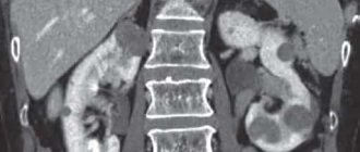
During ultrasound examination, it is possible to identify inflammatory, degenerative diseases of the urinary system, cysts, abscesses, and neoplasms of various etiologies.
Kidney ultrasound can be prescribed both for diagnostic purposes and to monitor the effectiveness of the therapy used or surgery performed. For patients undergoing surgical treatment of diseases of the urinary system, ultrasound may be prescribed to clarify the extent of surgical intervention, the presence of stones, the position of organs and the degree of damage to their tissues.
How to prepare for the procedure?
In order for an ultrasound examination with Doppler to go smoothly and its results to be highly informative, the patient needs to prepare. The most important condition for Doppler ultrasound is to reduce gas formation in the intestines. This allows for better visualization of the kidneys. For ultrasound examination of the renal arteries, the preparation required is quite simple.
Diet
In preparation for the study, a few days before ultrasound examination of the renal arteries (kidney vessels), you should follow a diet. Products that provoke increased gas formation in the intestines are excluded from the diet: bread, cabbage, legumes, raw fruits and vegetables, soda, sweets.
To additionally cleanse the intestines on the days of preparation, the patient is prescribed an enterosorbent (the most famous of them is activated carbon).
Ultrasound scanning on an empty stomach
Ultrasound of the kidneys with Dopplerography of the renal vessels must be performed on an empty stomach (preferably in the morning). The minimum interval between the procedure and the last meal is 6 hours.
It is permissible not to fully comply with the preparation rules for patients suffering from diabetes, coronary artery disease, arterial hypertension and who are forced to constantly take medications. It is not allowed to perform an ultrasound with Doppler after fibrogastroscopy or colonoscopy. After these procedures, air enters the intestines - this will complicate the ultrasound examination.
Preparing for an ultrasound of the kidneys
Patients who are about to undergo an examination for the first time often wonder whether they need to prepare for a kidney ultrasound. Obtaining accurate and informative results of a kidney ultrasound is impossible not only without the knowledge and experience of a sonologist, but also without the interest of the patient. After issuing a referral, the doctors of our medical center will definitely explain how to prepare for an ultrasound of the kidneys and what the preparation involves. Before the study, the patient must adhere to the following recommendations:
- The study must be performed strictly on an empty stomach. The time interval between the last meal and the ultrasound should be at least 8, and preferably 10-12 hours. This limitation is explained by the fact that after eating, intestinal motility is activated, which can significantly reduce the quality of visualization of the genitourinary system. Young patients can eat 5-6 hours before the test.
- If the gastrointestinal tract is not functioning stable, the patient suffers from constipation, on the eve of the study you can take a laxative, give a cleansing enema, or use a modern medicinal microenema.
- To improve the functioning of the gastrointestinal tract, you can take enterosorbents, activated carbon, and enzymatic agents, which are available in pharmacies without a doctor’s prescription.
- You need to be especially careful when drinking. During preparation for ultrasound, the patient must observe an increased drinking regime. This allows you to increase the evacuation capacity of the intestines, which means it helps to cleanse it. Before the test, the bladder must be completely full. To do this, you need to drink clean still water 1-2 hours before the procedure. As a rule, for adult patients, the amount of water drunk should be at least 1 liter. You should not urinate before an ultrasound.
- If the patient is forced to take drug therapy, it is necessary to warn the doctor conducting the study about this.
- If the patient must undergo an X-ray examination with contrast and ultrasound, then the interval between the examinations should be at least 5-7 days: the contrast agent introduced into the patient’s body during X-ray examination can significantly affect the information content of the kidney ultrasound.
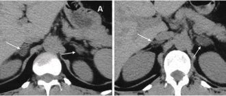
Interpretation of indicators: norms for blood vessels and blood flow
When scanning the kidney vessels, the doctor should evaluate the following indicators:
- Artery diameter. Normally, for the main trunk it should be 3.3-5.6 mm, for segmental arteries - 1.9-2.3 mm, for interlobar arteries - 1.4-1.6 mm, for arcuate arteries - 0.9- 1.2 mm.
- Systolic velocity of blood flow in the renal arteries. Normally, for the main trunk it should be 47-99 cm/sec., for interlobar arteries – 29-35 cm/sec.
- Diastolic blood flow velocity in the renal arteries. Normally, for the main trunk it should be 36-38 cm/sec, for interlobar arteries – 9-17 cm/sec.
The doctor also evaluates other indicators of the condition of the kidneys: shape and size, clarity of contours, location, and so on.
Why dopplerography is needed
Doppler ultrasound of the renal arteries (USDG) is a technique for examining blood vessels and the features of their work: diameter, structure, blood flow velocity in them. A change in the speed or direction of blood flow in a vein or artery is possible due to vascular spasm, thrombosis, atherosclerotic plaques, loss of tone and elasticity of the vessel wall and other reasons. 1,700-2,000 liters of blood pass through the kidneys per day, and abnormal renal arteries often cause hypertension, so it is important to identify vascular disorders in a timely manner.
Using ultrasound of the renal vessels, the following are assessed and identified:
- Impaired blood supply to the kidneys due to blood clots, plaques, stenoses and other reasons.
- Inflammation or other pathologies of the kidney.
- Localization and features of neoplasms.
- The presence of hypoplasia (changes) in the kidney.
Benefits of Sonography
Non-invasive (no needle or injection)
To conduct an ultrasound scan, it is not necessary to make punctures or incisions on the patient’s skin. There is also no need to use a contrast agent to improve visualization.
Get results quickly
Recording of results and assessment of blood flow in the renal vessels are carried out during the examination. After completing the examination, the patient receives the results with a transcript.
Allows you to identify pathology at the time of examination
At the time of examination, ultrasound makes it possible to make accurate diagnoses. It is important how correctly the patient prepared for the procedure. Also, of course, the technical capabilities of the ultrasound machine and the qualifications of the doctor are important.
How is the ultrasound procedure performed?
Ultrasound waves provide data on the condition of internal organs. The results of the study help the attending physician confirm, clarify or refute the diagnosis and evaluate the effectiveness of the chosen treatment method.
An ultrasound of the kidneys is performed as follows:
- The patient lies on the couch on his side, then on his stomach;
- A special gel is applied to the lower back;
- The sensor touches the skin and slides over its surface;
- From the sensor, ultrasonic waves travel to the internal organs, and the reflected signal is displayed on the computer screen;
- The doctor takes the necessary measurements and prints out the examination results.
The procedure lasts from 15 to 20 minutes.
What does a kidney vascular study reveal?
Ultrasound methods make it possible to obtain a complete picture of the location of the vessels supplying the kidneys, determine their exact location relative to the organ, measure the speed of vascular blood flow through the vessels, the diameter of the arteries, and also identify various pathologies.
Based on the results of the study, the attending doctor receives comprehensive data on the detection of arterial stenosis, thrombosis, atherosclerosis, the condition of the walls of arteries and veins, changes in their diameter, the presence of deformed areas, deviations of the vessel from the standard anatomical route, etc.
Dopplerography at the Kutuzovsky Children's Center
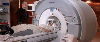
In the clinic you can quickly undergo any diagnostic procedures at a time convenient for you: laboratory tests, MRI, X-rays, ultrasound and others. Consultation with doctors of various specializations, comprehensive diagnostic programs and other medical services without queues and at affordable prices. For your convenience, we work seven days a week from 8:00 to 21:00 on weekdays and from 9:00 to 20:00 on Saturday and Sunday.
You can find out the cost of Doppler ultrasound of the renal arteries, ask questions, and sign up for an examination by calling the clinic or using the online registration form.
Interpretation of kidney ultrasound results
In order to make a diagnosis and decide on further tactics for patient management, the doctor compares the results of ultrasound diagnostics and normal indicators according to a number of criteria, which are presented in our table below:
| Criteria | Normal indicators |
| Kidney shape | Bean-shaped |
| Organ outlines | Clear, with smooth edges |
Dimensions:
| The same:
|
| Capsule thickness | 1.5 mm |
| Calyxes and pelvis | Not rendered |
| Basilar artery resistance index | 0,7 |
| Resistance index of interlobar vessels | Not less than 0.30 and not more than 0.75 |