The internal structures of the human body have different structures and densities of the fibers from which they are composed. Elastic, dense formations for the most part are located between the skeleton and the outer skin. Such tissues include layers consisting of fat cells, muscle and ligamentous apparatus, cartilage layers, vascular network, lymphatic accumulations, etc. The most informative and safe method capable of detecting and classifying pathological processes in soft fibers is magnetic resonance imaging.
The cervical area of the body is a very complex zone in structure and functionality, since its structure includes not only bone structures, the connection of the mobile (upper vertebrae) and static (occipital bone) parts of the spine, but also elastic formations - muscles, ligaments, blood vessels , cartilage, lymphatic system, glandular formations and nerve fibers. Pathology of any of these structures is fraught with the occurrence of not only pain in the neck, but also in the upper limbs, spine, head, and affects the normal functioning of all internal organs.
The most common type of diagnosis in modern medical practice is MRI. Nuclear resonance screening is one of the most universal methods, as it is able to identify and show even microscopic abnormalities in places that are difficult to reach by other clinical methods. Investigations for diseases in the upper parts of the spinal column are prescribed very often today, since when such pathologies occur, it is important to promptly, quickly and most effectively determine the original source of the disease. If this is not done, complications that are critical for a person may develop, and recovery from them will be much more difficult.
Benefits of MRI of the cervical spine
Unlike other research methods, MRI allows you to assess the work and condition of any structure of the human body. Regarding the cervical spine, MRI has the following advantages:
- X-rays and CT scans can only examine bone tissue;
- The use of Doppler allows one to evaluate blood flow in the area being examined;
- Ultrasound is only applicable to soft tissue.
While magnetic resonance imaging is used for any tissue, showing their structure in three-dimensional image mode.
About the capabilities of the method
A unique study of soft tissues in the Yauza hospital on a high-field magnetic resonance imaging scanner allows the doctor to examine and accurately assess the condition of the smallest structural parts of the patient’s tissues - individual muscle bundles, intermuscular septa, tendon fibers. No other method of radiation diagnostics provides such contrast of structures and tissues.
An examination using a Philips Ingenia 1.5T tomograph allows our specialists to accurately diagnose soft tissue pathologies even at the earliest stages of development if they seek medical help in a timely manner.
When is an MRI of the cervical spine prescribed?
An MRI scan is prescribed if there is pain in this area, as well as with functional disorders in the brain. In addition, the following reasons are worth noting:
- Suspicion of the presence of blood clots in the vessels;
- Diagnosis of the inflammatory process;
- Examination of the neck area for the presence of tumors and metastases;
- Diagnosis of osteochondrosis;
- Spinal stenosis and other abnormalities.
Make an appointment now!
Direct indications for use
Magnetic resonance imaging is prescribed for people with the following symptoms:
- Headache;
- Fainting;
- Frequent causeless dizziness;
- Feeling of neck tightness;
- Painful sensations in the scalp area;
- With numbness of the limbs, goosebumps;
- Pain in the back of the head and neck.
The doctor can accurately diagnose or deny vascular thrombosis or suspect disorders in the circulatory system. In addition, the study is necessary in the case of spinal cord injuries or suspected tumors; it is prescribed to select treatment tactics for spondylitis and osteomyelitis, as well as for various lesions of the spinal body.
Contraindications
The tomograph creates a magnetic field, so patients who have an insulin pump, pacemaker, hearing aid, or cardioverter-defibrillator in their body do not undergo the procedure. Scanning is not performed for people who have metal objects in their bodies (Ilizarov apparatus, staples and wires, stents on blood vessels). The procedure with contrast is contraindicated for pregnant women and nursing mothers. Contraindications include renal failure. For technical reasons, the study is not possible if the patient weighs more than 120 kg.
When is MRI of the cervical spine contraindicated?
MRI of the neck has a clearly defined list of contraindications. The study is not carried out if:
- The patient has metal implants, fragments, stimulators, etc. in his body;
- The patient suffers from a panic fear of being in a confined space;
- If a person has diseases related to the nervous system;
- If the subject cannot ensure long-term immobility;
- MRI is contraindicated in pregnant women;
- Scanning with the introduction of a contrast agent is contraindicated in the presence of individual intolerance to one of the components;
- Patients suffering from renal failure are allowed to undergo MRI only with the permission of a doctor, who is usually present when it is performed.
Are there any prohibitions for performing MRI of soft tissues of the neck?
Any medical procedure has both indications and prohibitions, which apply to certain categories of patients. Despite the fact that MRI diagnostics is one of the safest methods, it is prohibited in the following cases:
- The initial period of pregnancy, including the first three months of the term. For natural reasons, no studies have been conducted in medical practice aimed at identifying the harmful effects on the intrauterine fetus. Since the baby in the womb is in the active stage of growth and formation of all vital systems, doctors try to exclude any external influence during this period, including exposure to a strong magnetic field.
- The presence of metal elements in the body in the form of implants, staples, pins, etc. It is especially dangerous to fall into the field of action of a strong magnet if there are such objects in the neck and head. Under the influence of the tomograph, ferromagnetic parts become very hot and deformed, which leads to displacement of the implants and damage to internal biological structures. The presence of such elements inside the body must be reported in advance to both the attending physician and the radiologist on duty at the diagnostic center.
- MRI with contrast enhancement is prohibited for allergy sufferers who have immune rejection of the components of the staining drug. If there is a rejection reaction of the body to drugs, a thorough study is carried out to determine the immune reaction to the contrast agent. People who have not previously had allergies are also required to undergo rapid testing by administering a minimum dose of dye. If testing shows a response, the procedure is completely prohibited.
- Chronic urinary dysfunction with planned use of contrast. The drug itself is safe and does not stay in the body for more than two days. Patients suffering from chronic fluid retention due to kidney pathologies are prohibited from using dyeing, since with an unnatural long-term retention of it in the body, general intoxication can occur.
- Uncontrollable fear of confined spaces. If you get into the inside of the tomograph, emotional patients may experience a panic attack, which will not allow you to undergo the procedure and will negate all the results. During the session, you must be in a state of complete rest and remain motionless; the quality of the images depends on this.
- Neurological and mental abnormalities accompanied by involuntary movements.
- Excess body weight exceeding 120-200 kg. Obese clients do not have the opportunity to fit in the inside of the device, since its dimensions are not designed for such volumes. In this case, you can turn to open configuration installations that have no weight category restrictions.
What diseases can MRI detect?
An MRI of the cervical spine is performed for diagnostic purposes, that is, as a result, the doctor can confirm or refute a particular diagnosis. Magnetic resonance imaging can detect the following diseases:
- Osteochondrosis of the cervical spine;
- Spondylosis or spondyloarthrosis;
- Abnormal structure or location of the vertebrae;
- Developmental defects;
- Intervertebral hernia;
- Tumor processes;
- Destruction of the myelin sheath or multiple sclerosis;
- Arachnoiditis and myelitis;
- Damage to the structure of the spinal cord;
- Impaired blood flow in the cervical spine.
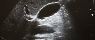
Scan result
As a result of the MRI scan, the doctor sees a complete picture of the cervical spine, upon evaluation of which he makes a diagnosis. A normal MRI result looks like this:
- The vertebrae have no roughness, the edges are smooth;
- The height and other parameters of the vertebrae are the same;
- The joints are localized in the intervertebral space;
- The structure of the spine is symmetrical, the location of the structures is correct;
- The images clearly visualize the spinal cord and its structures;
- There are no neoplasms, tumors, cysts, hernias, polyps, anomalies or pathologies, etc.
If the patient has a vertebral fracture, the image will show a fracture line, as well as fragments or a deformation zone. However, it is worth noting that in the case of fractures, magnetic resonance imaging is not indicative; it is recommended to carry out a standard radiographic examination. However, if you do an MRI of the spine, this will allow you to answer the question with maximum probability whether the spinal cord was affected by the fracture. If there is a hernial process in the intervertebral space, the doctor will notice a change in the location of the disc, as well as its deformation; it will protrude beyond the edges of the vertebra. If the patient has osteochondrosis, then the picture will show the growth of the edges of the vertebrae, and their surface will be rough and uneven due to the degenerative process.
What do specialists see on an MRI of the soft tissues of the neck?
The basic basis of the method is the mechanical generation of a microwave magnetic field. Such a field does not have a destructive or negative effect on biological compounds. A response resonance occurs in the fiber molecules, which is recorded by equipment, processed by a computer program and sent to the monitor in the form of black-and-white images in a color gamut of more than 500 shades. MRI images are taken step by step with an interval of several millimeters to a centimeter; the equipment allows you to produce not only static images of neck structures, but also combine them into a multidimensional projection, which can be rotated at the desired angle and bring the area required for study as close as possible.
The study is carried out only on high-power equipment (from 1.5 Tesla), which allows you to see the smallest details of the anomaly, identify neoplasms of various nature and assess the degree of their spread to adjacent areas. Magnetic resonance imaging detects not only inflammatory processes in elastic joints, but also shows the condition of bone fibers and shows the cause of the disease, which is important for a correct diagnosis. The patient gets the opportunity to quickly undergo an examination and receive a timely treatment plan.
Screening using the nuclear resonance method visualizes the condition of the spinal cord, cartilaginous layers between the vertebrae, pinched nerve fibers, the presence of tumor processes, obstacles in thrombosed vascular channels, infected areas, internal hemorrhages and destruction of the walls of the vascular network. Diagnostics shows:
- protrusion of the cartilaginous layer, intervertebral hernia;
- damage to the bone structure;
- benign and cancerous formations, migration of cancerous processes, the degree of their spread and ingrowth into adjacent areas;
- foci with inflammatory processes;
- congenital disorders;
- post-traumatic anomalies, chronic injuries;
- changes in glandular tissue, pathology in the larynx, vocal cords, lymphatic collections.
Since the neck and head areas are located in close proximity, simultaneous diagnosis of these departments can be prescribed. In some cases, this is necessary to identify the impact of the degenerative process on adjacent areas.
Preparing for the study
Magnetic resonance imaging of the cervical spine does not require any special preparation. You do not have to adhere to a diet or make adjustments to your usual medication intake. There is a standard protocol that you should follow to protect yourself from possible problems during the scan. Immediately before the procedure you need to:
- Notify the doctor about pregnancy, if any, and be sure to indicate the due date;
- Tell your doctor about your tendency to allergies if you plan to administer contrast;
- If you experience discomfort or fear while in a confined space, be sure to tell the medical staff;
- Before you begin the scanning procedure, you must remove all metal objects, otherwise they may become very hot.
Separately, it should be said that the patient must endure a five-hour fast from food if an MRI with contrast is performed.
What are soft tissues?
When talking about “soft tissue” in relation to MRI, this term usually means muscle tissue, subcutaneous fat, ligaments, tendons, fascia*, blood vessels, nerves and lymph nodes.
*fascia is a connective tissue membrane covering organs, blood vessels, nerves, muscles
MRI of soft tissues helps the doctor, for example, find out the cause of compression of a nerve or vessel. Quite often, an MRI of the soft tissues of the neck is performed to determine pathologies of the pharynx, larynx, and thyroid gland.
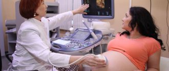
How does the procedure work?
It was previously said that magnetic resonance imaging is a 100% safe procedure. During its implementation, the patient is not exposed to negative radiation and does not experience pain, so it is recommended even for children at an age when they are able to remain motionless for a long time. The procedure usually takes from 40 minutes to 1 hour, not counting the time for intravenous contrast administration. The usual algorithm for performing an MRI of the cervical spine looks like this:
- The patient changes into disposable clothing or undresses down to his underwear;
- The doctor asks you to take a horizontal position on a special platform of the device;
- A contrast solution is administered;
- Medical workers firmly fix the head with bolsters, the limbs are immobilized with belts;
- The panel with the person being studied slides inside the device;
- The scanning process starts, and the person inside sees a ring moving around him;
- After the required time has passed, the doctor lets the patient know that the procedure is completed, the platform moves out and the device turns off. At this point, the scanning is considered complete.
It is worth noting that the operation of the device is almost silent; only a slight noise is felt, which is created by the device during the movement of the ring.
Main features of the procedure
In order to obtain a large amount of data for making a diagnosis, you need to use a closed-type MRI machine. The standard solution is the use of a high-voltage tomograph with a power of 1.5 Tesla.
It is best to go for examination with a doctor's referral. It should indicate what format of examination is needed - with or without contrast enhancement.
No special preparation is required for the procedure. All you need to do is remove all metal jewelry, as well as clothing that contains such accessories.
During the examination, the person is placed on a special table, and an MRI coil is placed around the neck. After this, the table with the person lying on it is placed inside the tomograph. The inside is quite spacious, there is good ventilation, as well as a device for communicating with a doctor. It is important to remain still throughout the entire process to avoid blurry images.
The average duration of an examination without the use of contrast enhancement is up to half an hour. The doctor examines the data received from the tomograph and studies the images. All results are provided to the client not only in the form of a snapshot, but also recorded on a special medium. A written conclusion is also prepared based on the results. On average, it takes up to 40 minutes.
The need to maintain constant immobility means that MRI may not be suitable for everyone. When a person has strong nervous tics, there is a possibility of a panic attack, claustrophobia, and he will have to refuse to visit the office with a tomograph.
Features of MRI with contrast
The use of contrast can improve the information content several times, and is also used in case of suspicion of the presence of a tumor process, as well as determining their structure, size and boundaries. In addition, the injected contrast allows the state of the vascular system to be assessed with maximum accuracy and the blood flow in the cervical spine to be analyzed.
Due to the fact that tumors or areas of inflammation are characterized by an increased concentration of vascular branches, the contrast agent will accumulate in these areas, which will indicate the exact location of the tumor or inflammation. Currently, gadolinium or drugs based on it are used as a contrast agent. Its use is explained by its safety, low likelihood of developing an allergic reaction, and rapid spread through the vascular network. It is safe to say that contrast-enhanced MRI is much more effective and informative than the standard procedure, but, at the same time, it is a more complex procedure and takes more time to complete.
- MRI of the back muscles of the spine.
- MRI of the lumbar region.
- Spinal examination - MRI or radiography.
What does an MRI of the neck show?
The study is prescribed by a doctor to clarify the diagnosis and obtain the most detailed idea of the state of the human body. There are several aspects to the final evaluation of images:
- Skin condition. The use of MRI helps to find various types of changes in the skin, as well as in the subcutaneous tissue layer.
- Symmetry of the cellular spaces of the neck. The resulting image helps to obtain the most accurate, detailed information and take measurements.
- Neoplasms. They can be found regardless of location. The method is considered one of the best for early determination of the growth of tumors, especially in the patient’s oropharynx and tonsils.
- Assessment of the size of the salivary glands. In addition to the size itself, other parameters are also studied - structure, position. The submandibular and parotid glands are checked.
- Condition of muscle tissue. You can find even the most imperceptible changes in the structure, and also draw conclusions about the basis on which they begin to form.
And this is only part of the likely changes that MRI shows. The information content factor in this area is very important because it gives the doctor maximum opportunities for assessment.