Computed tomography is based on ionizing x-rays. Scanning with a tomograph with the ability to create 3D reconstructions of internal organs, vessels and bones is a highly accurate examination method, preferable in a number of difficult situations: after strokes, pneumonia, suspected cancer. However, such examination cannot be done frequently.
In this article we will look at what the harm of X-ray radiation is and how to reduce its impact if the permissible limit has been exceeded.
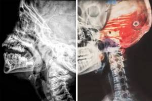
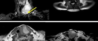
What is the difference between CT and MRI?
CT is an x-ray examination method, and the operating principle of MRI is based on the action of a magnetic field. As a result, CT and MRI have different contraindications. In addition, CT has radiation exposure and therefore cannot be used repeatedly.
CT is best used to diagnose the condition of the lungs, bone tissue, and lymph nodes; using contrast – to identify pathology of blood vessels, tissues, and internal organs.
MRI is better at diagnosing pathology of cartilage, ligaments, muscles, brain, some internal organs, and blood vessels.
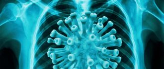
Lung damage due to coronavirus on CT
When a coronavirus infection is suspected, patients are most often concerned about the following questions: how is the degree of lung damage determined, when is hospitalization necessary, and is it possible to restore the lungs after pneumonia? Let's look at this topic in more detail and see how computed tomography of the lungs will be useful.
Manifestations of coronavirus
According to the observations of Chinese scientists and doctors, summarized in the “Handbook for the Prevention and Treatment of COVID-19,” chest pain with coronavirus already indicates a progressive (about 10 days) disease. With mild pneumonia at an early stage, discomfort does not bother you. Therefore, it is very important to listen to your body (especially your breathing) and measure your temperature. And if you have been in contact with a sick person or people from epidemiologically unfavorable countries, you need to do a test for COVID-19 and a CT scan of the lungs. Only tomography reveals the initial stages of lung damage due to coronavirus, when it is easiest to cure pneumonia and preserve the function of the respiratory organ.
Lung tissue cannot hurt much because it has very few nerve receptors. Discomfort in the lungs with coronavirus is a consequence of inflammatory edema. When the alveoli of the lungs fill with fluid or fibrous tissue, the pleural membrane stretches. This causes dull pain. Along with this, with coronavirus, the patient may feel:
- Pressure in the chest, severe distension;
- Unpleasant sensations during a deep breath, with a sharp breath there is a strong, prolonged cough;
- Discomfort in the neck, collarbones, between the ribs.
However, similar symptoms are characteristic of other respiratory diseases. However, in order to exclude coronavirus or detect it in time and prevent the development of distress syndrome.
ARDS (acute respiratory distress syndrome or “shock lung”) is an acute and severe condition that is characterized by bilateral infiltration and pulmonary edema with severe hypoxemia. An extensive inflammatory process sharply causes respiratory failure, heart problems, and pulmonary vascular spasm in the patient. In some patients, it progresses to fibrosis, after which complete recovery of the affected lungs is sometimes impossible. ARDS is the leading cause of death due to coronavirus.
What does lung damage look like on a CT scan?
Lung damage caused by a new type of coronavirus is indicated by so-called “frosted glasses” - relatively light areas resembling plaque, which indicate tissue compaction. Normally there should be no “frosted glass”. The peculiarity of computed tomography is that such signs of coronavirus pneumonia are visible quite clearly, even if the lungs are affected by 5% or less. Traditional radiography and other hardware diagnostic methods have lower resolution and can therefore give ambiguous results.
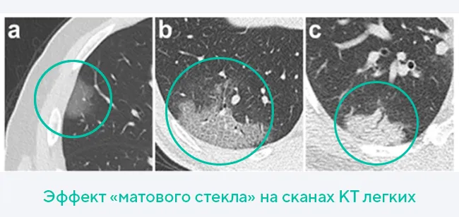
In pneumonia associated with COVID-19, areas of ground glass are located in both lungs: in the lower and lateral sections, around the bronchi or closer to the pleura. CT scan allows you to determine the extent of lung damage due to coronavirus. Less than three “ground glass glasses” correspond to a mild degree of the disease, more than three indicate moderate lung damage. If their consolidation is observed, the patient’s condition is assessed as moderate, with widespread compactions - severe.
To estimate the degree of damage as a percentage, the lungs are divided into 5 lobes: three in the right and two in the left. The radiologist examines each lobe and rates how damaged each lobe is on a five-point scale, with a score of 1 representing 5% obstruction or less and a score of 5 representing more than 75%. Next, all points are added up and multiplied by 4. The resulting number will express the degree of lung damage due to coronavirus as a percentage. If the respiratory organ functions at 50% or less, this is already grounds for hospitalization.
In addition to “ground glass” on a CT scan of the lungs in patients with coronavirus, the doctor will see other clinically significant signs of pneumonia:
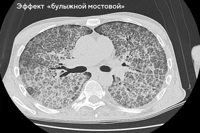
- “Cobblestone” or “patchwork” syndrome - when the compaction extends to the septa between the lobules of the lungs (approximately on the third day of pneumonia), the texture of the lung tissue on CT scans becomes similar to paving stones.
- Consolidation of “ground glass” - as the disease progresses (usually on days 5-8), the lung tissue becomes more dense and transmits X-rays less well, while there are fewer functional areas involved in gas exchange.
- Reverse halo symptom or rim syndrome - areas of compaction around the source of infection ("ground glass"), similar to rings. Occurs in more than 50% of coronavirus patients.
- A symptom of an air bronchogram is the presence of air in the lumen of the bronchi along with pronounced consolidation of ground glass.
Why do you need to use a contrast agent for a CT scan? Does it harm the body?
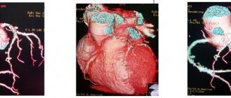
A contrast agent is administered during angiographic studies of the vessels of the brain and vessels of the neck, neck organs, chest organs, abdominal cavity and pelvis. Using a contrast agent, you can obtain visual information about changes in organs and tissues, the size of tumors and their boundaries.
The contrast agent can have a nephrotoxic effect if there is impaired renal function, and also affect the thyroid gland in hyperthyroidism. It is eliminated from the body within a few days. In order to speed up the elimination of the substance from the body, you need to drink more water.
If it is necessary to conduct a repeat study with intravenous administration of a contrast agent containing iodine (used in CT) and/or a paramagnetic agent (used in MRI), it is necessary that at least 3 days pass between studies.
Some people may have an allergic reaction to the contrast agent. If during or after the examination you experience itching, swelling in the throat area, or develop a rash, tell your doctor immediately.
If you have previously had an allergic reaction to contrast media or iodine, be sure to inform your doctor before the test.
Is it necessary to do a CT scan of the lungs for coronavirus?
During the first wave of the COVID-19 epidemic in April 2021, Russian doctors noted that 45.5% of those infected had no clinical manifestations of the disease—in such patients, the coronavirus infection developed asymptomatically. Today, CT scan of the lungs is considered the main method for diagnosing viral pneumonia, the main consequence of which is partial damage to the lungs.
Alveoli are small air cells in the lung. When their volume is reduced due to fibrosis or fluid accumulation, critical respiratory failure occurs. A person experiences shortness of breath, coughs with sputum (sometimes with blood), and body temperature rises.
With pneumonia caused by COVID-19, the most common concerns are:
- Chest pain and discomfort;
- Shortness of breath and lack of air;
- Dry cough;
- Loss of smell;
- Fever.
Laboratory blood tests aimed at identifying and identifying a viral pathogen sometimes give a false negative result. In this regard, doctors made a conventional decision according to which the presence of signs of lung damage on tomograms, despite a negative PCR, should be regarded as a probable coronavirus infection until an alternative diagnosis is made.
Another method of radiological diagnosis of pneumonia due to coronavirus is a standard chest x-ray. X-ray is a more affordable and widespread examination method. However, it is significantly inferior to computed tomography in terms of information content. The fact is that x-rays do not show lung damage of degrees I and II. This method has other disadvantages, for example, when shadows from large organs are superimposed on each other, artifacts are created that can be misinterpreted.
According to current clinical guidelines, during a pandemic, any compaction (infiltration) of the lung tissue and signs of inflammatory changes on radiographs should be considered suspicious for COVID-19. This means that after the x-ray the patient is sent to have a CT scan. To avoid unnecessary radiation exposure, if coronavirus is suspected, it is advisable for the patient to immediately undergo a CT scan of the lungs.
How harmful is a CT scan to the body?
The procedure involves exposing the human body to x-rays. It must be taken into account that such radiation accumulates in the body during frequent examinations.
In just one scan, a person can receive radiation in a dose of 3 to 10 MeV. The amount of radiation dose directly depends on the area being examined, as well as the type of equipment.
This radiation exposure is equivalent to the background radiation that affects a person for several years.
Moreover, such radiation doses are several times higher than those that a person can receive as a result of undergoing conventional radiography.
Clinical studies have confirmed the dangers of CT scans for pregnant women, since the procedure can cause the development of various pathologies in the unborn baby. If it is necessary to conduct an examination using contrast, it is important to consider the likelihood of allergic reactions.
What kind of CT scan of the lungs is done for coronavirus?
Today, the “gold standard” for CT scans for coronavirus is considered to be a slice-by-slice 1-2 mm scan on a multislice tomograph or MSCT of the lungs. Diagnostics on such modern devices takes only a minute and allows you to obtain images in the highest possible resolution. The procedure takes place in the most comfortable conditions for the patient, so it is suitable even for patients in serious health conditions (with artificial ventilation). A CT scan of the lungs for coronavirus is performed without contrast, and its diagnostic value is superior to X-rays, MRI, and ultrasound.
In the specialized department, patients undergo a CT scan of the lungs on a new generation multislice tomograph Siemens Somatom go.Now with reduced radiation exposure and immediately receive a recording of the study (CT scans) on a DVD.
When should a CT scan of the lungs be done during coronavirus?
According to the accepted classification of identified pathological changes, standard “CT1” corresponds to less than 25% lung damage, “CT2” - 35-50%, “CT3” - 50-75%, “CT4” - 75% or more. The peculiarity of pneumonia caused by the new coronavirus COVID-19 is that the complication progresses quickly to a more severe form.
Unlike X-rays, CT will show lung damage of 5% or less - the radiologist sees even single areas of infiltration with a diameter of 4-5 mm. Pneumonia corresponding to CT1, and sometimes CT2, cannot be determined from an x-ray. If you have characteristic symptoms, even mild ones, and test positive for COVID-19, there is no need to wait for the infection to spread more intensely and cause damage to large areas of the lungs.
CT scan of the lungs is indicated for:
- Temperature 38 degrees;
- Respiratory rate > 22 per minute;
- shortness of breath/cough/chest pain;
- Blood saturation
A CT scan for coronavirus is done even if the test for COVID-19 shows a negative result, and the x-ray does not reveal significant changes in the lung tissue (the lesions may still be small, there may be artifacts and shadows in the image) - while the patient is concerned about the above symptoms , contact with patients in the past is not excluded.
CT scan of the lungs after coronavirus
A CT scan of the lungs for coronavirus is done not only to assess lung damage, but also to monitor the recovery process as part of therapy. The first is done three days after the start of treatment, if it does not produce results and the patient does not recover. The next tomography can be repeated after a week if the patient’s condition does not improve.
With favorable treatment during the rehabilitation period, a CT scan of the lungs can be performed twice (interval - 2-3 weeks) to monitor the dynamics of lung recovery after coronavirus. In total, it is recommended to do no more than 5 CT scans per year.
Lung tissue is elastic and capable of regeneration. If the pathology is detected in time and treatment measures are taken, the patient’s body can cope with the infection within 1 month, and after rehabilitation, the functionality of the lungs will be completely restored.
If a patient was admitted to a medical facility with more than 50% lung damage, suffered severe pneumonia or acute respiratory distress syndrome, then fibrosis may develop. The consequences of pulmonary fibrosis resemble scars, and such pathological changes may be irreversible. However, if small areas are affected, then from a functional point of view they are easily compensated by healthy ones and are not felt throughout life. The feasibility and number of repeated computed tomography scans during the rehabilitation period are determined by the doctor.
Is there any harm?
There are two radically different opinions regarding the security of computer scanning. Some argue that the method is harmless, while others claim that radiation exposure causes irreparable harm to the body.
The truth, as studies show and experts say, is somewhere in the middle.
A CT scan of any organ or part of the body does involve x-ray radiation, that is, there is a potential risk, but the dose is very small, so there are no health problems. Naturally, provided that the person is not examined too often.
Regular exposure to radiation causes irreversible changes at the cellular level, leading to serious systemic diseases, including cancer.