Magnetic resonance imaging is a modern and highly informative examination method used for a thorough examination of internal organs and blood vessels.
A magnetic resonance examination can also be prescribed by specialists for a number of otolaryngological diseases, including examination of the nasal sinuses.
A magnetic tomography session is absolutely painless and is suitable for examining even small children. What an MRI can show helps a doctor diagnose any health problem or confirm the absence of one.
What are paranasal sinuses
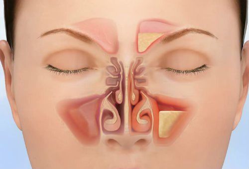
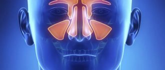
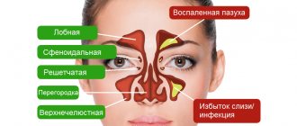
The paranasal sinuses are bony cavities located around the nose. They are coated with a mucous membrane and play a very important role - humidifying and warming the inhaled air. There are 4 pairs of sinuses in the human body:
- frontal sinuses;
- maxillary (maxillary);
- lattice;
- basic.
During a visual examination by an ENT specialist, the nasal passages are first examined. If the specialist was unable to identify the cause of the pathological process, for example, when comparing complaints and the clinical picture, the presence of a cyst may be suspected, but the doctor still finds it difficult to make a diagnosis, then the patient will be referred to undergo radiography or magnetic resonance imaging.
Is the procedure safe?
MRI of the paranasal sinuses is a safe diagnostic method for the health and life of the patient in the absence of contraindications to scanning. The most important advantage of all modern tomographs is that they do not emit harmful radiation during operation. The magnetic field does not have any negative effect on the subject, so tomography can be performed as many times as necessary to establish and clarify the diagnosis.
Gadolinium is used to perform an MRI of the nose using a contrast agent. In most cases, it does not cause any negative effects. The only exceptions are individual intolerance to the body, as well as disorders of the liver and kidneys.
Specifics of operation of a magnetic resonance imaging scanner
The human body consists largely of water, and water contains elementary particles called protons. Essentially, the human body is a source of protons. Elementary particles have one interesting feature - spin.
This is the angular momentum of the proton, which is equal to ½. If you place a proton in a strong magnetic field, it will begin to emit radio waves with a certain frequency and intensity (depending on the location). This strong magnetic field is what MRI equipment is used for. It has a built-in software and hardware complex that not only creates a magnetic field, but records information, processes it and turns it into a high-quality three-dimensional image.
The computer picks up radio waves and determines their frequency/intensity. Using complex mathematical algorithms, the system localizes the proton, which emits radio waves.

Once all the protons are found, the tomograph creates a complete picture of the scanned area with all kinds of sections, the ability to enlarge, and so on. Diagnostics (including scanning and processing of results) takes an average of 20-40 minutes.
Indications for MRI of the sinuses
This type of diagnosis is prescribed, as a rule, in cases where it is not possible to identify the cause of the disease for quite a long time. These include:
- systematic pain in the nasal sinuses, in which there are signs of a pathological course on x-rays;
- periodic headaches for no apparent reason, which even painkillers do not always help relieve;
- frequent nosebleeds;
- copious nasal discharge, uncharacteristic of a cold;
- any significant facial injuries;
- a sharp decrease in the sense of smell, a drop in visual acuity;
- systematic congestion of the nasal passages;
- in the presence of symptoms of various diseases, which include polyposis, sinusitis, sinusitis, cyst proliferation.
Most often, specialists prescribe MRI for sinus cysts and sinusitis, and the procedure is performed on patients multiple times. In addition, MRI for cysts allows in many cases to avoid the need for surgical intervention if treatment is started in a timely manner.
Where to get a tomography?
You can find out where it is best to undergo these and other examinations using 36go.ru. Here you can easily sign up for diagnostics, as well as select a medical center by location and cost of procedures. The service also contains all the information about current discounts, promotions and other advantageous offers. A CT scan of the nasal sinuses can cost from 2,900 to 18,900 rubles, and an MRI of the nose costs from 1,900 to 25,600 rubles.
You can make an appointment at the chosen clinic or get help in choosing from a specialist by calling the phone number posted on the main page of the site.
What does the diagnosis show?
Many patients who are scheduled to undergo this type of examination wonder what the tomography shows? There may also be serious concerns that multiple cysts may develop in the sinuses. However, such serious diseases are quite rare, so magnetic resonance imaging is not always prescribed only for cysts.
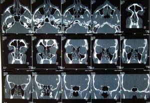
MRI of the nose can detect almost any pathology of the nose and sinuses. In addition, on high-precision images, a medical specialist can always see the fluid formed in the sinuses and accurately determine the location.
If necessary, magnetic resonance imaging is performed very often if progressive inflammatory processes and the presence of multiple cysts are suspected. MRI for a cyst provides the opportunity to determine the most optimal method of treating benign formations and tumors in the nasal sinuses at a very early stage.
It is difficult to underestimate the benefit of scanning in such a serious pathological process as sinusitis. After all, this disease cannot be delayed and the success of further treatment depends on correct diagnosis. In case of sinusitis, the ENT specialist very often prescribes a referral for tomography and his recommendations cannot be ignored. Regardless of the stage and form of sinusitis, the patient needs a comprehensive examination, which includes an MRI.
MRI of the sinuses, carried out in a timely manner at the very beginning of the onset of the disease, helps prevent further development and reduce the risk of complications.
2.Indications
It is impossible to list all the situations of establishing, clarifying, and differential diagnostics in which an MRI examination is necessary and often irreplaceable. Only the main groups of such indications can be distinguished:
- traumatic brain injuries;
- the need to exclude/confirm neoplasms (oncopathology), incl. “invisible” for other diagnostic methods;
- diagnosis (including early) of demyelinating diseases - multiple sclerosis, leukoencephalopathies, myelopathies, etc.;
- establishing the nature, location and scale of heart attacks/stroke;
- monitoring dynamics in the rehabilitation period after neurosurgery;
- diagnostics of vascular condition (MR angiography).
Additional examination of the nasal paranasal sinuses is usually prescribed:
- for acute allergic reactions;
- if there is a suspicion of oncological pathology and/or the presence of a cyst, if the results of other studies (for example, radiography) are not informative enough and leave doubts;
- for injuries;
- with anomalies of anatomical development.
Advantages:
- the study is not associated with any radiation exposure (therefore it allows repeated procedures and removes contraindications for many categories of patients);
- in many cases there is no alternative in terms of the nature and completeness of diagnostic information;
- when studying the brain and maxillofacial structures does not require special preparation;
- Thanks to the use of modern computer technologies, it allows you to recreate a three-dimensional image of an organ and/or monitor its functioning in real time.
Visit our Neurology page
Contraindications for diagnostics
Despite the fact that MRI is considered a completely safe diagnosis, during which the patient does not experience any radiation exposure, there are still certain contraindications to its use:
- MRI of the paranasal sinuses is unacceptable for patients with fixed metal prostheses in the body;
- The examination should not be carried out on women expecting the birth of a baby. To date, studies on the impact of this type of diagnosis on the developing fetus have not been carried out in full. For this reason, tomography is prescribed to pregnant women in exceptional cases, when the expected benefit of diagnosis for the mother outweighs the potential risk to the fetus. For example, an MRI for a pregnant woman’s cyst will most likely be prescribed, since treatment of the disease should begin as quickly as possible.
- MRI is not possible for patients with limb tremor. After all, this will cause low-quality images to be obtained, as a result of which specialists will not be able to decipher them correctly.
- MRI with contrast should not be performed on people with an allergic reaction to the contrast agent.
- Removable dentures (they can be removed if necessary).
- Fear of closed spaces (before undergoing an MRI of the nose, doctors may offer sedatives.
- Scanning using contrast during the lactation period. The contrast agent passes into breast milk and may have a toxic effect on the nursing infant. Therefore, if the diagnosis is still carried out, the woman will have to give up breastfeeding for 2-3 days.
general information
MRI is a modern, informative and safe method for the patient’s health for diagnosing diseases of the paranasal sinuses and adjacent soft tissues.
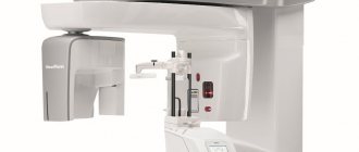
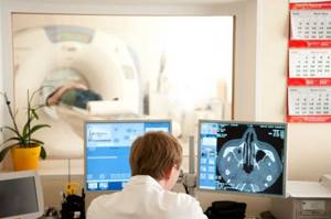
The device for conducting the study is called a tomograph. During diagnosis, it delivers high-intensity radiofrequency pulses. At the same time, the magnetization of the patient’s body tissues changes, and the effect of nuclear magnetic resonance occurs. After the tomograph stops operating, the magnetization becomes the same.
The impulses that are reflected from the body tissues are processed by a special computer program and displayed on the screen in the form of a layer-by-layer image of the area under study.
MRI of the paranasal sinuses using contrast
For certain indications, specialists may prescribe an MRI of the nose using a contrast agent. For example, an MRI for a cyst is almost always performed with contrast.
Before such a scan, a substance is injected into the patient’s body that stains the vessels, making it possible to visualize them in detail. Such a diagnosis has a number of advantages: for example, an MRI will necessarily be performed in case of a cyst, because only enhanced tomography can provide the most complete picture of the course of the disease. MRI of the sinuses with contrast takes much longer.
Before contrast is administered, the patient should undergo a contrast sensitivity test. If the results show that a person has signs of an allergic reaction, an MRI of the paranasal sinuses will be performed without the use of contrast.
Contrast MRI
Contrast agents are used to improve visualization of the scanned area. The substance literally illuminates the cavities of the human body, which helps make the final image as informative, clear and detailed as possible.
Most MRI contrast agents contain gadolinium. The substance is administered intravenously after the first (native) scan, after which, quite often, the need for an additional contrast stage is determined. After administration of the contrast agent, a second scan begins. The contrast penetrates inside, interacts with hydrogen protons, enhances the signal emanating from them and increases the contrast of the image.
Preparing for diagnostics
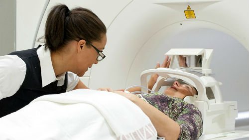
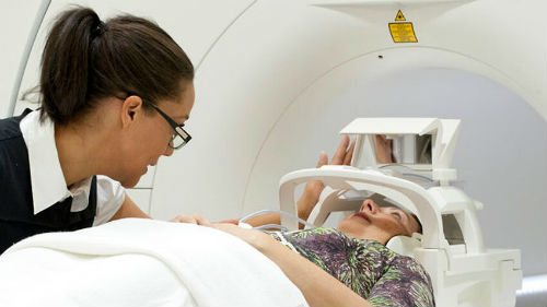
MRI of the nasal sinuses is a diagnostic method before which the patient does not need to comply with preparatory measures, which is a significant advantage of the procedure. However, before an MRI of the nose, the doctor will conduct a detailed consultation, where he will tell the patient the essence of the examination, and also give useful tips on how to behave correctly during the scan.
Each patient must comply with the following recommendations:
- the clothes in which he will undergo the examination should be spacious without any metal parts;
- It is unacceptable to come to the scan with makeup;
- You will have to leave all your jewelry outside the office: rings, earrings, piercings, hairpins, bobby pins, brooches, as well as a mobile phone.
Any metal objects can have a negative impact on the operation of the tomograph, resulting in unclear images.
Those patients who are prescribed MRI of the paranasal sinuses using contrast will still have to adhere to some restrictions:
- such an examination must be carried out on an empty stomach, therefore it is unacceptable to eat food at least 4 hours before the tomography;
- If there are allergic reactions to contrast, the radiologist must be aware of this before performing an MRI of the nose.
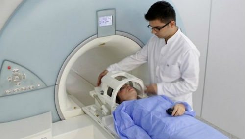
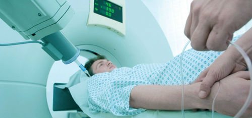
How are CT and MRI performed?
It works on the principle of an X-ray machine, but is a more “advanced” procedure.
During the examination, the head is found inside a huge ring. To do this, a person lies down on a bed and moves inside the device. Next, the specialist turns on the tomograph, and a fan of X-rays is directed into the desired area of the body. Reflecting from the cells, they “go” to ultra-sensitive sensors. From there, the signal is sent to the tomograph monitor in the form of an image of the nose and sinuses in different projections. MRI of the nasal sinuses is also quite informative. But the examination is carried out using a magnetic tomograph. The patient is placed on a bed that is slowly pushed into the tunnel. Next, the equipment is turned on, and strong magnetic radiation is created around the body. Hydrogen-containing cells begin to “respond” to the emerging field. They oscillate at different frequencies, which is reflected in the final images.
Diagnostic features
The nasal sinuses contain a huge amount of bones and cartilage, which complicates diagnostic measures. All patients must consider the following points before examination:
- you need to empty your bladder to avoid the urge to urinate during the scan;
- the examination will take place in a confined space, so you need to be prepared for the fact that you will have to experience certain inconveniences for a certain period of time;
- The device makes specific noises during operation, there is no need to be afraid of this - this is quite normal. You can use earplugs or ask the medical staff before the scan to give you headphones with music - this is normal practice in many clinics;
- It is unacceptable to move your body during scanning, this may have a negative impact on the quality of the images;
- If an MRI of the sinuses is performed using contrast, it is important to pay close attention to your health: slight dizziness, rapid heartbeat, and sweating may occur. More severe symptoms may prompt you to report changes in your health to your doctor. It is possible to contact him even during the procedure by using a special button.
- The duration of a scan using a contrast agent is about 1 hour, without the use of contrast – 15-30 minutes. As soon as the tomography is completed, the radiologist will begin to interpret the results.
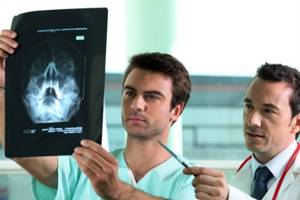
If the patient cannot wait to receive the results, he can ask to have the conclusion sent to him by e-mail, and after the conclusion is received by e-mail, it must be shown to the attending physician.
How the research works
First you need to sign up for a tomography. To undergo an MRI in our clinic, call us by phone or leave a request on the website. After this, you need to go to the medical center at the appointed time. No special preparation is required for diagnosis. It is enough to leave prohibited items in a specially designated place.
Prohibited items include bank cards, equipment and electronics, as well as all metal objects: jewelry, watches, wardrobe items.
The patient is placed in the device. You must lie still throughout the procedure. The process lasts about 10-20 minutes. When contrast is administered - approximately 20-30 minutes.
After scanning the tissues, you need to wait for the tomography results. The patient receives pictures, a radiologist’s report and a diagnostic protocol. You need to contact an otolaryngologist with documents.