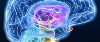
Brain electroencephalogram (EEG)
– a procedure that is performed to detect electrical activity in the brain. It is carried out in order to find out whether the patient has a disease such as epilepsy. This disease is very dangerous and manifests itself in seizures and convulsions, during which the patient loses consciousness.
This is a very common chronic disease. It goes without saying that a motorist suffering from such an illness can pose a threat to his own life, as well as the lives of other road users.
That is why, if a citizen has this disease, he is not allowed to drive a vehicle. The procedure for passing the medical examination was changed several times.
Currently, passing an EEG is mandatory for driver candidates and drivers with categories C, D.
The rest can be sent for research at the discretion of a psychiatrist. If the doctor deems it necessary, he writes out a referral.
In such a situation, until the patient undergoes an EEG, he will not be issued a certificate stating that there are no contraindications for driving the vehicle. This means he won’t be able to get a driver’s license.
What does the EEG show?
The electroencephalogram shows disturbances and pathological changes in the functioning of the brain. Based on the results of the study, neurological disorders are diagnosed and the scale of organic lesions is assessed. EEG records:
- causes of loss of consciousness;
- the nature of panic attacks;
- epileptic foci and their localization in the lobes of the brain;
- changes in electrical activity before attacks;
- changes that allow you to evaluate the effect of drug therapy and rehabilitation (EEG is taken before and after the course).
Characteristics of the main EEG parameters
EEG is based on the following concepts:
- average oscillation frequency;
- their maximum amplitude and phase;
- assessing the differences in curves on different channels and their time dynamics.
The EEG is recorded between two points on the scalp using one of two methods: bipolar or monopolar. When using the bipolar technique, the difference is between two active electrodes, which are located at electrically active points on the scalp. In a monopolar study, electrodes are placed at different points on the head.
The dominant rhythm of the human EEG at rest is the alpha rhythm. It is closely related to the mechanisms of perception and memory. The role of the alpha rhythm is a kind of functional stabilization of the brain and ensuring readiness to respond.
At rest, other components are also observed on the EEG. The delta rhythm is absent in healthy people at rest and is recorded in the fourth stage of sleep. Theta rhythm is closely related to mental and emotional stress. During mental work, both theta and delta activity increases.
The transition from a state of rest to tension is accompanied by a desynchronization reaction. Its main component is high-frequency beta activity. Mental work is accompanied by an increase in the power of the beta rhythm. The second important characteristic of electrical potentials of the brain is the amplitude of oscillations. It is closely related to their frequency.
The EEG pattern is called a "pattern". It differs from individual people in appearance and individual characteristics. In pregnant women, an individually specific pattern is normally recorded, which has developed by the age of 15-18 years. Changes in the pattern may be evidence of pathology.
Who is assigned to the study?
EEG is a safe and painless type of diagnosis. Examination using an electroencephalograph is prescribed for both children and adults. Allowed during pregnancy and breastfeeding. Can be done in a dream. Indications for diagnostic testing are extensive:
- traumatic brain injuries;
- sleep disorders;
- neurosurgical interventions;
- dizziness or frequent headaches;
- VSD;
- diencephalic crises;
- panic attacks;
- attacks accompanied by convulsions;
- endocrine pathologies;
- inflammatory diseases of the nervous system;
- brain tumors;
- problems with blood vessels of the cervical and head;
- neurological pathologies, developmental delay in children;
- any brain damage that develops in pregnant women and women who have recently given birth.
EEG is a convenient, modern and safe method for studying the brain
Electroencephalography is a convenient and simple method for assessing brain function. Like all methods based on recording biopotentials from the surface of the human body, EEG of the brain is completely safe, and is performed even on pregnant women at any stage. It is known that our nerve cells - neurons, in the process of their work, create, conduct and change the electrical current passing through them. In some ways, the way the brain works is reminiscent of the way the electrical appliances around us work. Only the current generated by neurons is extremely small, and if the current “in the socket” is measured in volts, then the current of nerve cells is measured in microvolts (which adds another “five decimal places”). Not so long ago, very large devices were used to record such small currents, and the person being examined had to be placed in a special shielded room in complete darkness, so that even the slightest influence “from outside” would not “distort” the recording of brain EEG potentials. Moreover, the first electrodes used to record EEG almost half a century ago were stuck into the skin like pins! Modern devices are so sensitive and most importantly “smart” that EEG can be recorded in almost any, almost “home” environment. It is important that a person can relax, sit (lie down) comfortably and close his eyes. Of course, everyone present during this procedure turns off their mobile phones and creates a “zone of increased silence,” however, it is no longer necessary to isolate a person in a cell; just close the window and turn off the lights.
What happens during the immediate procedure for recording an electroencephalogram ? As we agreed, a chair/chair and position are selected so that the person is as relaxed as possible. A special helmet made of silicone threads is placed on the subject’s head. It should fit tightly onto the surface of the head. The electrodes that are most often used for recording EEG in outpatient settings are called bridge electrodes; they are conveniently located, fixed using silicone threads of the helmet on the surface of the head like chess pieces. Since the brain has a fairly large surface, the researcher’s task is to record potentials evenly from the entire working (it is called convexital) surface of the brain. In order to successfully register such a weak current, the part of the electrode that is in direct contact with the skin is made of silver, and the contact site is moistened with saline solution. This is probably the only “most unpleasant” fact in the entire electroencephalography procedure: when something cold and wet is placed on the head. Although on hot summer days, subjects are usually very happy with this stage.
Next, the recording of the electroencephalogram itself begins. Depending on the clinical tasks, it can last from 5 to 20 minutes. A good rule is not only to record with your eyes closed at rest, but also to record so-called functional tests. There are three of them:
- A test with opening and closing the eyes demonstrates the brain’s reaction to an increase in the flow of information, since the person who opens the eyes involuntarily begins to examine and analyze the surrounding environment.
- A test with rhythmic photostimulation of white or red color consists of blinking a special bright lamp at a distance of 30 centimeters at different frequencies, from usually 2-4 Hz (that is, blinks per second) to 20-30 Hz. Using this test, when performing an EEG, it is possible to “catch” the so-called photoparoxysmal reactions. For example, if a person has an epileptic focus that periodically causes seizures, to prove its existence, in order to record the activity of this focus on the EEG, it must be provoked by such a flickering of light.
- Three-minute hyperventilation test - during which the subject is asked to breathe deeply, evenly (but not often!) for three minutes. With such deep breathing, even a relatively healthy person may begin to feel a little dizzy, because the brain vessels reflexively dilate, and the brain begins to experience a slight “non-dangerous” hunger. Many pathological modes of brain functioning do not manifest themselves in ordinary situations, namely under stress (fatigue, illness, emotional shock) during which, just like with three minutes of deep breathing, the brain begins to experience “slight hunger.”
Then, when the recording is completed, a functional diagnostics doctor (neurophysiologist) begins working with it. By studying the characteristics of electrical signals and their changes over time (frequency, amplitude, severity in one point of the head and “unexpressiveness” in others), the doctor can draw conclusions not only about the absence or presence of epilepsy (because EEG has long been considered the gold standard for diagnosis in epileptology) , but also about the peculiarities of brain function: for example, about the balance of excitation and inhibition, the presence of inconsistency between various brain structures, the degree of maturity of the cortex in children, and much more. Objective markers of the characteristics of the electrical work of the brain on the EEG during anxiety, nervous tension, depression, stuttering, and cerebral circulatory disorders have long been described in the literature. That is why, going beyond epileptology, EEG is successfully used not only in neurology, but also in psychotherapy and psychiatry. The standards of many examinations (when conducting medical examinations for workers in hazardous industries, when issuing driver’s licenses, when a patient consults a psychotherapist, at the beginning of a neurologist’s examination of a person with a headache, etc.) include EEG of the brain as one of the first diagnostic methods.
EEG does not require any special preparation other than a “clean head” (without hairspray or other hair styling products)!
An EEG of the brain can be done for a fee at our center every working day. Tel for inquiries and appointments 246-11-51
How to prepare for the procedure
When preparing for an EEG, you must strictly follow your doctor's instructions. He must stop taking certain medications at a certain time before the procedure. Also, 12 hours before the test, you should give up caffeine-containing foods and drinks - tea, coffee, chocolate desserts, store-bought energy drinks. Before the procedure itself, it is advisable to eat two hours before the start.
Hair must be clean. The use of varnishes and other styling products, as well as oils, masks and conditioners is not allowed. Additional hair processing will worsen the contact of the electrodes with the skin.
Usually the EEG is performed in a calm state, try to relax and avoid stress. In some cases, the doctor may ask you to stay awake for a certain period of time before the examination.
Alcoholic epilepsy consequences
Alcoholic epilepsy has serious consequences for the patient’s brain:
✔ as a result of the long-term toxic effects of ethyl alcohol in neurons, the intracellular transport of calcium and chlorine, the work of glutamate-dependent channels and gamma-aminobutyric acid receptors are disrupted, and the integrity of the membranes of nerve cells is disrupted;
✔ inhibition processes in the brain become ineffective, neurons become overexcited, rapid depletion of the nervous system occurs and some nerve cells die;
✔ each convulsive attack leads to cerebral edema, metabolic disorders and, as a result, irreversible death of a significant number of neurons.
✔the result of such processes in the brain is a rapid decrease in mental abilities, attention, memory (alcoholic dementia), the occurrence of severe alcohol withdrawal states, complicated by delirium tremens or Gaye-Wernicke encephalopathy.
How to do an electroencephalogram
The procedure takes from half an hour to two hours. A special cap is placed on the patient’s head (sometimes additionally secured to the chin). It allows you to attach electrodes to the skin. They capture vibrations, amplify them and convert them into waves that are transmitted to the device. The doctor sees and evaluates the functioning of the brain on the monitor in real time. There are devices that immediately record the result on paper, like an electrocardiogram.
Diagnostics are carried out in a sound-insulated room. Before the procedure, turn off the lights. The prepared patient, wearing a cap with attached electrodes, lies down on a couch or sits in a chair. During the EEG, one or more functional tests are used:
- photostimulation - a bright sharp flash of light;
- monotonous alternation of lights on and off;
- hyperventilation (deep rapid breathing);
- audio stimulation – turning on loud sounds;
- falling asleep (naturally or under the influence of sleeping pills).
Research methodology
Brain diagnostics are carried out in a specially designed room. The subject sits on a couch or sits in a comfortable chair. A special helmet with a network of built-in electrodes is first put on. During the examination, the doctor may be near the patient or outside the room.
Before conducting the study, the degree of technical errors is assessed, the doctor asks the person to open and close his eyes a couple of times. At the 1st stage, a resting EEG is recorded. It is important to exclude any movement during the time allotted for diagnosis.
At stage 2, the brain’s reactions when exposed to stressful situations are recorded. For this purpose, the following provocative tests are carried out, which can cause increased electrical activity in the brain:
- Hyperventilation tests. The diagnostician asks the patient to breathe as deeply as possible for 3-4 minutes.
- Rhythmic photostimulation. Assess how the brain reacts to flashes of bright light. The test uses a strobe effect where the lamp flashes approximately 20 times per second.
EEG is a completely painless, fast, effective and safe method. Immediately after electroencephalography, the patient can resume taking medications that were discontinued before diagnosis.
Electroencephalogram in children
The study is prescribed to children from birth. Babies can sleep during the diagnostic period. Adult, active children sit on their mother’s lap during the procedure. The child is distracted with toys and books; you can bring them from home.
A small patient does not experience pain or discomfort during an EEG, but may be scared. Therefore, at home, it is advisable to explain to the child what will be done to him and to rehearse the situation during games.
At the medical center, you will undergo an electroencephalogram without queues, in a comfortable environment. If necessary, a specialist will make a full interpretation of the results. It is also possible to make an interpretation of an EEG previously recorded elsewhere. Consultations with a neurologist and specialists are available at the clinic.
Interpretation of EEG in pregnant women
When interpreting an EEG for pregnant women, a functional diagnostics doctor pays attention specifically to the rhythms of the brain. The following indicators are considered standard results:
- the predominance of alpha and beta waves in a state of wakefulness;
- the same pattern of electrical activity in both hemispheres of the brain;
- absence of abnormal bursts of electrical activity.
During pregnancy, EEG abnormalities may be observed due to hormonal imbalance. Sudden bursts of brain activity or slower rhythms may be signs of a tumor, injury, or epilepsy. The pattern of electrical activity of the brain in the hemispheres varies significantly. Changes in activity that are observed simultaneously in different parts of the brain may be signs of infectious diseases, metabolic disorders or poisoning. Delta waves are characteristic of deep sleep; with some injuries or damage to the brain, they can be observed in awake people. They have a frequency range from 1 to 4 Hz. It cannot be zero, since such a condition means the death of the brain.
Get an EEG by calling. Experienced functional diagnostic doctors take into account the physiological characteristics of the pregnant woman’s body when interpreting the results of the study. Neurologists and neurophysiologists not only conduct research in accordance with European recommendations, but also advise patients and give recommendations on further management of pregnancy.