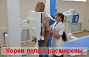
The roots of the lungs are expanded - a phrase that can often be observed on the radiologist’s report. It indicates a number of pathological diseases that develop not only in the lungs, but also in the cardiovascular system, and sometimes in organs more distant from the bronchopulmonary system.
The term “roots of the lungs” refers to the area of the gate or entrance/exit of the lungs. Roots are analogous to a tree, which is what a bronchial structure (bronchial tree) looks like on an x-ray. But from an anatomical point of view, the roots of the lungs included the main bronchus, as well as the pulmonary artery and pulmonary veins (upper and lower).
The structure of the roots also includes lymphatic vessels and nodes along with the nerves of the pulmonary plexus.
For reference. It should be noted that not all elements of the roots (extrapulmonary) are visible on the radiograph. Hence the discrepancy between the concept of the anatomical “root of the lung” and the radiological one.
The roots of the lungs are expanded - what is it?
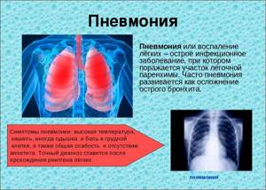
The lung is a parenchymal organ that consists of alveolar tissue, airways, as well as blood and lymphatic vessels. All these structures are responsible for adequately ensuring the function of external respiration.
The root of the lung is an anatomical formation that includes the main bronchus, pulmonary artery and vein, as well as the lobar vessels and bronchi that extend from them. These structures are surrounded by fiber in which lymph nodes and lymphatic vessels lie. A change in any of these anatomical formations should be considered as a pathological process in the root of the lung.
For reference. The roots of the lungs are expanded - this is a morphological sign that is most often detected on x-rays and fluorograms, less often on computed tomography or magnetic resonance imaging.
It is not a specific diagnosis and does not have specific symptoms or treatments. The roots of the lungs can be expanded for many reasons, which should be clarified after establishing this fact.
As a rule, changes are detected as follows: the patient has complaints characteristic of pathology of the respiratory system or simply undergoes a preventive examination. He is prescribed an x-ray or fluorography.
After developing the film, the doctor writes a conclusion: the roots of the lungs are expanded or changed. Next, the patient goes to a therapist, who conducts an additional examination in search of the cause of the identified changes.
For reference. Thus, enlarged roots of the lungs are a symptom that is a manifestation of pathology of the cardiovascular or bronchopulmonary system.
Main reasons for expansion
The expansion of the roots of the lungs is accompanied by a noticeable change in breathing in children. But in adults this condition is less pronounced: when listening to the organ, percussion dullness may be absent.
In addition, the following symptoms may occur:
- Very severe cough, especially when lying down.
- Pain in the region of the ribs, where the roots of the organ are located. They can be aching, but more often they are short-term and acute in nature.
- Dyspnea. It is strong and appears even at rest.
The above signs may indicate a variety of pathologies. Therefore, if they occur, you should immediately consult a doctor. Self-treatment can cause serious complications.
An x-ray is required. Based on its results, it is determined how expanded the lungs are. In this case, the increase can be one- or two-sided.
There may be several reasons for this condition:
- Congested lungs. The enlargement of the pulmonary roots occurs as a result of dilation of the veins. In this case, there are no clear boundaries, but the expansion of the lungs is externally different from tumor processes. Towards the periphery, the darkening becomes less pronounced. Uniform bilateral expansion is observed. Auscultatory symptoms are also observed - wheezing is heard in both lungs. The size of the heart may increase or the organ may move a little, causing cardiac abnormalities to appear - arrhythmia, signs of defect, and others.
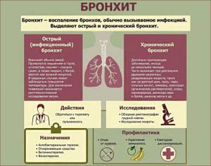
- Chronical bronchitis. Regardless of the etiology of the disease, it is almost always accompanied by an increase in the roots of the lungs. Lymph nodes located near the roots are poorly defined. Most often, this condition is observed with asthmatic bronchitis, less often with bronchiectasis.
- Tuberculosis of the lymph nodes. The clinical picture differs depending on the duration of the disease. If a person once suffered from tuberculosis and healed it, in this case the lesion may be faintly visible in the image or may be completely absent. If the disease appears for the first time, then the root nodes have sharp, clear boundaries. In this case, the lesion can be either unilateral or bilateral. The clinical picture of this disease is similar to sarcoidosis. Therefore, differential diagnosis is required, usually the diagnosis is made by exclusion. For tuberculosis of the lymph nodes, the Mantoux test is positive.
Fluorography may also be prescribed to make a diagnosis. This diagnostic method is slightly safer than x-rays, but less informative.
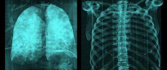
Roots of the lungs on x-ray
X-ray shows the shadows of the chest organs and their location relative to each other. Denser organs create a larger shadow, airy ones create a smaller one.
Since X-ray film is a negative of an actual photograph, light structures that give a large shadow are called darkening, and dark air structures are called clearing.
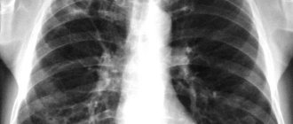
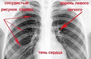
The root of the lung looks like a darkening against the background of the lung tissue. It can be found at the border of the lungs and mediastinum at the level of the I-IV rib.
On an x-ray, most of the shadow of the root is created by the arteries, since they have a greater density compared to other elements. The bronchi contain air, therefore they are distinguished by stripes of enlightenment against the background of the vascular pattern.
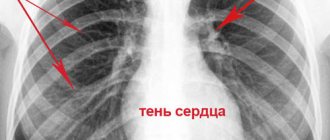
Darkening of the root zone
Darkening of the lungs is characterized by a white shadow, which can spread over the entire surface of the organ or be limited - it occupies only part of the lung (for example, the root area). Shading can be one-sided or two-sided. This may indicate toxic pulmonary edema or heart disease.
However, this condition can be very dangerous . Darkening is observed in cancer, complicated forms of tuberculosis and pneumonia. If a darkening is detected in the image, the radiologist must clarify its exact location. It can be inside the organ or be extrapulmonary.
If the patient has early stage cancer, surgery may be performed to remove a lobe of the lung. Such a person can be completely cured. Therefore, when making a diagnosis, you should not immediately panic.
Structure of the roots of the lungs
In conclusion, radiologists usually write: “The roots of the lungs are structural” or “The roots of the lungs are not structural.”
For reference. This phrase has an important diagnostic value, since each element in the structure of the roots has its own function and changes under the influence of certain reasons.
Each lung root has a head, body and tail. The “head” is the part that goes to the upper lobe. It includes the upper lobe pulmonary artery, the vein of the same name and the bronchus. “Tail” is the name given to the corresponding lower lobe formations. “Body” is the place where the head meets the tail and their transition to the base of the root.
All elements of this structure should smoothly transition into one another. If they are not distinguishable from each other, we can talk about pathology of the roots of the lungs.
In addition, the blood vessels are clearly distinguishable from the bronchial tree. Arteries are visible on x-rays as dark stripes that are divided dichotomously. Normally, they become narrower from the root to the periphery.
For reference. The bronchi are visible as stripes of clearing. They are practically invisible against the background of the lung tissue, but the main and lobar bronchi stand out among the arteries and veins of the root. Veins cannot be distinguished from arteries on x-ray.
The structural root must have a head, body and tail, as well as clearly distinguishable main and lobar bronchi and vessels. If these components are not visible or cannot be distinguished from each other, they speak of a structureless root.
General characteristics of roots
The roots of the lungs are a system that includes a number of elements. Based on the state of the elements, conclusions can be drawn about the state of the entire system. Significant characteristics include the following.
Extension
Normally, the width of the lung root corresponds to the width of its arterial component and in the area of the root body is 15 mm. Typically, the roots of the lung expand on both sides, but the right one is easier to measure than the left. Due to the fact that the structures of the left lung on the x-ray are partially covered by the shadow of the heart, left-sided pathology is detected later.
Often the roots of the lungs are expanded due to an increase in their vascular component. This is observed in left ventricular heart failure, when the pressure in the pulmonary veins increases due to stagnation of blood in them. In the pulmonary artery, pressure may increase due to lung diseases such as emphysema or atelectasis.
Diseases of the bronchial tree also lead to root expansion.
For reference. First of all, the air-bearing component of the root expands during peribronchitis. At the same time, infiltrates around the bronchi make their shadow wider than it should be normally.
A dangerous sign is a polycyclically expanded root. As a rule, such pathology is observed on one side. The polycyclicity of the root is given by the lymph nodes, which are located along the large vessels and bronchi.
Enlarged lymph nodes are most often observed during active tuberculosis, but there are also more rare causes: sarcoidosis and metastases of a malignant tumor.
Attention. At the same time, it is worth remembering that young people are more likely to have tuberculosis , and older people are more likely to have a tumor process.
Seal
As a rule, compaction of the roots of the lungs is observed together with their expansion and may be due to the same reasons.
For reference. Induration is a more pronounced than normal intensity of the shadow. In rare cases, the roots of the lungs are compacted with a normal diameter of their shadow.
An isolated compaction indicates a chronic process. Most often, this pathology occurs with chronic inflammation of the bronchial tree, when part of their wall is replaced by connective tissue. In this case, the shadow will not expand, but it will become more intense.
heaviness
The heaviness of the roots of the lungs on the radiograph looks like an intensification of one of the components of the root, its isolation from the others in the form of a cord. As a rule, this sign indicates chronic pathology.
For reference. It occurs in long-term vascular diseases, when the walls of arteries or veins become significantly thicker.
The heaviness of the bronchial tree indicates chronic lung diseases, most often obstructive bronchitis and bronchial asthma .
As a rule, heaviness of the roots of the lungs is a bilateral sign and develops on both sides simultaneously.
Fibrosis
Fibrosis is the proliferation of connective tissue within parenchymal or hollow organs. Root fibrosis occurs when connective tissue fibers tighten the root structures and deform them.
For reference. Typically, fibrosis develops in the lung parenchyma and then spreads to the roots.
The causes of this pathology can be long-term pneumonia, atelectasis, chronic bronchial diseases, chronic forms of tuberculosis.
Enhanced pulmonary pattern
Enhanced pulmonary pattern is the visibility of the pulmonary vasculature at a distance of more than 15 mm from the root of the lungs. In a healthy person, the vessels of the lungs are divided dichotomously, decreasing in diameter from the root to the periphery.
At a distance of 10-15 mm they become so thin that they are no longer visible on an x-ray. They can be distinguished if arterial or venous congestion occurs in the vessels of the small circle. For example, heart failure can lead to such a pathology.
Severe inflammatory disease of the bronchial tree also leads to increased pulmonary pattern. But in this case, not because of the vascular, but because of the bronchial component.
Arteries can be distinguished from bronchi on an x-ray by the fact that the latter extend from each other at right angles. When arteries divide, an acute angle is formed.
For reference. The cause of increased bronchial component of the pulmonary pattern can be acute and chronic bronchitis.
Diseases for which a doctor may prescribe fluorography
Pleurisy
The presence of the wording “sealed sinus”, as well as a note about changes in the diaphragm in the fluorographic report most often indicate a history of pleurisy.
Lung cancer
The interpretation of “strandy roots”, as well as a note of changes in the diaphragm in the fluorographic report may indicate that the patient has lung cancer.
Acute bronchitis
The interpretation of the fluorographic conclusion “increased pulmonary (vascular) pattern” is observed in acute inflammation of any origin, including bronchitis. Increased pulmonary pattern in inflammatory diseases, as a rule, disappears within a few weeks after the illness.
Pulmonary tuberculosis (miliary)
The location of focal shadows (foci) in the image (shadows up to 1 cm in size) in the upper parts of the lungs, the presence of calcifications (round-shaped shadows, comparable in density to bone tissue) is typical for tuberculosis. If there are a lot of calcifications, then it is likely that the person had fairly close contact with a patient with tuberculosis, but the disease did not develop. Signs of fibrosis and pleuroapical layers in the image may indicate previous tuberculosis.
Acute respiratory viral infection
The interpretation of the fluorographic conclusion “increased pulmonary (vascular) pattern” is observed in acute inflammation of any origin, including ARVI. Increased pulmonary pattern in inflammatory diseases, as a rule, disappears within a few weeks after the illness.
Pulmonary tuberculosis (focal and infiltrative)
The location of focal shadows (foci) in the image (shadows up to 1 cm in size) in the upper parts of the lungs, the presence of calcifications (round-shaped shadows, comparable in density to bone tissue) is typical for tuberculosis. If there are a lot of calcifications, then it is likely that the person had fairly close contact with a patient with tuberculosis, but the disease did not develop. Signs of fibrosis and pleuroapical layers in the image may indicate previous tuberculosis.
Acute obstructive bronchitis
The interpretation of “increased pulmonary (vascular) pattern” in a fluorographic report can be observed in acute inflammation of any origin, including bronchitis. Increased pulmonary pattern in inflammatory diseases, as a rule, disappears within a few weeks after the illness.
Danger of pathology
Changes in the roots of the lungs indicate that the pathology of the respiratory or cardiovascular system has reached the stage of decompensation. This applies to chronic inflammatory lung diseases and chronic heart failure.
In addition, changes in the roots in combination with characteristic foci in the pulmonary parenchyma make it possible to diagnose tuberculosis.
Sometimes polycyclically altered roots are the first manifestation of cancer metastases.
Attention. Tumors often very quickly begin to metastasize to the lungs and lymph nodes of the root, so such a finding becomes the first step in diagnosing cancer.
Detection of altered roots of the lungs should alert the attending physician and the patient. It is important to find the cause of these changes in time.
In addition, respiratory failure can result from changes in the roots of the lungs. The fact is that the roots represent the largest vessels and bronchi of the lungs. If the pathological process affects these structures, the breathing mechanism changes significantly.
What does the seal indicate?
If the roots of the lungs are compacted, this may indicate various diseases. The diagnosis is made not only on the basis of fluorography. The patient must undergo a blood and urine test; a CT or MRI may be necessary.
Most often, compaction leads to expansion of the pulmonary structures. Sometimes only local compaction is observed. This indicates a chronic disease. In this case, the compaction is a consequence of excessive accumulation of connective tissue.
Simultaneous expansion and thickening of the roots of the lungs may indicate chronic bronchitis or pneumonia. The same phenomenon can be observed in other diseases, accompanied by additional changes - the presence of lesions in the lungs, cavities, etc.
Prevention and treatment
Prevention of changes in the roots of the lungs consists of early diagnosis of those diseases that can lead to such an outcome.
Attention. In this regard, the annual fluorographic examination of the population provides invaluable assistance to doctors. The fluorogram shows all changes in the lungs, and sometimes signs of cardiac pathology.
It is impossible to treat enlarged roots of the lungs. This is a symptom that requires treatment of the pathology that led to its development. With successful treatment of the underlying disease, the roots of the lungs acquire a normal shape.