According to statistics, up to 80% of children suffer from perhaps the most common parasitic disease – enterobiasis.
Cost of testing for enterobiasis (scraping for pinworm eggs): 300 rubles.
Hygienic skills in children, as a rule, are not yet sufficiently developed, so it is during childhood that the incidence of helminthic infestations peaks. Most often, enterobiasis affects young children attending preschool institutions.
The causative agent is the pinworm (Enterobius vermicularis). This is a small helminth that lives in the large intestine. The source of infection is a sick person. The female pinworm, usually at night, crawls out of the rectum and lays eggs in the folds near the anus.
At the same time, children are bothered by itching in the perianal area, which can have varying intensity and duration, depending on the degree of the disease and the number of pinworms. Scratching the anus by a patient leads to abrasions on the skin, secondary bacterial infection, and dermatitis, which aggravates the course of the disease. Also, parasite eggs accumulate under the child’s nails, which contributes to the spread of infection, and if swallowed again, it ensures re-infection (reinvasion).
Girls may experience a specific complication of enterobiasis - vulvovaginitis due to the penetration of pinworms into the genital tract.
Abdominal pain is a common symptom of enterobiasis. Sometimes acute abdominal pain may be the reason to seek surgical help.
Allergization of the body and suppression of the immune response is a common pathological effect inherent in all helminths.
Hand washing and short-cut nails in children are the two most basic rules for preventing enterobiasis.
Detailed description of the study
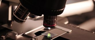
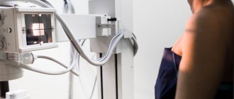
Laboratory research is carried out using microscopy and allows you to detect pinworm eggs in the perianal fold. The analysis is carried out for differential diagnosis of enterobiasis, as well as examination of persons who have been in contact with a sick person.
Enterobiasis is caused by pinworms, helminths that parasitize the human intestines. They belong to the class of roundworms and have a length of 2–13 mm. Infection occurs from a sick person during ingestion of pinworm eggs. The route of transmission is contact and household. Causes of infection:
- insufficient hand hygiene;
- contamination of household items, including children’s toys, with helminth eggs.
On the surface of objects, pinworm eggs remain invasive for up to a month. They are resistant to treatment with disinfectants, not all of which are capable of killing helminth eggs.
Enterobiasis is the most common helminthic infestation disease in Russia: its share accounts for 76.2% of infection cases. Most often, children in organized groups - schools and preschool educational institutions - get sick. The detection of pinworm eggs in scrapings for enterobiasis is associated with their life cycle. Pinworms parasitize the lower part of the small intestine and the entire length of the large intestine. At night, females leave the human body through the rectum and lay from 5 to 15 thousand eggs, which require oxygen for development, in the perianal fold. Eggs mature in 4–6 hours. The person experiences itching and discomfort and tries to get rid of it by scratching the area of infection. So the eggs get under the nails, then into the oral cavity and again into the intestines. Inside the body, they mature, turning into an adult in 10–12 days, after which the cycle repeats again. Helminth eggs can be easily detected by microscopy of an imprint obtained from the perianal fold. They are practically absent in feces. If there are symptoms of the disease, but the test result is negative, it should be repeated after 3-5 days. With intense helminthic infestation, the itching becomes painful, which causes sleep disturbance. The person becomes irritable and unrestrained. Against the background of intense enterobiasis, children are capricious a lot, eat poorly, and lose weight. Sometimes pinworms emerging from the anus migrate to the female genital organs, causing vulvitis and vulvovaginitis. In rare cases, helminths penetrate the uterus and fallopian tubes, forming granulomas. At a low level of invasion, enterobiasis can be practically asymptomatic.
How to understand that a child has enterobiasis, and what to do now?
Sometimes parents discover pinworms themselves - in the child’s pot or on his body. In this case, additional diagnostics are only of an auxiliary nature. All that remains is to make an appointment with a pediatrician as soon as possible to start treatment.
However, there are also symptoms that may indicate helminthiasis. The main and most specific of them is itching in the anus, especially at night and in the morning. The rest are only indirect in nature: nausea, loss of appetite, lethargy, irritability, abdominal pain, frequent cystitis, urethritis, and in girls - vulvitis and vaginitis. With any of them, it is necessary to immediately show the child to a qualified pediatrician: infection with pinworms significantly reduces immunity, the gastrointestinal tract suffers from them, in addition, the little patient himself experiences severe discomfort.
Separately, it is worth mentioning the widespread belief that because of worms, children grind their teeth in their sleep. This is a myth that has no basis. Bruxism (teeth grinding) can be caused by a physiological reason, that is, an incorrect bite or jaw structure, or a neurological reason - a high level of stress, which causes a spasm - but certainly not by parasites.
References
1. Clinical recommendations (treatment protocol) for providing medical care to children with enterobiasis. FSBI NIIDI FMBA of Russia, 2014.
2. “MUK 4.2.3145-13. 4.2. Control methods. Biological and microbiological factors. Laboratory diagnosis of helminthiasis and protozoa. Guidelines" (approved by Rospotrebnadzor on November 26, 2013).
3. R. S. Arakelyan, N. A. Sergeeva, V. Sh. Sangadzhieva, O. V. Konnova, A. N. Zagina, A. A. Obukhova. Clinical and epidemiological aspects of the course of enterobiasis in school-age children. Children's infections. 2018; 17(1): 50-53.
4. Infectious diseases: national guidelines / Ed. N.D. Yushchuka, Yu.Ya. Vengerova - M.: GEOTAR-Media, 2010. - 1056 p.
5. V.P.Sergeev Atlas of clinical parasitology and tropical medicine. Authors Academy. Partnership of scientific publications KMK, 2010
Is it possible to get rid of pinworms on your own?
If you are sure that you have discovered enterobiasis, but have not yet made an appointment at the children's clinic, the first thing you need to do is to switch to an increased level of hygiene for the time until the child is cured, with the whole family: change bed linen daily, wash all linen at temperature above 55 degrees and iron, wash more often, wash hands, toys, and carry out wet cleaning in the apartment. It is recommended to take carpets, pillows and bedspreads out into the cold for 2-3 hours and then shake them out (in the summer, on the contrary, in the hot sun). You will have to live in this mode for at least a week.
There are many traditional ways to combat worms - for example, garlic enemas. However, self-medication is fraught with unpleasant consequences and, first of all, relapses. Enterobiasis should be treated with the whole family, comprehensively and under the supervision of a doctor.
There are several common anthelmintic drugs that are most often prescribed to infected families by pediatricians. These are Pyrantel, Wormil, Nemozol, Vermox. The drug and dose are selected individually for each patient, depending on height, age and weight. At the same time, the doctor prescribes local remedies that stop itching and gives hygiene recommendations.
How informative is the test for enterobiasis?
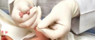
Parents are faced with the need to get tested for enterobiasis more often than with the disease itself. It is required to obtain a medical card for kindergarten, for a certificate that the child can attend the pool and some other sports sections. To do this, a scraping is taken from the anus area with a cotton swab, spatula or special adhesive tape, and then examined in the laboratory to detect pinworm eggs. It is done strictly in the morning, before the skin in the area of the perianal folds is worn out.
However, many parents are faced with the fact that it is not informative enough.
Worms lay eggs cyclically, not every night, and they may not be detected, even if the child has parasites, which means there is a possibility of a false negative result. Therefore, it should be treated only as a screening method. Additional information can be provided by a blood test and, of course, a direct examination by a competent pediatrician. MAKE AN APPOINTMENT PRICES
Diagnosis of enterobiasis
In addition to the typical patient history, which includes the cardinal symptom of intermittent perianal itching, examination of clothing and underwear, the anal area, and stool can provide diagnostic information. Moving, worm-like parasites can sometimes be seen on underwear, sheets, or directly on the edge of the anus. In cases of severe infestation, worms may be passed in the stool. Sometimes individual adult worms are visualized during proctoscopy or colonoscopy. A macroscopically identified worm reliably indicates infection.
Many patients do not experience visible pinworms. Because oviposition occurs on the anal folds, worm eggs, which are not visible to the naked eye, can be detected by using commercially available cellulose tape (“Scotch tape”) in the morning before bowel movements and before washing the genital area (Scotch tape test). To do this, repeatedly press the sticky side of the tape (pre-cut to size, for example, 10 × 2 cm) to the anal and perianal area with the buttocks separated. The tape is then attached to a suitable slide, sticky side down. Microscopic detection of characteristic worm eggs confirms infection. The slides do not need to be stored, prepared or preserved in any particular way. Even ready-made diagnostic kits have appeared in microbiological laboratories and pharmacies. An alternative is to clean the anal area with a cotton swab and then place it in a saline solution. The solution can then be used for microscopic examination for worm eggs (or in molecular genetic detection methods). This diagnostic method should be performed on three different days to increase its sensitivity (from approximately 50% in the case of a one-time tape test to approximately 90% if performed in three sessions). Stool microscopy is not a useful diagnostic tool because worm eggs are laid outside the intestine. Serological methods also have no diagnostic value. Likewise, neither blood eosinophilia nor elevated immunoglobulin E levels are usually expected due to the low invasiveness of the worms.
Head of the pediatric department, pediatrician of the first category of children's medical
Infection with pinworms E. vermicularis (enterobiasis) remains a pressing problem, despite the fact that the level of infection has decreased significantly with the improvement of housing and sanitary and hygienic living conditions in Russia. Although pinworm infection occurs without consequences or even asymptomatically in approximately 40% of cases, enterobiasis is often associated with significant psychological stress for both babies and their parents. Lack of knowledge about the spread of parasites and how it can be prevented, unsuccessful attempts at treatment, as well as a high rate of relapses and autoinfection can lead to resignation to the disease on the part of patients or their parents.
The estimated rate of infection with E. vermicularis worldwide is more than one billion people. Pinworm infection is common in temperate climates and industrialized countries, where it occurs at all levels of society. On average, about 18-25% of children aged 4 to 10 years can be infected with pinworms.
What are pinworms?
Enterobius (Oxyuris) vermicularis or pinworm is a pathogenic intestinal parasite of humans belonging to the genus Nematoda. Symptomatic pinworm infection is called enterobiasis (older term: oxyuriasis). The first description of worm eggs was made by the Swedish scientist Carl Linnaeus in 1758. It can be assumed that E. vermicularis has been successfully parasitizing the human body since ancient times. These worms have a characteristic "thread-like" appearance. Females are 9–12 mm long and approximately 0.5 mm in diameter, while males are shorter, 3–5 mm long, but visible to the naked eye due to their greater thickness. They are colored a bright whitish-beige color, have a round shape and move by energetic crawling. The extensive reproductive system of a fertilized female worm is often completely filled with eggs (>10,000 eggs per worm). Like all nematodes, E. vermicularis has a thick outer protective covering (cuticle). Their eggs are two-layered, elongated oval, asymmetrical and translucent, similar to a loaf of bread. The first larval stage in the egg can often be clearly visualized. Parasite eggs can remain viable for up to five days outside the body.
Risk factors and methods of transmission of pinworms
A number of studies have identified the main risk factors for pinworm infection: children aged 4–11 years, and sometimes adult men, are especially affected. A large percentage of sick children attend kindergarten or primary school. Close social contact, putting toys or writing utensils into the mouth, and nail biting (onychophagy/perionychophagy) play important roles in exposure to E. vermicularis at this age. Scratches in the perianal area, uncontrolled contact of the anus with fingers, poor personal hygiene, and failure to wash hands before eating are all factors associated with significantly higher rates of infection.
The significant transmission potential of E. vermicularis is due to the strength and adhesive properties of the eggs, which are particularly adhering to hands and retained under fingernails, thereby easily maintaining the chain of transmission. At room temperature, eggs cease to be infectious after 5 days. Since E. vermicularis is a purely pathogenic parasite of humans, domestic animals do not act as natural reservoirs of infection.
How many days does the analysis take?
The testing time depends on the capabilities of the laboratory of a particular medical institution. As a rule, private clinics perform tests much faster than public ones. The average time to receive the result of a scraping study is 1 business day.

Regardless of the time of delivery of the material, its examination is carried out within 8 hours
The certificate has its own validity period. After submitting the material for analysis and receiving the result, it will be considered valid only for the next 10 days. Then, if the certificate turns out to be unclaimed, the study will need to be repeated.
If the child does not have pinworms, then no worm eggs will be found in the material submitted for diagnosis. If the result is positive, the baby will be prescribed drug therapy. After completing the course of treatment, the scraping will be taken again.