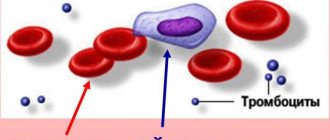
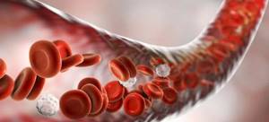
Erythrocytes or red blood cells are the most numerous of the highly specialized blood cells. The functions of red blood cells are extensive, but the main one is that they saturate the tissues of the body with oxygen, returning carbon dioxide back to the lungs.
What are red blood cells?
Even those who are far from medicine sometimes ask questions: what are red blood cells in the blood? What are they needed for? Along with platelets and leukocytes, these blood cells are formed in the red bone marrow of vertebrates, including humans. They are the most numerous and participate in the life of all systems, facilitating the movement of oxygen through tissues and organs. Because of their shape and unique plasticity, red blood cells can easily move through capillaries, facilitating gas exchange.
The structure of red blood cells
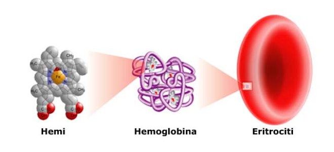
The structure and functions of red blood cells make them plastic and easily deformable. The liquid contents of cells - the cytoplasm - is rich in hemoglobin, which contains a divalent iron atom that binds oxygen. The same pigment gives the bodies their red color. Red blood cells are disc-shaped and do not have a nucleus, which is lost during maturation. The composition of red cells is as follows:
- reticular stroma;
- hemoglobin-filled cell;
- dense shell.
The structure of human red blood cells is simplified: inside there is a membrane resembling a mesh, while the plasma membranes of leukocytes and platelets are more complex. The membrane of red cells is special - it is impermeable to cations (with the exception of potassium), but it allows chlorine anions, oxygen molecules and carbon dioxide to pass through well.
How are red blood cells formed in the blood?
How are red blood cells formed? Tissue growth occurs through the multiplication of one cell, called proliferation. After this, the stem cells, as the ancestors of hematopoiesis, form a large body with a nucleus, which is lost as the red blood cell grows. Once in the bloodstream, the body is transformed into a ready-made red blood cell. The process takes up to 3 hours, and red cells are formed in the body without interruption.
Every second, more than 2 million red blood cells are formed in the bone marrow of the spine, skull and ribs, in addition - in the endings of the arms and legs (in children). Circulating in the blood for 3-4 months (about 110 days), red blood cells are absorbed by macrophages and destroyed in the spleen and liver. A small part of them undergoes phagocytosis - capture by solid particles of cells - in the vascular bed. The transfer of oxygen throughout the body and participation in the transfer of carbon dioxide are the central functions of red blood cells. Cell production begins in the fifth month of fetal development.
What do red blood cells look like?
The structure of red blood cells is related to the function they perform, and in appearance they differ from other blood cells circulating in the body. They have a different – special – shape and size. By nature, blood cells are endowed with peculiar features - tiny size, flattened disk shape, absence of a nucleus. This is necessary in order to quickly cope with the transport of gas in the blood.
Shape of red blood cells
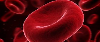
Red blood cells are a flattened, biconvex disc (discocyte). The intracellular space is increased due to the lack of membrane partitions and a nucleus, which mature erythrocytes of all mammals lack. The shape of human red blood cells also increases their total surface area. Inside the bodies there is an increased volume of the protein pigment hemoglobin, which binds oxygen and carbon dioxide molecules.
The specific form increases the efficiency of the basic function of all red blood cells. However, the entire mass of blood cells is heterogeneous. Along with the cells of the regular shape of the biconvex disc, there are others, their percentage of the total number is small (less than 10%). This:
- flat-surfaced squamous cells;
- aging types of these cells - echinocytes;
- spherical spherocytes;
- dome-shaped stomatocytes.
Red blood cells - sizes
The diameter of blood cells varies from 6 to 8.2 micrometers (µm). The maximum thickness is only 2 microns. The tiny size makes it easy to move through microscopic capillary vessels. Modern medicine calls the phenomenon when the normal size of red blood cells increases in one direction or another: macrocytosis and microcytosis. The diameter of healthy cells is 7-9 microns, they are called normocytes. Everything below is microcytes, and everything above is macrocytes.
What function do red blood cells perform?
Blood cells play an important role in the human body.
In addition to carrying oxygen to tissues from the lungs, the functions of red blood cells in the blood include:
- Reverse transport of carbon dioxide to the lungs from tissues.
- Transfer of useful amino acids on its surface.
- Delivery of water from tissues to the lungs. It is released in the form of steam.
- Isolation of erythrocyte coagulation factors.
- Regulation of blood viscosity, which, due to the participation of red cells, is smaller in small vessels compared to large ones.
Respiratory function of red blood cells
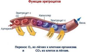
The acid-base state, that is, the ratio of hydroxyl and hydrogen ions in the biological environment, is regulated by red blood cells. They transport O2 and CO2 from tissues to the lungs. Gas exchange is the main function of red blood cells.
How it works:
- Inhaled oxygen enters the lungs. Blood cells squeeze through narrow vessels and tiny capillaries there.
- The iron in hemoglobin captures oxygen, causing the pigment to change color from blue to red. And red blood cells carry the collected oxygen throughout the body.
- Hydrogen is oxidized by body cells, and at the same time carbon dioxide is formed. Most of it returns through the lungs, but some molecules remain on the red blood cells.
Nutritional function of red blood cells
When answering the question what function red blood cells perform, they mention transport. But they “transport” not only oxygen and carbon dioxide, but also useful substances. Essential amino acids and lipids are concentrated on the surface of red cells, getting there from the plasma, and are transported to tissue cells. This is the nutritional function of red blood cells.
Protective function of red blood cells
An important function of red blood cells is to protect the body from harmful substances. On the surface of red blood cells there are proteins of a protein nature. Thanks to them, red blood cells are able to bind some toxins and neutralize them, acting as a protector against poisons. In addition, red cells take part in blood coagulation, hemostasis (vascular-platelet) and fibrinolysis - the process of dissolving blood clots.
Enzymatic function of red blood cells
Red blood cells are carriers of various enzymes. This is another transport function of red blood cells in human blood. All enzymes in blood cells can be divided into three types:
- regulating oxygenation and dioxygenation;
- facilitating the performance of transport functions;
- providing biological processes with energy.
Transfusion
When determining your blood group, you should never make a mistake. Knowing the blood group is especially important when transfusing it. Not every one is suitable for a certain person.
Extremely important! Before blood transfusion, it is necessary to determine its compatibility. You cannot give incompatible blood to a person. This is life-threatening.

When incompatible blood is administered, red blood cell agglutination occurs . This occurs with this combination of agglutinogens and agglutinins: Aα, Bβ. In this case, the patient develops signs of transfusion shock.
They can be like this:
- Headache,
- Anxiety,
- Flushed face
- Low blood pressure,
- Rapid pulse,
- Tightness in the chest.
Agglutination ends with hemolysis, that is, red blood cells are destroyed in the body.
A small amount of blood or red blood cells can be transfused as follows:
- Group I – into the blood II, III, IV,
- Group II - to IV,
- Group III – to IV.
Important! If there is a need for transfusion of a large amount of fluid, only blood of the same group is infused.
Blood hemolysis
Red cells live no longer than their prescribed period - 110-120 days - and are continuously destroyed in the blood, releasing hemoglobin. The process is called hemolysis, and its types differ in nature, mechanism and place of occurrence. So endogenous hemolysis occurs in the body, and exogenous hemolysis occurs outside it, for example, in a heart-lung machine. In addition, the destruction of red blood cells occurs:
- Intracellular
- in the spleen, liver, bone marrow. - Intravascular
- in blood plasma.
By nature, physiological and pathological breakdown of blood cells is distinguished. Red blood cells perform the transporter function assigned to them and die in the blood plasma or tissues. In the latter case, the destruction of the bodies is provoked by negative factors and pathological conditions, such as:
- anemia;
- rheumatic diseases;
- kidney pathologies.
There are several types of hemolysis:
- Temperature
, arising due to exposure to cold. - Chemical
, which is promoted by the action of alcohols, ether, alkali, acid, which dissolve lipids in the membrane. - Biological
, caused by natural factors such as poisons of insects, snakes, bacteria or transfusion of incompatible blood to a person. - Mechanical
- occurs when membranes rupture. - Osmotic
, which occurs when red blood cells enter an environment where the osmotic pressure is lower than blood pressure. Water enters the bodies, they swell and burst.
Red blood cells and blood groups
Normally, every red blood cell in the bloodstream is a cell that is free to move. With an increase in blood acidity pH and other negative factors, red blood cells stick together. Their bonding is called agglutination.
This reaction is possible and very dangerous when blood is transfused from one person to another. In order to prevent red blood cells from sticking together in this case, you need to know the blood group of the patient and his donor.
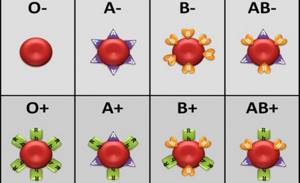
The agglutination reaction served as the basis for dividing human blood into four groups. They differ from each other in the combination of agglutinogens and agglutinins.
The following table will introduce the characteristics of each blood group:
| Blood type | Availability |
| agglutinogens | agglutinins in plasma | |
| I | 0 | αβ |
| II | A | β |
| III | B | α |
| IV | AB | 0 |
What is ESR?

Laboratory tests show the number of red blood cells in the blood, their size, shape, and changes. But there is a special ESR analysis (erythrocyte sedimentation rate), reflecting the ratio of plasma protein fractions. To do this, the blood is placed in a test tube containing substances that prevent it from clotting. The weight of blood cells is higher than plasma (1.080 to 1.029), and they settle at the bottom. By measuring the time during which this happens, the ESR is calculated.
If the indicators are abnormal, doctors consider this as an indirect sign of a current inflammatory disease, for example:
- pancreatitis;
- appendicitis;
- adnexitis.
The norm of red blood cells according to this study varies depending on age and gender:
- The speed of movement of red cells in newborns is 1-2 mm/h.
In the period from a month to six months, it sharply increases to 11-17 mm/h, but then comes to 1-8 mm/h. - ESR in men does not exceed 2-10 mm/h.
- In women, this indicator: from 3 to 15 mm/h
, in pregnant women it is higher - with the approach of childbirth it reaches a maximum value of 55 mm/h.
Interpretation of results
There are no reference values for the results of this analysis. All data obtained is analyzed by the attending physician in conjunction with the results of other laboratory and instrumental studies. The patient's medical history and other factors are also taken into account.
The morphology of red blood cells is assessed individually for each patient. Violations in the shape, size or color of these cells, the presence of inclusions can indicate various hematological diseases and severe intoxication.
The test is used for diagnosis in combination with other studies. After the diagnosis is established and treatment is prescribed, the analysis is prescribed at certain intervals. The obtained results allow you to monitor the patient’s condition, assess the severity of the disease, and the effectiveness of treatment.
The norm of red blood cells in the blood
The presence of pathological conditions is also indicated by the concentration of red cells in the blood. To count their number, they use a special apparatus - Goryaev’s camera. The biomaterial is placed in a mixer and diluted with a 3% chloride solution - ratio 1:100. A drop of the mixture is delivered into a chamber with square grids; when they are filled, laboratory assistants examine the results under a microscope and calculate the number of red blood cells in 1 μl of blood.
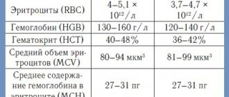
The average value of the norm is 3.8 to 5.10 x 10¹²/l, i.e. several million cells per microliter. The numbers also vary by age and gender.
Red blood cell count for different categories:
- 4-5.1 million/µl in men;
- from 3.7 to 4.7 million/μl in women and from 3 to 3.5 million/μl in pregnant women;
- in children from one year to 12 years: 3.8–5 million/μl and 3.9–5.9 million/μl in newborns.
The functions of red blood cells in human blood are not limited to the transport of oxygen and carbon dioxide. Highly specialized cells are important in the life of the body, and by determining their quantity and quality (appearance, thickness and speed of movement), doctors conduct laboratory tests that help determine the presence of various pathologies.

