Angiography is one of the modern diagnostic methods. This is a contrast X-ray examination of blood vessels. Angiography is used in fluoroscopy, radiography, computed tomography, hybrid operating rooms (operating theater for medical imaging). Diagnostics determines the functionality of blood vessels, the state of collateral blood flow, the extent of the pathological process and its features. What do you need to know about diagnostics, what are the indications/contraindications, and can you decipher the diagnosis yourself?
General characteristics of the study
Angiography is a method for diagnosing blood vessels, which is based on the properties of x-ray radiation. The study can be general or selective. Using general angiography, you can examine all the vessels of the human body. A selective method is necessary for contrast and visualization of specific vessels.
Content:
- General characteristics of the study
- Applications of angiography
- Indications and contraindications for the procedure
- Features of preparation for the procedure
- Diagnostic principle
- Decoding the diagnosis
- Possible side effects
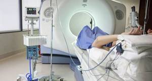
There are two techniques for performing angiography - computed tomography and magnetic resonance imaging. CT allows you to get a clear picture of blood vessels and assess the nature of blood flow. The obtained information is analyzed using special image reconstruction algorithms. With the help of MRI, which, unlike CT, does not use X-rays, images of blood vessels are also obtained, but the device reveals both the functional and anatomical features of the blood flow. The selection of a diagnostic method depends on the patient’s health characteristics and the degree of damage to the body.
The study is divided into two types - puncture and catheterization. What does this mean and which method should you prefer? The puncture is used for superficially located vessels. The contrast agent can be injected directly through a special syringe.
A contrast agent is a drug that is injected into an organ/bloodstream/body cavity. It provides contrast enhancement (staining) for radiological examination. The substance helps to visualize the vascular bed, the internal relief of blood vessels.
There are two large groups of contrast agents: iodine-containing (water- or fat-soluble) and water-insoluble. Angiography uses iodinated contrast agents. They are divided into ionic and nonionic substances. Non-ionic contrast agent is suitable for intravascular administration and is considered the safest. It is superior to ionic drugs in terms of lower risk of adverse reactions and lower cost.
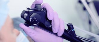
The second type of angiography is catheterization. The method is used if the artery or vein that needs to be examined is located deep under the skin. Before starting the diagnosis, the doctor administers anesthesia to the patient. Then an incision is made in the skin/subcutaneous tissue, the desired vessel is found and the introducer is inserted into it. The introducer is a thin plastic tube with a diameter of 10 centimeters or less. A catheter and other necessary instruments are placed inside the tube. The introducer is used to protect blood vessels from various damages. The catheter itself looks like a long thin hose. Through it, a contrast agent is supplied to a specific vessel.
As soon as an iodine-containing drug is ingested, it spreads through the bloodstream in the following sequence: large arteries - small arterioles - capillaries - small venules - large veins. While the substance moves through the human body, the device takes a series of X-ray images. It is these images that are the purpose of diagnosis - from them the doctor determines the norms and pathologies of blood vessels. The speed at which the contrast agent spreads indicates the speed of blood flow. All information received is recorded on a digital medium and transmitted to the patient. The results of the study may also be needed by other specialists.
The doctor tries to perform the angiography as quickly as possible to reduce the patient's exposure to X-ray radiation.
Features of cardiac MR angiography
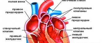
Veins, vessels and arteries are capable of absorbing X-rays, as a result of which their condition cannot be assessed using classical images.
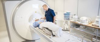
For such an examination, magnetic resonance equipment was created. MR angiography, which is performed on the heart, shows detailed visualization of the structure of blood vessels using the injection of a radiopaque agent. The examination is carried out using special equipment in a separate room, where there is:
- tomograph for studying blood vessels;
- installation for video recording with x-ray;
- high-speed fluorographic camera.
The clinical technique is used to identify various lesions that are associated with blood vessels, heart and kidneys. Using diagnostics of this format, you can identify:
- stenosis;
- aneurysms;
- cysts;
- tumors of various types.
Typically, such a heart test is considered routine. However, if a patient is being treated in a hospital and signs of increasing angina are detected, screening is carried out promptly.
Applications of angiography
Diagnostics is used in the following areas of medicine:
- oncology (to identify cancerous tumors/their metastases in which a branched capillary network is formed);
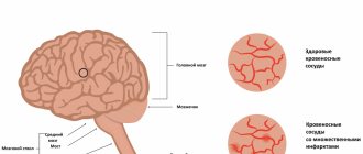
- neurology (for detection of aneurysm, hematoma, oncology, bleeding in the vessels of the brain);
- phlebology (to determine the location of narrowing/blockage of veins, their congenital diseases, atherosclerotic pathologies or blood clots);
- pulmonology (to identify lung malformations or sources of bleeding);
- vascular surgery (used during preparation for operations on blood vessels to clarify their structure, location and features).
Indications and contraindications for the procedure
| Indications | Purpose of diagnosis |
| Brain examination | |
| Aneurysm | Preventing vascular pathologies that can lead to stroke |
| Angioma | Detection of cancer |
| Hemorrhagic stroke | Determining the source of cerebral hemorrhage |
| Heart attack | Monitoring the condition of the organ, determining the need for thrombolysis |
| Hematoma | Identification of the size, features, location and degree of threat of hematoma |
| Cancerous tumors | Monitoring the size, condition, nature of the tumor, identifying possible risks and further therapeutic manipulations |
| Traumatic brain injury | Examination of the general condition of the organ and the consequences of traumatic brain injury |
| Vascular malformation | Search for the source of bleeding, localization of pathology |
| Extremity examination | |
| Thrombosis | Identifying the location of a blood clot, collecting information about its size and the degree of narrowing of the arteries |
| Atherosclerosis | Determining the degree of vasoconstriction |
| Diabetic foot syndrome | Identification of the nature and extent of vascular damage |
| Dissecting arterial aneurysm | Collection of information about the localization of the defect, the length of the dissection and the general condition of the artery |
| Mechanical injury | Confirmation or refutation of vascular deformation, selection of further medical procedures |
| Post-surgical examination or checking the effectiveness of drug therapy | Evaluation of treatment results, determination of further therapeutic or rehabilitation course |
| Examination of coronary vessels | |
| Heart attack | Determining the location of blood flow disturbances and the degree of arterial blockage |
| Atherosclerosis of the coronary arteries | Identification of the degree, spread of pathology and further medical intervention |
| Ischemia | Assessment of the degree of vascular damage |
| Congenital diseases of the coronary vessels | Determining the type of disease, its characteristics and the need for surgical intervention |

Doctors recommend replacing CT angiography with MRI angiography or ultrasound only in cases where the procedure could seriously worsen the patient's condition or cause complications. X-ray diagnostics are strictly contraindicated for pregnant women, since radiation can cause fetal development abnormalities and disruptions in the normal course of pregnancy. Contraindications also include:
- Acute infectious/inflammatory diseases. With angiography, the risk of bacteria and viruses entering the blood is extremely high. This will increase the risk of vascular infection and suppuration at the point of catheter insertion.
- Severe heart failure. Diagnostics affects blood pressure and can provoke sharp jumps, which is undesirable for heart disease. Additionally, the patient may become agitated, which will cause interruptions in cardiac activity.
- Renal/liver failure (including decompensated). The iodine-containing drug affects the kidneys, more precisely, the excretion of urine, while the contrast agent is retained inside the body. Moreover, the combination of stress and a contrast agent can be fraught with hepatic coma.
- Individual intolerance to the drug. A patient with an iodine allergy may experience a severe allergic reaction such as toxic bullous dermatitis, anaphylactic shock, or angioedema.
- Blood clotting disorder. With increased blood clotting, there is a high risk of blood clot formation, and with decreased blood clotting, there is a risk of bleeding.
- Mental pathologies. During angiography, the patient remains conscious. Moreover, he must follow some of the doctors’ instructions and constantly report on his own well-being and sensations. A mentally ill person will not be able to do this, and additional stress will only worsen the situation.
- Thrombophlebitis. The contrast agent can intensify the inflammatory process in the vein and cause blockage of the vessel/blood clot.
How to prepare for cardiac MR angiography?
The body scan presented is considered an invasive clinical technique, so the patient's condition is monitored both before and after diagnosis. In some situations, doctors recommend hospitalization; screening is done in an inpatient setting. Before the session, the patient needs to undergo tests:
- general blood analysis;
- Analysis of urine;
- biochemical blood testing;
- fluorography;
- ultrasound examination;
- coagulogram;
- determine blood groups;
- identify the Rh factor.
If the subject takes medications that affect the coagulation of blood cells, the medication is discontinued several days before the examination. Additionally, drinking alcohol is prohibited for two weeks before the start.
Doctors advise taking a test for intolerance to an additional amplifier a couple of days before the start of the diagnosis. If signs of an allergic reaction occur, the procedure is canceled. An alternative in this case is magnetic resonance screening without the use of contrast.
Features of preparation for the procedure
Before diagnosis, the presence of contraindications is excluded, fluorography and electrocardiography are performed to monitor the patient’s condition. Two weeks before angiography, it is necessary to completely avoid alcohol so as not to distort the overall result. In some cases, your doctor will hydrate you (filling the body with fluids) to dilute the contrast agent, making it easier to remove from the body and minimizing the harmful effects on the liver.

To reduce the risk of an allergic reaction, the doctor prescribes antiallergic medications immediately before the procedure. 4 hours before angiography you should not eat food or drink any liquids. Before the examination, the patient must remove jewelry and remove all metal objects, as they interfere with the free passage of X-rays. The specialist then determines the area of the passage, shaves the hair on the skin of this area and cleans the intervention area.
Before starting angiography, the doctor is required to obtain written consent for the procedure from the patient.
Advantages of angiography at the Innovative Vascular Center
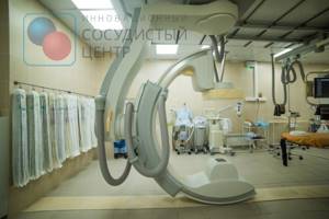
Arteriography of the lower extremities is performed on a stationary Philips Allura Xper FD20 angiographic complex, or on a Veradius mobile angiograph if performed intraoperatively. The use of modern angiographic systems makes it possible to conduct research at any required frame rate, which gives an objective picture of the nature of blood circulation in the limb.
Our clinic uses CO2 angiography without the use of iodine contrast. This method is well suited for patients with iodine intolerance and allows any operation to be performed using the endovascular method.
To reduce the risk of complications, we perform angiography most often through a peripheral radial approach on the arm, which allows the patient to begin walking immediately after the procedure.
Diagnostic principle
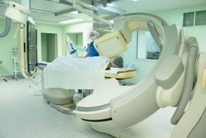
Angiography, regardless of the type and area of diagnosis, is carried out according to a single scheme. First, the doctor administers a tranquilizer and an antihistamine intramuscularly. They are necessary for a general reduction in anxiety and prevention of a pathogenic reaction to a contrast agent. The area of skin (under which the required vessel is located) is treated with an antiseptic drug, a painkiller is injected subcutaneously, and then a small incision is made.
An introducer and catheter are inserted into the incision. A special solution is first injected into the vessel being examined to prevent spasm and minimize the irritating effect of the contrast agent. The catheter is advanced through the introducer to the desired vessel under the control of an x-ray machine. Once the goal is achieved, a contrast agent is injected through the catheter and the image is taken. In some cases, this step is repeated 2-3 times.
After collecting information, the catheter and sheath are removed from the body. Then the doctor stops the bleeding (it opens due to trauma to the vessels by the tube), applies a sterile pressure bandage and gives instructions. You should remain in bed for the next 6-10 hours after the procedure to minimize the risk of blood clots.
What happens during the study
The x-ray surgeon performs the examination only in a hospital setting in a special x-ray operating room. 30-40 minutes before the examination, the patient is given an intramuscular injection in the department. This is a premedication, which consists of a mild sedative and antiallergic drug. The patient will be slightly relaxed but alert and will be able to communicate with the x-ray surgeon throughout the procedure.
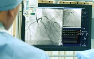
The study is performed on a special angiographic table in the supine position. Metal electrodes will be placed on the arms and legs, which will transmit information about each heartbeat and its frequency - to record an ECG. The area of the artery puncture will be treated with special and aseptic solutions. The patient is covered with a disposable, sterile surgical linen and local anesthesia is performed, during which a slight prick and numbness of the skin in the area of arterial puncture is felt. Then the x-ray surgeon will puncture the artery with a thin needle (less than 1.0 mm) and then through a thin and long tube (catheter) will introduce contrast into the lumen of the leg arteries being examined (see Figure).
This will allow you to see blocked or narrowed areas of the artery. When contrast is administered, the patient may feel heat in the groin for 10-15 seconds. On average, the study lasts no more than 30-40 minutes.
After completing the study, the x-ray surgeon will stop the bleeding at the site of arterial puncture within 10-15 minutes and apply a pressure, aseptic bandage, after which the patient is taken to the ward on a lying gurney.
Decoding the diagnosis
| Symptoms | Manifestations | Pathology |
| Brain examination | ||
| Narrowing of the venous sinuses | The lumen of the venous sinuses (the internal space between the dura mater) is excessively narrowed | Result of traumatic brain injury, thrombosis |
| Pathological changes in blood vessels | The contrast agent passes from the artery directly into the vein without passing through the capillaries | Congenital vascular disease |
| Inability to visualize an artery or part of it | The picture of the vessels is unclear, abruptly interrupted | Thrombosis or atherosclerosis |
| Hemorrhage into brain tissue | An iodine-containing drug escapes into the environment through a burst vessel | Aneurysm, hemorrhagic stroke, hematoma, or traumatic brain injury |
| Decreased blood flow, narrowing of some blood vessels | The rate of spread of the contrast agent is uneven - it passes through some vessels more slowly than through others | Atherosclerosis, compression of blood vessels (with cerebral edema), ischemia, consequences of traumatic brain injury or inflammation |
| Irregular contours of arteries | The image indicates uneven vessel walls (bulging) | Aneurysm, atherosclerosis, mural thrombus formation, congenital vascular disease |
| Extremity examination | ||
| Stenosis | The lumen of the vessel is narrowed by 30-90% | Atherosclerosis, ischemia, presence of hematoma or cancer, thrombosis |
| Occlusion | The contrast agent cannot spread through the vessel because its bed is blocked | Thrombosis or thromboembolism |
| Vascular malformations | Contrast material flows into another artery/vein/lymphatic vessel because other vessels are too tortuous or branched | Congenital disorders of vascular structure |
| Vasodilation | The vessel twists, areas of its expansion or contraction are visible, the vascular wall is protruded | Aneurysm, varicose veins, congenital vascular diseases |
| Examination of coronary vessels | ||
| Aneurysm | The artery wall is bulging | Atherosclerosis, endocarditis, fibromuscular dysplasia, result of chest trauma |
| Calcinosis | The lumen of the vessel is narrowed due to excessive amounts of calcium (Ca) deposits on its walls | Endocarditis, one of the side effects of atherosclerosis |
| Developmental anomalies | Excessive narrowing, widening, tortuosity or arrangement. An iodine-containing drug passes from one vessel to another | Heart disease, aneurysm, vascular malformations |
| Ostial stenosis | The lumen of the vessel is narrowed within 3 millimeters | Thrombosis, arteritis, atherosclerosis |
An experienced specialist - a radiologist - should interpret the results of angiography. In controversial cases, consultation with another specialist – a vascular surgeon – is necessary. Do not prescribe treatment yourself or try to diagnose the condition from information on the Internet. Strictly follow the doctor's instructions and follow the therapeutic course.
What is angiography of the arteries of the lower extremities (LAN)
Angiography of the arteries of the lower extremities (LAN) is a minimally invasive study that is performed in a specially equipped cath lab. ANC is performed using ultra-low doses of X-ray and a special contrast agent, which is injected into the arteries of the lower extremities through the lumen of the catheter.
ANC allows you to determine the state of the blood supply to the legs and will help to develop the most optimal and effective further treatment tactics for the patient.
ANC will identify areas of narrowing of the leg arteries and/or areas of completely closed (occluded) arteries.