Authors: Hérbene Jose Figuinha Milani, Enoch Quindere de Sá Barreto, Renato Luis da Silveira Ximenes, Carlos Alberto Raimundo Baldo, Edward Araujo Júnior, Antonio Fernandes Moron
Objectives of the study
Diagnostics are carried out in order to
- Identify the presence of defects and developmental anomalies
- Find out the gestational age
- Set the number of fruits (one, two or more)
- Determine the location of the placenta
- Diagnose possible processes in the pelvis and their negative impact on the fetus.
Anomalies and defects, their identification, description, and the issue of prolongation are the main goal of doctors.
In the case of serious defects that cannot be compatible with life, it is still possible to terminate the pregnancy in the 2nd trimester without particularly negative effects on the mother’s health.
A study performed by a qualified specialist helps to identify thanatophore dysplasia, ectopia cordis, anencephaly, holoprosencephaly, myelomeningocele, etc.
Pathologies also include:
- Holoprosencephaly - there is no division of the brain into hemispheres; it is represented by a hemisphere with one cavity.
- Anencephaly – The cerebral hemispheres are absent (partially or completely). There are no soft tissues or bones of the cranial vault.
- Tanatorphoric dysplasia (a lethal form of chondrodysplasia) – shortening of the limbs.
- Meningocele is a spinal hernia, a type of spina bifida, developing due to pathology of the neural tube.
- Ectopia of the heart is its abnormal, atypical location.
Procedure and preparation for screening
On the eve of the procedure, you should not limit yourself in eating habits.
A long gestational age allows for an informative study without any preliminary measures. Screening takes about 40 minutes and takes place in a specially equipped ultrasound diagnostic room. There is such an ultrasound room in every branch of the Medok clinic network.
The patient takes off her clothes and lies down on the couch in a comfortable position. To check fetal activity, the doctor may recommend that the pregnant woman touch her stomach or lightly tap it with her fingers.
Ultrasound indicators that clarify the deadline
The length between the parietal bones is called the biparietal size of the head.
The gap between the outer contours of the occipital bone and the frontal - the fronto-occipital size of the head.
Abdominal circumference - measured in a plane perpendicular to the axis of the body. The location of the sensor is as follows: the gas bubble of the stomach and the middle part of the hepatic vein should be displayed on the screen (the place where the hepatic vein enters the umbilical vein should not be captured). The upper poles of the points should be behind the ultrasound area. The sensor is parallel to the fetal spine and behind the location of the heart. Then it is turned 90 degrees
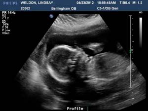
Image of a fetus at the twentieth week of adus, holding perpendicular to the abdominal wall of the fetus. Measurements are carried out several times until several identical results are obtained.
Femur length - ultrasound measures the body of the femur, excluding the epiphysis, since it is less ossified and poorly visualized. This indicator is the shortest length from the proximal to the distal points of the diaphysis. An adequate interpretation of the results obtained is necessary, because normal indicators can range from minus or plus 4-5 weeks from the average value.
Determine if the fruit is the same.
With an ultrasound at 20 weeks of pregnancy, the indicators of the number of fetuses are visualized quite clearly. If the pregnancy is multiple, each one is carefully examined. If monoamniotic twins are identified, they need to be diagnosed for the presence of adhesions. As a result of ultrasound diagnostics, it is possible to detect the death of one of the fetuses.
How is the placenta located?
During pregnancy, the uterus develops unevenly, so it happens that the placenta moves. It is important that it is correctly localized. Ultrasound can distinguish complete or partial placenta previa
. Then the patient has a risk of bleeding, the risk of intrauterine fetal death, and the pregnancy may be terminated.
Diagnose possible pelvic processes, they can negatively affect the further course of pregnancy.
Adequate blood supply to the placenta can be impaired by fibroids in the uterine wall. They can also cause intrauterine growth retardation. Volumetric processes occurring in the pelvis can lead to obstruction of the birth canal, so ultrasound screening requires identifying them, clarifying their size, and finding out their location.
What does an ultrasound show?
Ultrasound at 20 weeks of pregnancy
(we’ll look at the normal indicators in the decoding tables below) very informatively and reliably shows how the fetus develops and how comfortable it is in the womb. The correspondence of the fetal size to the established gestational age is assessed.
Important points of the second ultrasound are the examination of the internal organs of the fetus and the identification of their pathologies.
Examination of fetal organs
By the 20th week, the unborn child has already formed all the internal organs. Using ultrasound, the doctor assesses their condition, structure and degree of maturity. He examines the heart, kidneys, liver, brain, stomach. Attention is also paid to assessing the development of the fetal limbs, its spine, facial bones, and so on.
A detailed examination of internal organs using ultrasound allows one to identify heart defects of various types and predict the further course of pregnancy, as well as assess the level of viability of the child after birth.
Basic fetal indicators
There are a number of indicators by which the level of fetal development is assessed:
- BPP is the biparietal size of the fetal head. At 20 weeks of pregnancy, it should be approximately 36 mm (plus/minus a few mm).
- LZP – fronto-occipital size. At week 20, this parameter should normally be 56-68 mm.
- Fetal abdominal circumference. At 20 weeks, this figure should average 144 mm (plus/minus a few mm).
- Fetal head circumference. At the 20th week of pregnancy, the normal OG can be 154-186 mm.
- Length of the femur (normal 26-38 mm).
- Cephalic index, that is, the ratio of BPR to LZR. This index allows you to accurately identify the type, size and structure of the fetal head.
Ultrasound also evaluates the size of the fibula, tibia, radius, forearm bones and other parts of the body.
Placenta
An ultrasound examination necessarily evaluates the condition of the placenta. First of all, you need to determine the degree of its maturity. At 20 weeks and up to the 27th week, as a rule, the degree of maturity is assessed as zero. Then comes the first stage - until the 32nd week. Before the 34th week, the second stage of placental maturation should normally be diagnosed, then the third stage begins. The longer the pregnancy, the older the placenta becomes, the more difficult the process of transferring nutrients to the child is.
Ultrasound helps to see where the placenta is attached to the walls of the uterus. The norm is attachment to the back wall.
An important parameter is the thickness of the placenta. This figure should constantly increase. For example, if in the early stages the placenta may have a thickness of 10-11 mm, then by the end of the third trimester (by the 36th week) its thickness may be 35 mm.
Premature aging of the placenta, its thinning, and detachment become alarming signals, and in such situations a decision is often made about premature delivery.
Umbilical cord
The second ultrasound involves a detailed examination of the umbilical cord, through which the baby receives intrauterine nutrition.
It is necessary to examine her vessels - normally there should be three of them. There is another, more rare, but not pathological option - when the umbilical cord has only two vessels (vein and artery). In the second case, monitoring of pregnancy should be enhanced, since it is likely that in the last weeks of pregnancy the fetus will lack the oxygen that reaches it through the two vessels of the umbilical cord.
At the second ultrasound, they also pay attention to the number of umbilical cord loops and check whether there is any entanglement.
Indicators
General data of the ultrasound report
- How many fruits (the number and relative position are indicated).
- Presentation (which large part of the fetus is closest to the plane of exit from the uterus: the head/pelvic/presenting part is absent. The baby is still small, so in the future it can change its position).
- Fetal position (longitudinal/oblique/transverse).
Longitudinal position of the fetus - the axis of the fetal body coincides with the axis of the uterus. Transverse position of the fetus - the axis of the fetus is perpendicular to the axis of the uterus. The oblique position of the fetus is average between the longitudinal and transverse positions.
- Condition of the muscular layer of the uterus (is there hypertonicity, is there a possible threat of interruption).
- Is the fetal neck entwined with the umbilical cord?
- Condition of amniotic fluid (signs of high or low water levels, presence of suspended matter in amniotic fluid
- Localization of the placenta (the lower edge is located at a distance of at least 7 cm from the internal os)
- Cervical length.
- External and internal pharynx, condition (normal: at week 20 both are closed).
Ultrasound, table
| Indicators | Standard |
| Weight | 315–400 g |
| Height | 16–24 cm |
| Biparietal size | 43–52 mm |
| Head, circumference | 155–186 mm |
| Belly, circumference | 124–160 mm |
| Femur, length | 30–37 mm |
Doppler
| Indicators | Norm |
| Heart rate | 120–160 rpm |
| Placental resistance index | 0,38–0,7 |
| Resistance index in umbilical cord arteries | 0,65–0,84 |
| Systole-diastolic ratio in the umbilical artery | 3,86–3,95 |
| Systole-diastolic ratio in the uterine artery | 1,92–1,98 |
At the end of the study, the condition of the organs is described in detail: the brain, heart chambers, lungs, number of fingers, development of the kidneys, intestines. An appropriate conclusion is drawn.
What will your doctor see during your third trimester screening?
Diagnosis in the last trimester of pregnancy is important in preparing for childbirth. In the process of scanning with an abdominal sensor, the obstetrician-gynecologist receives a lot of valuable information about the placenta, amniotic fluid and the patient’s readiness for childbirth in general. Ultrasound of the 3rd trimester provides data on:
- The amount of amniotic fluid (oligohydramnios, normal, polyhydramnios)
- Maturity of the placenta and the condition of the blood vessels in it
- The condition of the internal organs of the fetus (heart, lungs, liver, kidneys, etc.)
- Fetal parameters: chest diameter, abdominal diameter and circumference, spine parameters, limb length (femur and humerus)
In addition to ultrasound, the doctor may prescribe standard studies for pregnant women such as Dopplerography and cardiotocography. These diagnostic procedures are designed to identify the speed and nature of blood flow in the placenta and assess the state of the fetal cardiovascular system.
Determining the sex of the baby
Determination of gender and its accuracy depend on the period at which the woman is and the experience of the physician. 20 weeks is the time when the doctor finds out whether there will be a boy or a girl, almost 100% correct.
A boy's sexual characteristics are based on ultrasound visualization of the scrotum and penis, and a girl's - the labia. Mistakes are also possible when fingers or loops of the umbilical cord are mistaken for the penis. In girls, swelling of the labia is possible: it goes away over time, but sometimes contributes to incorrect gender determination. The doctor perceives the swelling as the scrotum. Sometimes there are difficulties in determining gender due to the “modesty” of the baby, who clench his legs tightly and make it difficult to make it clear who he is.
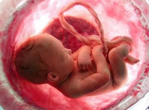
Image of a fetus during the 2nd trimester
Difficulties can also be caused by inactivity of the fetus, which is turned away from the sensor.
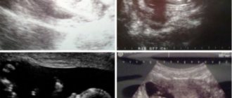
Decoding the main ultrasound artifacts and signs
Ultrasound artifacts can appear during diagnostics regardless of the device. An example is a mirror artifact; it is often perceived incorrectly by the device, resulting in an incorrect interpretation of the research results. On the screen, the specialist sees a picture that is deeper than it actually is. This occurs because the sound beam undergoes multiple reflections when colliding with a highly reflective object. The mirror image appears distorted, blurry, and hypoechoic.
When performing an ultrasound at 20 weeks of pregnancy, due to a mirror artifact, a single pregnancy can be interpreted as a multiple pregnancy (twins). Swelling of the rectosigmoid colon sometimes contributes to the appearance of a picture simulating a second fetus or an abdominal heterotopic pregnancy. The reflective surface was created due to the interface between the distended rectosigmoid colon and the posterior wall of the uterus. It is important to correctly decipher the image.
Ultrasound at 20 weeks of pregnancy
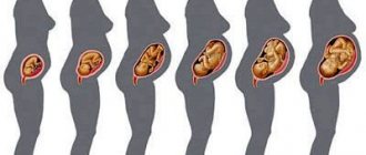
The second trimester (14-26 weeks) of pregnancy is considered the easiest period when bearing a child. The expectant mother is no longer tormented by toxicosis, the baby in her stomach begins to move slightly, which, of course, pleases her.
During this trimester, monitoring of fetal development continues, and ultrasound is included in the list of mandatory diagnostic procedures. The recommended time for the second screening ultrasound is 20 weeks of pregnancy.
Why is an ultrasound performed at 20 weeks of pregnancy?
Ultrasound at 20 weeks of pregnancy
carried out for the purpose of:
- Assess fetal development parameters and compare them with normative ones.
- Identify/refute the presence of chromosomal or genetic disorders in the fetus. Perhaps the second screening will show defects that were not noticed during the first ultrasound examination.
- Determine the sex of the child. At this time, you can find out who will soon be born with almost 100% probability (although mistakes still happen).
- Assess the length of the cervix and identify possible prerequisites for premature birth (if the length of the cervix is less than normal).
- Determine the amount of amniotic fluid. At 20 weeks, low or polyhydramnios can be diagnosed.
- Assess the condition of the umbilical cord, see the number of its vessels.
- Assess the functioning of the placenta, its maturity, measure its thickness.
- Assess blood flow in the feto-placental and utero-placental complex. This study is called Doplerometry.
- Identify pathologies of the female reproductive system (for example, fibroids, etc.).
Anomalies, deviations and pathology
20 weeks is the time when ultrasound will help find the most important deviations.
Brain abnormalities.
The first plane of study is the lateral ventricles. The second is the frontal and occipital horns of the lateral ventricles of the embryonic brain. The third plane is the cerebral peduncles, the visual thalamus and the cavity of the transparent septum. Here the fetal head is assessed. To evaluate the brain regions located in the posterior fossa of the skull, the transducer is deployed and shifted posteriorly from the third scanning plane. The cerebellum, hemispheres and cistern magna are studied.
The following pathologies are identified:
- Anencephaly - the bones of the skull and soft tissues of the brain are absent.
- Acrania – no bones of the cranial vault.
- Holoprosencephaly - the brain is not divided into hemispheres, but is represented by a hemisphere with one cavity.
- Ventriculomegaly - the cavities of the ventricles of the brain are enlarged. If at 20 weeks the ultrasound diagnostician’s report showed ventriculomegaly, then at the next screening, with an increase in the ventricular cavity, hydrocephalus can be judged.
Ultrasound also reveals developmental anomalies of the spinal column: spina bifida - occurs when the closure of the neural tube is disrupted.
At the second screening, the doctor examines the baby’s face, assesses its formation, and notices anomalies: cleft lip without cleft palate, cleft lip and palate. They are possible from different angles
At 20 weeks, the heart and kidneys of the fetus are almost formed. If heart defects and dilated pelvis are detected, the specialist may suspect Down syndrome.
It is important to examine the gastrointestinal tract and identify its pathology. The stomach and esophagus must be visualized.
Sometimes there is no cavity in the stomach or, conversely, a large expanded cavity is visible, which indicates pathology. If the stomach is visualized, then you need to look at its connection with the esophagus and intestines. There may be no esophagus (esophageal atresia). If the fetus has swallowed blood, then this fact will be noticeable to an experienced specialist. If there is a suspension of hyperechoic inclusions in the child’s intestines, then this is possibly a genetic pathology (Down syndrome, cystic fibrosis, etc.).
Sometimes an ultrasound scan at 20 weeks can detect a teratoma or hemangioma.
Teratoma is a benign tumor. As a rule, it is localized in the sacrococcygeal region. Hemangioma is a benign vascular tumor.
Fetal ultrasound: indications for performance, types, interpretation - MEDSI
Table of contents
- The main types of ultrasound of the fetal organs and how they are performed
- Interpretation of ultrasound of fetal organs
- Advantages of carrying out the procedure at MEDSI
Ultrasound examination (ultrasound)
is a type of examination that is carried out using a special device - a sonograph, emitting high-frequency waves. This analysis allows you to determine the presence of pathologies of various organs.
Since ultrasound is one of the safest types of examination, it can be used to examine pregnant women.
During the period of gestation, up to 5 planned ultrasound examinations can be performed at different periods, measured in weeks:
- At 5–7 weeks: allows you to diagnose pregnancy
- At 11–13: fetal development, the presence or absence of pathologies, and the condition of the placenta are assessed
- At 19–21: the size of the fetus, the development of its heart, the presence of amniotic fluid in the placenta, and the sex of the child are determined
- At 32–34: the following parameters are important here: the degree of development of the umbilical cord, the weight and size of the fetus, the proportionality of the size of the birth canal and the baby’s head
- Before childbirth (during the first contractions or at the time of amniotic fluid discharge): with this ultrasound, the likelihood of complications during childbirth is determined
The main types of ultrasound of the fetal organs and how they are performed
There are two main types of ultrasound used to examine fetal organs:
- Abdominal
- Transvaginal
There are also such types of ultrasound modifications as:
- Doppler
- 3D ultrasound
- 4D research
Abdominal (transabdominal) ultrasound is performed as follows:
- Woman lies on her back
- The doctor smears her stomach with gel, which ensures better signal permeability
- Then the specialist moves the sensor over the abdomen, and the image is projected on the screen
For transvaginal ultrasound, it is necessary to insert a sensor inside the vagina, so the woman assumes a position as in a gynecological chair. An individual condom is placed on the sensor, then it is lubricated with gel for the procedure. In some cases, this type of ultrasound is more informative, but it cannot be used in the second or third trimester of pregnancy, since such an analysis can cause premature contractions of the uterus.
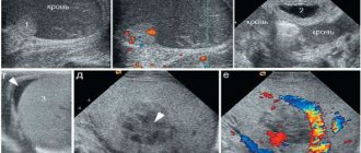
Doppler ultrasound allows us to examine the structure of the fetal circulatory system and the presence of pathologies in it, as well as the speed of blood flow and disturbances in the functioning of the placenta. The vessels are displayed in different colors on the ultrasound results. This analysis is recommended for use at the 12th or 20th week of pregnancy.
3D research allows you to make a three-dimensional spatial image of the unborn child. A special computer program is used for this. Such an analysis can help identify heart defects in cases where conventional ultrasound did not provide accurate information.
4D ultrasound of fetal organs shows a spatial image in real time. This way you can see and evaluate the correct functioning of the internal organs of the fetus.
If a pregnant woman experiences pain in the lower abdomen or bleeding, the doctor may prescribe an unscheduled ultrasound to prevent the development of complications.
Interpretation of ultrasound of fetal organs
To interpret the results of an ultrasound examination, special tables of values are used, since the parameters of the fetus at different stages of its development differ.
The main criteria are:
- Quality of placenta development:
- Maturity
- How is it attached?
- General state of development (presentation, etc.)
- Thickness
- Amniotic fluid volume
- BPD - biparietal head diameter (measured from temple to temple along the minor axis)
- FL—femur length
- HC - head circumference
- CRL - body length from crown to sacrum
Ultrasound examination allows us to identify pathologies in fetal development such as:
- Down syndrome - this disease occurs due to the presence of an extra chromosome in the genome, which causes developmental delays, as well as other disturbances in the functioning of the body
- Anencephaly - characterized by complete or partial absence of the cerebral hemispheres and cranial vault bones
- Heart disease is a congenital defect of the myocardial wall, as well as large vessels
- Spina bifida (including myelomeningocele, etc.) is a spinal condition that is characterized by incomplete closure of the neural tube in a partially immature spinal cord (spina bifida); with a form of the disease called myelomeningocele, the brain and nerve roots come out (spinal cord herniation)
- Hydrocephalus is an excessive accumulation of cerebrospinal fluid in the cranial cavity.
- Fusion (atresia) of the duodenum - characterized by the absence of a lumen in one of the sections of the intestine
It is important to remember that regular preventive ultrasound examinations during pregnancy will help identify possible pathologies and develop effective treatment tactics.
Advantages of carrying out the procedure at MEDSI
- The MEDSI network of clinics conducts more than fifty types of ultrasound examinations using modern devices such as Pro Focus 2202, Philips iU22
- Interpretation of the survey results is done by experienced specialists who have high qualification categories and regularly improve their skills
- To make an appointment, you need to call 8 (495) 7-800-500
- There are more than 20 clinics in Moscow, so it’s easy to find a convenient place for examination