directions
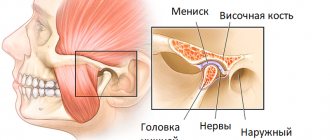
The elbow joint is a rather vulnerable place, subject to frequent loads, injuries and disorders. It has a complex anatomical structure and consists of the brachioradial, humeroulnar and radioulnar joints, connected in one capsule.
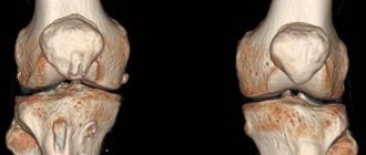
A traumatologist is mainly treated with dislocations, bruises, fractures of the elbow joint and with unpleasant symptoms that may be harbingers of diseases and pathologies. To determine the nature and extent of the damage and to make an accurate diagnosis, the doctor prescribes an x-ray of the elbow joint.
Currently, work is underway on the website to change the price list; for current information, please call: 640-55-25 or leave a request, and an operator will contact you.
Prices for services
- X-ray of the forearm, ulna and radius in direct posterior projection 900a
- X-ray of the elbow joint in direct posterior projection 900a
- X-ray of the elbow joint in axial projection 900a
- X-ray of the elbow joint in lateral projection 900a
- X-ray of the forearm, ulna and radius in lateral projection 1350a
The information and prices presented on the website are for reference only and do not constitute a public offer.
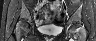
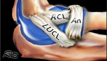
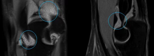
Lateral collateral ligament complex
Ulnar collateral ligament. Mark Anderson, University of Virginia Health Sciences Center, 10/5/2013
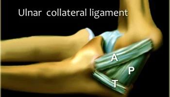
Mark Anderson, University of Virginia Health Sciences Center, 10/5/2013
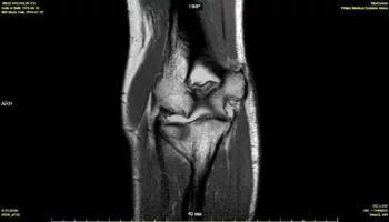
MRI, coronal slice, PD
Lateral ulnar collateral ligament (LUCL) - lateral ulnar collateral ligament
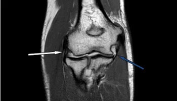
Radial collateral ligament (RCL) - radial collateral ligament (white arrow)
Ulnar collateral ligament (UCL) – ulnar collateral ligament, anterior bundle (blue)
MRI, axial slice, PD with fat suppression
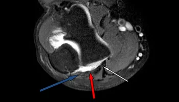
Ulnar collateral ligament (UCL) – ulnar collateral ligament, anterior bundle (white)
Ulnar collateral ligament (UCL) – ulnar collateral ligament, posterior bundle (red)
Ulnar nerve (blue arrow)
Our clinics in St. Petersburg
Structural subdivision of Polikarpov Alley Polikarpov 6k2 Primorsky district
- Pionerskaya
- Specific
- Commandant's
Structural subdivision of Zhukov Marshal Zhukov Ave. 28k2 Kirovsky district
- Avtovo
- Avenue of Veterans
- Leninsky Prospekt
Structural subdivision Devyatkino Okhtinskaya alley 18 Vsevolozhsk district
- Devyatkino
- Civil Prospect
- Academic
For detailed information and to make an appointment, you can call +7 (812) 640-55-25
Make an appointment
We draw your attention to the schedule of technological breaks in the CT and X-ray rooms.
X-ray of the elbow joint allows you to detect inflammation, tumors, various changes in the joint space and adjacent tissues, examine adjacent areas of the elbow joint, and determine the nature of the damage.
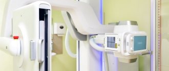
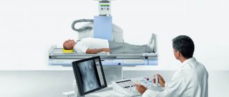
You can have an X-ray of the elbow joint done as quickly and efficiently as possible using the latest Italian equipment in the Medicenter clinic network. The clinics have a trauma department, where experienced and competent specialists will provide you with the necessary first aid for injuries and injuries, take an x-ray of the elbow joint, identify the presence of pathology and disease, and prescribe competent, effective treatment.
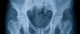
Bone tumors
Schematic representation of typical location and morphological characteristics of bone tumors
Bone tumor — Systematic approach and Differential diagnosis, Henk Jan van der Woude and Robin Smithuis, Radiology department of the Onze Lieve Vrouwe Gasthuis, Amsterdam and the Rijnland hospital, Leiderdorp, the Netherlands, www.radiologyassistant.nl 2010
Abbreviations
ABC - aneurysmal bone cyst, CMF - chondromyxoid fibroma, EG - eosinophilic granuloma, GCT - giant cell tumor, FD - fibrous dyspalsia, HPT - hyperparathyroidism with Brown's tumor, NOF - non-ossifying fibroma, SBC - simple bone cyst
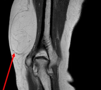
Lipoma of the inner surface of the lower third of the shoulder, expansive growth: pushing aside muscle bundles, no invasion of surrounding tissues. Also, the structure of the formation is homogeneous and is represented only by adipose tissue
Metastasis of small cell lung cancer to the distal humerus: invasive growth with bone destruction, invasion of surrounding soft tissues and joint capsule, reactive swelling of adjacent soft tissues
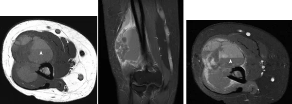
Schwannoma of the radial nerve of the lower third of the shoulder, a “target” sign on MRI
Soft-Tissue Tumors and Tumorlike Lesions: A Systematic Imaging Approach, Jim S. Wu, MD Mary G. Hochman, MD Radiology: Volume 253: Number 2—November 2009
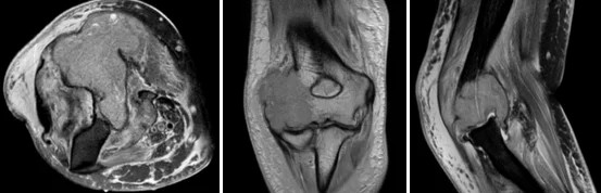
Aneurysmal bone cyst of the distal humerus, expansively spreading along the lateral sections, fluid levels in the cystic cavities (arrows)
Indications and contraindications for x-rays of the elbow joint
X-ray of the elbow joint is indicated for pain in the elbow area, clubhand (deviation of the hand from the longitudinal axis of the ulna and radius), impaired mobility, tumor processes, inflammatory-degenerative pathologies, injuries such as dislocations, fractures, bruises, subluxations, if various kind of pathology and disease, etc.
Contraindications to x-ray examination of the elbow joint are pregnancy, the patient's critical condition, the presence of mental and neurological diseases that do not allow them to remain at rest. For children under 15 years of age, any X-ray examination is carried out strictly according to indications with a referral from the attending physician.
Related and Complementary Research Methodologies
Modern digital X-ray equipment provides doctors with images on the screen of a connected computer, on paper or digital media.
At the same time, radiologists say that pathological conditions of the joints cannot always be fully displayed on an x-ray, so other examination methods are used for them:
- computed tomography of joints;
- ultrasonography;
- Magnetic resonance imaging.
Each of the above methods demonstrates to specialists certain tissues, including the soft tissues of patients - muscles, tendons and ligaments. The high accuracy of these diagnostic methods makes it possible to mutually complement information about the pathological aspects of various periarticular tissues.
To analyze the cavities inside the joints, it is necessary to puncture the synovial capsule. The procedure is carried out with the collection of joint exudate. Impurities are easily detected in this joint fluid, which may indicate a number of diseases. These impurities are studied in more detail in laboratories under a microscope, and on the basis of such conclusions, experts draw conclusions about the infectious or non-infectious nature of the disease.
If it is necessary to study the structure of the joints, another diagnostic technique is used - arthroscopy.
Arthroscopy is performed through two punctures, into the first of which an arthroscope with a special camera is inserted, and in the second, the necessary manipulations are performed with special instruments.
In this case, such a procedure is very informative and provides specialists with the opportunity to see firsthand the development of various pathologies.
Based on the clinical manifestations of diseases, doctors choose those diagnostic methods or their complex that can most accurately provide information about all the processes occurring in the human elbow joint in order to make a timely and accurate diagnosis and prescribe the necessary therapy.
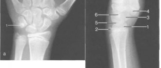
The procedure for radiography of the elbow joint
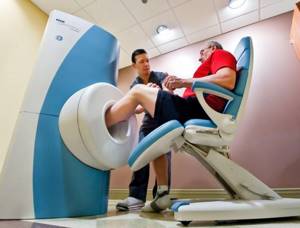
X-ray - a picture of the elbow is taken in three projections: direct, lateral and axial. During an X-ray of the elbow joint, the patient sits and his arm is placed on the table. In some cases, the patient is placed on his back or side, or the image is taken in a standing position.
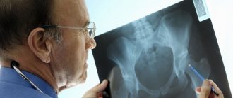
After receiving the images and their description, the patient is referred, depending on the identified injury, disease, pathology, to a traumatologist, orthopedist, surgeon, rheumatologist or more specialized specialists, such as an oncologist, endocrinologist, etc.
Three degrees of muscle damage
- 1st degree – corresponds to microtears at the level of the muscle-tendon junction, which is manifested by “feather-like” swelling, all muscle fibers can be traced
- 2nd degree - some of the fibers are not determined, or are located freely, locally within the muscle defect there is accumulation of fluid, swelling of surrounding tissues, the formation of intramuscular hematomas is possible
- 3 degrees - complete rupture, when within the fascial sheath there is no connection between fragments of the damaged muscle, or a small part of the fibers
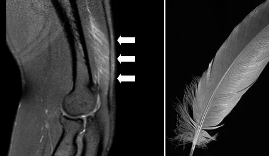
Damage to the triceps brachii muscle of the 1st degree - “feather-like” swelling of the musculotendinous junction
X-ray diagnosis of tumor lesions of bones
Radiography is the primary method for diagnosing tumor lesions of bone tissue. Therefore, at the first stage, it is important to assess the fact of invasive growth, the destruction of bones that has taken place, or to refute this and find reliable signs of a benign process.
The basis for such an assessment will be several important provisions:
- Boundaries of education:
- a. Clear contours with a sclerotic rim - usually a sign of benignity
- b. Clear contours are usually a sign of benignity
- c. Blurred contours - usually a sign of malignancy
- Periosteal reaction:
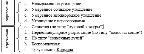
Periosteal Reaction, Rich S. Rana, Jim S. Wu, Ronald L. Eisenberg, AJR:193, October 2009
Solid thickening – benign periosteal reaction against the background of osteoma
Bone tumor — Systematic approach and Differential diagnosis, Henk Jan van der Woude and Robin Smithuis, Radiology department of the Onze Lieve Vrouwe Gasthuis, Amsterdam and the Rijnland hospital, Leiderdorp, the Netherland, 2010
Layered (onion-skin-type) periosteal reaction with intermittent contour – an aggressive reaction against the background of Ewing’s sarcoma
Bone tumor — Systematic approach and Differential diagnosis, Henk Jan van der Woude and Robin Smithuis, Radiology department of the Onze Lieve Vrouwe Gasthuis, Amsterdam and the Rijnland hospital, Leiderdorp, the Netherland, 2010
Osteochondroma of the humerus: presents as an exostosis (red arrow) with a cartilaginous cap (blue circle)
Vascular malformations
It is very important to distinguish between the terms “hemangioma” and “vascular malformation”, since specialists not directly involved in the treatment of such pathologies often confuse the terms, which leads to incorrect tactics for examining and treating patients, wasting time and money.
In 1846, the German scientist Robin Virchow coined the term “hemangioma” to describe hypervascular lesions on the skin based on their macroscopic structure, thus describing vascular malformations, vascular tumors, and hyperplasias. Having analyzed the etymology of the term hemangioma, one can understand that the scientific approach from the time of the beginning of knowledge of anomalies in the development of the vascular system classified vascular malformations as tumors. This approach involved treating hemangiomas as tumor formations.
With the development of morphological and clinical diagnostic methods, Virchow's classification underwent constant changes. The approach to the interpretation of vascular malformations was fundamentally changed in 1982, when Mulliken and Glowacki proposed a classification separating the concepts of vascular malformation and hemangioma. Vascular malformation does not imply tumor or primary hyperplastic tissue growth, only abnormal vascular networks with various combinations of vessels. Hemangioma suggests the presence of a tumor or hyperplasia that primarily developed from the tissue of the vessel. (Mulliken JB, Glowacki J: Hemangiomas and vascular malformations in infants and children: A classification based on endothelial characteristics. Plast Reconstr Surg 69:412-422, 1982 2.; Mulliken JB, Glowacki J: Classification of pediatric vascular lesions. Plast Reconstr Surg 70:120-121, 1982).
In 1996, at the Rome Symposium of the International Society for the Study of Vascular Anomalies, the world scientific community officially replaced the term “hemangioma” with “vascular malformation”, adopting a new classification.
The currently publicly accepted ISSVA classification is:
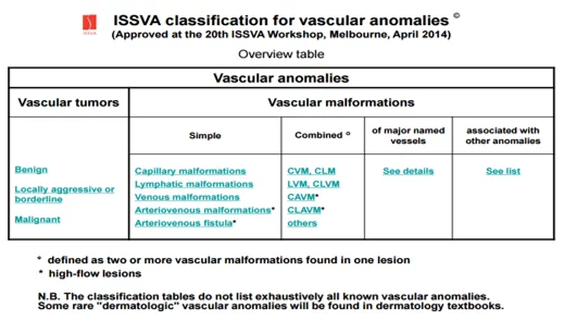
ISSVA classification for vascular anomalies (Approved at the 20th ISSVA Workshop, Melbourne, April 2014) https://www.issva.org/.
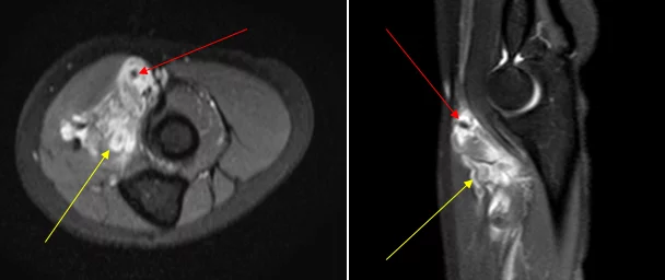
Venous malformation of the soft tissues of the ulnar fossa: pathological network of venous vessels (yellow arrow), phleboliths in the lumen of pathological vessels (red arrow), a characteristic sign of venous malformation