Author
Posysoeva Margarita Alekseevna
Leading doctor
Ultrasound diagnostic doctor
until September 30
Appointment with a doctor based on the results of instrumental diagnostics with a 50% discount More details All promotions
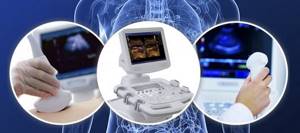
Abdominal ultrasound
is a modern and informative diagnostic method using ultrasound.
An abdominal ultrasound includes examination of the liver, bile ducts, gallbladder, pancreas and spleen. Ultrasound examination is absolutely safe and painless, which, combined with the accuracy of the data obtained and the simplicity of the procedure itself, makes this method in demand, and often simply irreplaceable.
Normal abdominal organs on ultrasound, how is it determined?
The abdominal cavity is an area on the human body that contains vital organs. This includes the stomach, liver, pancreas, and gall bladder. And it is important that all these organs function normally. As a preventive measure, it is recommended to do an ultrasound of the abdominal cavity once a year, which will help identify the disease at the initial stage of development. Let's consider what research indications are considered normal.
What does an abdominal ultrasound show?
Ultrasound can detect:
- changes in the size of internal organs;
- various types of neoplasms;
- inflammatory changes in organs, indicating the following diseases:
- hepatitis;
- cirrhosis of the liver;
- pancreatitis;
- cholecystitis;
- cholelithiasis.
In what cases is ultrasound examination prescribed?
In addition to routine preventive examinations, situations arise when a person requires this procedure. As a rule, the following symptoms may occur so that the patient is prescribed an ultrasound of the organs in Krasnoyarsk:
- Abdominal pain.
- Heaviness.
- Bitterness.
- Tingling in right side.
- Heartburn.
- Unreasonable vomiting.
- Suspicions of cancer.
During the study, they can check one specific organ that is causing concern, or they can conduct examinations of all organs located in the abdominal cavity:
- Liver.
- Pancreas.
- Gallbladder.
- Intestines.
Normal values are assessed by the following indicators: the anteroposterior size and thickness of the organ, echostructure, changes in the capsule when visualized. And also how neoplasms are visible, if any are present. Based on all indicators, a conclusion is drawn. Therefore, it is recommended that all people undergo an abdominal ultrasound in Krasnoyarsk once a year and not worry about their health in the future.
Ultrasound of the abdominal organs - what it shows, preparation and methodology
An abdominal ultrasound begins with visualization of the liver and right kidney. Their degree of echogenicity, uniformity of tissue structure, vascular pattern, and clarity of contours are determined. Then the anechoicity, shape and lumen of the gallbladder, the location and features of the walls of the portal vein and the common bile duct are assessed. Next, the structure of the pancreas, spleen, large veins and arteries is examined.
During the examination of the kidneys, their size, structure, contours and position are determined. In the retroperitoneal space, the adrenal glands, retroperitoneal tissue, lymph nodes and large vessels are visualized. The hollow organs of the gastrointestinal tract are not studied in detail, but fluid accumulation in the area of the small and large intestine may be detected. The following groups of pathologies are diagnosed using ultrasound:
- Liver diseases
. Ultrasound reveals signs of many liver diseases. Diffuse changes in tissues are observed in cirrhosis and hepatitis; focal – with tumor formations. Increased echogenicity is characteristic of fatty hepatosis and liver fibrosis. - Diseases of the spleen
. The most common secondary pathologies of the spleen are the result of systemic diseases, infections, and cancer. A homogeneous increase in echogenicity is characteristic of portal hypertension, disorders of erythropoiesis, and heart failure. With the pathology of lymphopoiesis and granulopoiesis, multiple myeloma, the echogenicity of the parenchyma decreases. Splenomegaly is defined as an enlargement of an organ that develops in systemic inflammatory diseases. Local changes are visible in calcifications and tumors. - Gallbladder diseases
. Diseases of the gallbladder diagnosed by ultrasound include kinks and constrictions, disturbances in the outflow of bile, blockage and dyskinesia of the bile ducts. In case of cholelithiasis, high-density echostructures and an ultrasound shadow behind them are found in the lumen of the bladder. A sign of chronic cholecystitis is inflammatory changes in the walls of the organ. - Diseases of the pancreas
. The main pathologies of the pancreas are pancreatitis, cysts, tumors, and fatty infiltration. Progressive fibrosis is manifested by a decrease in the size of the organ. Pancreatitis - general or local enlargement of the gland, uneven contours, alternating areas of normal and increased echogenicity. With prolonged pancreatitis, calcifications are formed - bright hyperechoic inclusions. Focal changes in echogenicity are detected in cysts and tumors. - Vascular pathologies
. The most significant vascular diseases are venous thrombosis, arterial stenosis, aortic aneurysm, and extravasal compression of the celiac trunk. With thrombosis, a violation of blood flow, damage to the walls of blood vessels, a thrombus with heterogeneous echodensity or a thrombus of homogeneous echogenicity is determined.
The results of sonography of the ABP are assessed in conjunction with complaints, data from an objective examination of the patient, and other instrumental and laboratory studies.
Features of the procedure
Ultrasound of the abdominal cavity is performed transabdominally, i.e. through the abdominal wall, and does not cause the patient any pain or discomfort. The patient lies down on the couch and exposes the abdominal area. The specialist applies a special water-based gel to the skin of the anterior abdominal wall, which is designed to ensure that there is no air gap between the device’s sensor and the patient’s body.
Next, the diagnostic specialist places the sensor on the skin and begins to move it over the abdomen, while simultaneously viewing the image on the monitor screen. The patient does not experience any unusual or uncomfortable sensations.
Interpretation of the results can be carried out directly by the specialist who conducts the ultrasound, or by the patient’s attending physician, who just needs to look at the images and measurement results to make a diagnosis.
Diagnosis of the gallbladder using abdominal ultrasound
An ultrasound diagnostic specialist, at the same time as examining the liver, also describes the gallbladder, because these are complementary organs: the gallbladder produces bile, which enters the liver and breaks down fats. Normally, on the monitor screen, the organ has a slightly pear-shaped shape with a length of 6-9 cm, a width of 3-5 cm with a wall thickness of up to 4 mm.
The diameter of the bile duct through which the fluid enters the liver does not exceed 6 mm. If the ducts are dilated, then the patient most likely has poor bile flow. It remains inside the organ and presses on its walls. This problem is fraught with digestive disorders and severe pain.
On the ultrasound machine screen, a healthy gallbladder has smooth and uniform contours without thickening. A thickness of more than 4 mm indicates an inflammatory process. In chronic cholecystitis, echogenicity increases. Areas with shifting echogenicity indicate the presence of sand in the gallbladder. Stones also move with changes in body position and have increased echogenicity. Ultrasound displays them as white areas. Polyps of the mucous membrane of the gallbladder have the same echogenicity as the organ itself. But they are clearly visible on the screen. If they are over 10 mm, they are removed. In other cases, the dynamics of growth are observed.
Diagnosis of lymph nodes using abdominal ultrasound
Ultrasound does not normally visualize lymph nodes. If they are visible on the monitor screen, this indicates the development of such a terrible disease as myeloid leukemia or lymphocytic leukemia. With this disease, the spleen also increases in size.
If lymph nodes are visible on ultrasound, but other organs are normal in size, the person may be developing an infection. The spleen filters the blood from bacteria and produces antibodies, so during an infectious infection, it is the internal lymph nodes that will increase in size. Depending on their location, the doctor identifies disturbances in the functioning of neighboring organs. The heterogeneous echostructure of the spleen indicates the destruction of its tissues, blood diseases and damage to the organ by pathogenic microbes and viruses.
How to eat properly?
Is it possible to eat before an ultrasound? Ultrasound examination is performed on an empty stomach. Therefore, you cannot eat for 8-10 hours and drink for 2-3 hours. For children, the duration of fasting is reduced. Infants are not allowed to eat for 3 hours, 1-3 year olds for 4 hours, and children aged 3-12 years for 6-8 hours. It is not recommended for children to drink water within an hour.
People diagnosed with diabetes should not fast for long periods of time. Therefore, before the session, they can drink tea and eat a piece of dried white bread. You need to eat little by little, but often (every 3-4 hours). You can't wash down your food. Drinking is allowed only 40 minutes after a meal or an hour before it. While eating, you should not rush or talk to prevent swallowing air.
During the day, you should drink about one and a half liters of liquid: purified or mineral still water, unsweetened lightly brewed tea, herbal teas, unsweetened compote, liquid jelly.
Dinner on the eve of diagnosis should be light; meat and fish dishes should not be consumed. You cannot have breakfast on the day of the diagnosis. If the examination is scheduled for the afternoon, then a light breakfast is allowed, but a couple of hours before going to the clinic you need to take enterosorbents.
What does a normal liver look like on ultrasound?
The optimal time to conduct the study is in the morning, on an empty stomach. The liver normally appears on ultrasound as an organ with smooth edges and a fairly homogeneous structure, in which bile ducts and vessels are distinguished, in particular the inferior vena cava.
The size of the liver in diameter is approximately 20-22 cm. The organ has right and left lobes.
If a heterogeneous structure or pathological formations are detected in the liver, additional examination is required to examine the cellular structure: a biopsy of the pathological focus.
Diagnosis of the pancreas using abdominal ultrasound
Using ultrasound diagnostics, both the body and the head of the pancreas are examined. This organ regulates metabolic processes and also secretes enzymes involved in the digestion of food. Without this organ, a person cannot live a day, and any disturbances in the functioning of the gland significantly reduce a person’s quality of life. Normally, the organ has the shape of the letter S. It is difficult to determine the entire pathology using an ultrasound machine. The specialist sees only isolated areas, as well as indirect signs of the problem. If they are noticed, the doctor refers the patient for a more detailed examination.
The gland is 14-22 cm long and weighs 70-80 grams. The main duct has a diameter of 1 mm, but with pancreatitis or neoplasm it increases, and with stones it decreases. Inflammation changes the size of the organ or its individual parts. In 60% of cases, malignant tumors are localized in the head of the gland; it reaches a length of up to 3.5 cm. Cysts also lead to an increase in size; they make the contours convex.
The contours of a healthy organ should be clear and uniform. Vagueness indicates inflammation, although reactive edema is sometimes a chain reaction to a stomach ulcer or gastritis. Stones and cysts have clear contours, and neoplasms have blurred boundaries. But only an experienced and attentive specialist can see this on the monitor screen of an ultrasound machine.
Increased echogenicity is typical for stones and chronic pancreatitis, and in acute pancreatitis it is reduced. White spots on the monitor screen indicate cysts and abscesses. Mixed echogenicity occurs when there is a change in the structure of the organ (for example, with pathology of the islets of Largehans, which produce insulin).
Diet before ultrasound examination
You need to prepare for an ultrasound three days in advance. During this time, it is necessary to reduce the load on the mucous membrane and gradually normalize the function of the digestive tract. Diet is the basic preparation, but it is not limited to it. Avoid foods that cause gas and reduce fiber intake.
The number of meals should be at least five times a day in small portions, every 3.5–4 hours. The last dose is no later than 4 hours before bedtime. Liquids should be consumed 1.5–2 liters per day, a little less for children. If the examination is scheduled for dinner, then the last meal taken in the evening is a light dinner. If the procedure is in the afternoon, then a light breakfast, including a couple of eggs and green tea. You can have breakfast depending on the appointed time at 8–10 am (6–8 hours before the procedure). After this, it is advisable not only not to eat, but also not to drink. You should not eat candy or chew gum during this time.
specialist
Our doctors will answer any questions you may have
Chikov Sergey Vladimirovich Ultrasound Doctor
Ask a Question
Prohibited Products
Gas formation in the intestines greatly interferes with ultrasound examination. Therefore, three days before the procedure, you need to exclude from your diet a number of foods that cause flatulence and minimize fiber intake.
Legumes
The fruits of legumes are rich in vegetable protein and complex carbohydrates. This combination leads to intense gas formation. Peas, vetch, beans, lentils, and beans are prohibited when preparing for an ultrasound.
Carbonated drinks
Carbonated drinks contain carbon dioxide, also known as carbon dioxide. Sometimes it is formed as a result of the natural fermentation process, for example, in kvass. Therefore, all these drinks are prohibited.
Flour products and pasta
These foods contain gluten as well as polysaccharides, which when combined cause bloating. This also includes semolina.
Whole grain cereals
These include corn, rice and millet cereals. All of them are rich in fiber and can cause gas formation.
Onion and garlic
These foods contain fructans, a polymer of fructose. Even in small quantities, onions and garlic can cause bloating.
Raw vegetables
It is recommended to exclude cabbage, as well as radishes, radishes, daikon and watercress. These foods irritate the intestinal walls, contain a lot of fiber and can cause gas.
Dairy
Flatulence can be caused by milk. It contains milk sugar - lactose. A lack of the lactase-breaking enzyme can cause gas.
Alcoholic drinks
Alcohol irritates the walls of the stomach and intestines and provokes an inflammatory reaction. Beer also contains fermentation products, including carbon dioxide.
Fruits and berries
First of all, fruits cause bloating. Containing large amounts of simple sugars - fructose. These include grapes, melon, bananas. Berries and fruits with sourness cause gas formation to a lesser extent.
Sweets
Candies, chocolate and other sweets containing simple carbohydrates that promote gas formation are excluded from the menu.
On the eve of the study, you should also refrain from fatty foods, including rich soups with meat broth, fatty fish, lard, and fatty meat.
Authorized Products
Three days before the ultrasound, it is recommended to switch to food with easily digestible proteins and other components that do not cause putrefactive processes in the intestines and gas formation. All dishes should be eaten in moderation.
The following dishes are recommended:
- porridge made from buckwheat and oatmeal cooked in water (rolled oats);
- low-fat broths, soups cooked with vegetables;
- dietary meat with easily digestible protein (turkey, chicken, rabbit);
- boiled or steamed vegetables (carrots, cucumbers, pumpkin, zucchini);
- quail and chicken eggs;
- low-fat fish (any river fish, cod species);
- mashed potatoes.
The following drinks are allowed the day before:
- still water (mineral or artesian);
- lightly brewed tea;
- dried fruits compote;
- fruit drinks;
- herbal infusions;
- fruit juices.