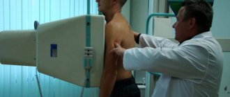
Fluorography is an x-ray examination that examines the ability of tissues to absorb radiation differently.
Many hardware diagnostic methods require preliminary preparation. Often, a person needs to follow a diet and nutrition regimen.
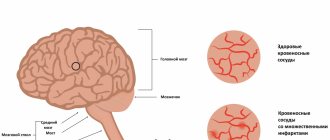
On this page, read what an MRI with contrast is and when this examination is prescribed.
What does fluorography reveal?
Fluorography is necessary when detecting lung cancer and tuberculosis in the first stages. The photographs show:
- lungs;
- heart with arteries and veins;
- rib cage;
- collarbone;
- shoulder blades;
- vertebrae;
- bronchi.
The organs of the gastrointestinal tract are also viewed, but not their contents. Using fluorography you can detect:
- cystic formations;
- bronchitis;
- skeletal and muscular changes;
- foreign bodies;
- hernias;
- inflammation;
- pneumosclerosis;
- tumors;
- pneumonia;
- bronchial obstruction;
- fibrosis;
- pneumothorax;
- ulcers;
- pathologies of the cardiovascular system;
- pleural changes (fusion or layering);
- the presence of infiltrates or gases in the lungs.
During the examination, pathological cavities are clearly visible - abscesses, calcific areas, echinococcal cysts, places of accumulation of pus. As can be seen from the above, food in the intestines will not interfere with the examination at all.
When preparation is needed
The results of fluorography can only be seriously affected by the presence of foreign objects in the sensor area of the device. Foreign objects can create an extra shadow on the final image, which can lead to an erroneous diagnosis or distract from the actual pathology.
To get accurate and reliable fluorographic images, follow these tips:
- Remove jewelry from your neck. Jewelry made of any metal will leave a shadow in the photo. To avoid such misunderstandings, remove the jewelry during fluorography.
- Remove all outer clothing and bra. The bra has wires and metal clasps that can interfere with getting an accurate shot.
- Pull your hair into a bun. If you have hair below your shoulders, take care of a hair clip or elastic band; long hair can also distort the picture of the study.
- Follow all instructions from the specialist. Try to follow the doctor’s instructions flawlessly - hold your breath for the strictly specified time, do not move during the photo.
Before you sign up for fluorography, check what type of device is installed in the clinic. Studies using a digital X-ray machine expose the patient to radiation in a dosage of 0.02-0.05 mSV. The film device gives a radiation dose equal to 0.1-0.3 mSV, therefore, examination with a digital X-ray machine is safer.
Indications for examination
Fluorography must be performed annually as a preventative measure. An examination is necessary if there is a suspicion of pathologies that can be identified.
In addition, fluorography is necessary when applying for a job, chest pain, if close relatives have had cancer, or the patient himself is a long-time smoker.
An examination is also necessary if a person has been in contact with a patient with tuberculosis.
Necessity of application
The main purpose of fluorography is to detect pulmonary tuberculosis in the early stages. X-ray allows you to find lesions even when wheezing is not yet observed when listening to the lungs. If a person is suspected of having tuberculosis, he will need to undergo a more thorough examination.
Most often, the study reveals the following problems:
- tuberculosis;
- areas with an inflammatory process;
- tumors;
- pneumofibrosis (connective tissue);
- presence of foreign objects
- abscess
- cystic cavities
- atelectasis
- parasitic lesions of the lungs
- hernia
- hydrothorax
- pneumothorax, etc.
Frequency of procedure
Fluorography in the clinic is usually performed once a year, unless there is a reason for it to be done more frequently. The radiation received during the procedure is safe and cannot cause harm to the body. If there are people with tuberculosis in a person’s close circle, it is worth undergoing fluorography more often, usually in such cases this is done 2 times a year. This makes it possible to detect the disease at the moment of its occurrence.
If certain suspicions and symptoms are present, then you should immediately undergo radiography, because fluorography has less specificity. Typically, fluorography is used to confirm the absence of disease. The procedure is not performed for pregnant women and children under 14 years of age (without clinical indications); if problems arise, a more accurate study is used. Women during the feeding period should also refuse fluorography; their use should be consulted with a specialist.
Contraindications for examination
Fluorography is not performed for children under 15 years of age. In this case, diagnosis is replaced by a more gentle method - mainly ultrasound examination, since X-ray irradiation negatively affects the health of a fragile organism.
Also, fluorography is not performed on pregnant women. In exceptional cases, it is possible to conduct such an examination only after the 25th week of pregnancy. In this case, the reasons for performing fluorography must be very compelling or vital.
Is it possible to eat and drink before fluorography?
The stomach not only does not participate in the diagnosis of the thoracic region, but also does not interfere, since it is separated from the scanning area by a diaphragm. Therefore, you can eat before fluorography.
Restrictions on nutrition are imposed by examination methods that require the administration of contrast fluid for a clearer picture. For some types of X-rays and CT scans, a person will have to stop eating and drinking for 8 hours before the procedure.
Is it done on an empty stomach or not?
The diametrically opposite question often arises about whether fluorography is done on an empty stomach. There is no need to worry: the condition of the stomach does not affect the examination results in any way. Whether or not you undergo the procedure on an empty stomach does not matter in this case.

Does food affect results?
As can be seen from the contraindications, they also do not indicate that the procedure is prohibited on a full stomach. Therefore, no diet is required. Before fluorography, it is not forbidden to consume food and water. The method is based on tissue permeability, so food has no effect on the results.
In addition, in the photographs, shadows from different organs overlap each other. As a result, some of them are not identified. For example, the esophagus is located behind the mediastinum. For this reason, it (and even more so its contents) is not displayed at all in the pictures.
The stomach is located under the diaphragm, is not visualized during scanning and does not interfere with the examination when diagnosing the chest. Many studies are carried out using contrast solutions.
In this case, it is necessary to first follow a diet or fast for several hours before the procedure. Since fluorography is not done with contrast, there is no need to change your usual diet or diet.
Fluorography
Fluorography is an x-ray examination that involves photographing a visible image on a fluorescent screen, which is formed as a result of the passage of x-rays through the body and uneven absorption by organs and tissues of the body. In a digital fluorograph, unlike screen-film technology, the energy of X-ray photons passing through the human body is perceived by one of the systems for digitizing the image.
Traditional film fluorography provides a reduced image of the object. There are small-frame (for example, 24×24 mm or 35×35 mm) and large-frame (in particular, 70×70 mm or 100×100 mm) techniques. The latter approaches radiography in diagnostic capabilities.
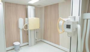
Digital fluorography provides a full-size image of the study object on the screen. Optical resolution and image size, however, depend on the type of detector and digital system. the AXIOM Iconos R200 at our disposal with the FLUOROSPOT COMPACT digital system produces images up to 240x300 mm in size.
The most common diagnostic method using the principle of fluorography is fluorography of the chest organs, which is used primarily to identify hidden diseases of the chest organs (respiratory tuberculosis, pneumoconiosis, nonspecific inflammatory diseases and tumors of the lungs and mediastinum, pleural lesions). Based on fluorographic studies, individuals with suspected diseases of the chest organs are selected. Patients who have changes in the lungs or heart undergo x-rays.
Digital fluorography has become a huge step forward compared to the classical film technique, and a logical continuation of the further use of all the advantages of classical fluorography over other x-ray diagnostic methods.
The main advantages of digital fluorographs are:
- high quality and informative images,
- minimal radiation exposure during examination,
- ease of archiving and retrieving data,
- no x-ray film or chemicals.
- High throughput of the equipment
- Low cost per examination
As for information content, digital fluorography has indeed become comparable to survey radiographic photographs. This means that the ability to detect pathology during mass examinations increases sharply. Doctors and researchers give different figures on this topic, but they all agree that, in comparison with traditional fluorography, even the simplest digital technology can increase the recognition of diseases by at least 15%.
Dose loads from different devices differ, but here we can confidently talk about at least a 5-fold reduction in dose from the simplest digital fluorographs in comparison with the best examples of traditional technology. Such a small dose makes it possible to expand the age group for X-ray prophylaxis of tuberculosis. Some devices (for example, with a linear silicon detector) are capable of delivering a radiation dose comparable to only a fraction of the human dose in just one day from natural radiation sources! To make it clear what level of radiation we are talking about, a little simple mathematics. The background radiation level in “clean” areas is 10-15 microroentgens per hour. This means that in 10 hours of a person’s life, about 100-150 microR will be formed. So, this is exactly the dose (150 µR) that is generated by the lowest dose digital fluorographs. For comparison, we note that a good film apparatus gives about 25,000 μR. As they say, comments are unnecessary.
Indications for fluorography
Currently, it is recognized that it is necessary for every healthy person to undergo examination once a year. Naturally, if complaints of cough, shortness of breath, or weakness appear, the patient should be examined immediately.
There are no absolute contraindications; severe general condition or other reasons that do not allow the patient to stand, severe shortness of breath, or pregnancy are considered relative. In any of these situations, only your doctor can decide whether a test is needed.
No special preparation is required. Perhaps we should only remind you of one thing. The results of the study are influenced by the position of the diaphragm, a thin muscle that separates the chest from the abdominal cavity. In this regard, the main unfavorable factor is high pressure in the abdominal cavity. Remember how you feel after going to a party where there was no opportunity to dance, and the table, according to Russian custom, was laden with all sorts of goodies. Surely, when you returned home, it was hard to breathe. Therefore, on the day of the study, you should limit yourself to a light breakfast. And if you suffer from constipation, then the night before it makes sense to take a mild laxative (regulax, bisacodyl, senade).
Diagnostic description
The fluorographic research technique is based on attenuation of X-rays when passing through tissue, and recording the result obtained on a CCD matrix or on a semiconductor linear detector.
Since the tissues of the formations have different composition and density, the radiation is slowed down and scattered, and an image of the organs appears on the sensitive surface in the form of shadows of varying intensity. This diagnostic method differs from radiography only in the size of the image - a reduced image is formed, which shows:
- diaphragm dome;
- the heart and adjacent blood vessels;
- bronchial tree (increased pattern);
- lungs (changes in the structure of lung tissue, the presence of foci of tuberculosis, neoplasms);
- pleural cavity (presence of fluid, air);
- mediastinal area (the space between the right and left lungs);
- part of the skeleton bones (ribs, vertebrae, collarbones, shoulder blades).
Fluorography reveals changes that occur in people with a long history of smoking. Such patients are at risk for developing lung diseases; they are recommended to undergo annual fluorography.
Other points of preparation for the procedure
There are no special preparatory measures for undergoing diagnostics. On this day you can lead an active lifestyle: play sports, do physical labor, etc. The results of the examination will not be affected by women’s periods or the use of medications. Even a slight cough will not be an obstacle.
However, despite the absence of special requirements for undergoing fluorography, contraindications still exist.
- Children under 15 years of age do not undergo fluorography, replacing it with a more gentle ultrasound procedure. This restriction is necessary to preserve the health of the child. X-ray radiation has a detrimental effect on the child's body. The consequences can be dangerous: the occurrence of cancer, decreased immunity, changes in the general condition of the body for the worse.
- Another restriction applies to pregnant women. Almost all types of diagnostics using x-rays are prohibited during pregnancy: cystography, x-rays, fluorography. Especially if you need to carry them out for preventive purposes. The exception is fluorography in late pregnancy, after approximately the 25th week. The reasons for the procedure must be compelling when the risk of fetal harm due to the disease outweighs the risk of radiation exposure. The procedure in this case will be carried out only as prescribed by a doctor.
People with poor health after serious illness, as well as patients with severe cough and shortness of breath and people who are unable to hold their body in an upright position are exempt from undergoing fluorography.

Is smoking and drinking alcohol allowed?
Experience shows that a cigarette smoked an hour or half an hour before the procedure will not significantly affect the diagnosis. The picture will always show an experienced smoker in whose lungs pathological changes have already occurred. Until then, the fluorographic apparatus will not determine whether a person smokes or not.
Alcohol will also not affect the test results. Here, the social factor is more likely to play a role: hospital staff and other patients may insist that an intoxicated visitor leave the office and even the hospital building.
Thus, before fluorography you can eat, drink, smoke and drink alcohol in moderation.
However, for the same social reasons, on the day of fluorography, it is better not to eat dishes with large amounts of onions and garlic. This behavior will demonstrate respect for the medical staff and will make the medical procedure mutually enjoyable.
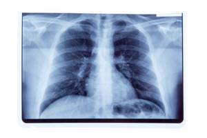
What can you see on fluorography?
In a fluorographic image, the doctor sees a black and white image of the chest and internal organs. The organs of the chest absorb radiation differently, so the picture on the image is heterogeneous. The heart and bronchi appear as light spots; a white shadow from the heart is visible in the center of the fluorogram. Hollow structures (lungs) appear dark, while dense ribs and clavicles appear light. If the lungs are healthy, their tissue looks homogeneous.
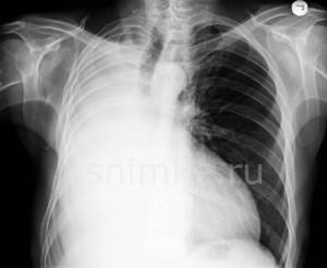
What can a doctor detect?
- An increased heart size may indicate heart failure or cardiomyopathy.
- The accumulation of fluid (effusion, blood) is an important symptom that requires further diagnosis.
- Bone fractures.
- Tumors.
- Granulomas may indicate tuberculosis.
- Pulmonary edema.
- Other pathologies.
Chest fluorography should be distinguished from x-rays. The last diagnostic method is used according to the doctor’s indications.
Both fluorographic and x-ray examinations of the lungs are a simple, fast and effective diagnostic method that has been helping doctors prevent and diagnose diseases for several decades.
Why do fluorography
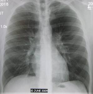
A fluorogram may be normal - in a healthy person who has not previously suffered from any lung diseases. The normal fluorography is transparent lung fields without any shadows.
There is a pathological fluorogram - when changes are detected that indicate a previous or existing disease: • various benign and malignant tumors; • focus of inflammation; • fibrous formation; • pulmonary tuberculosis; • foreign bodies; • necrosis of tissues of internal organs; • abscesses.
Of course, fluorography of the lungs will not provide the most accurate information about the pathology, but it allows timely diagnosis of changes in the lung tissues, the appearance of fluid and neoplasms. Until now, the procedure is the most popular in the preventive diagnosis of the lungs.