the whole list
Modern diagnostic techniques make it possible to identify the earliest pathological changes in the functioning of human organs and systems. It is difficult to imagine the development of medicine without the use of magnetic resonance imaging or computed tomography - non-invasive methods for diagnosing diseases. But, faced with the need to choose between one type of examination and another, many patients begin to wonder how CT differs from MRI, and which method is best to prefer depending on their health condition.
A person far from medicine may mistakenly think that these methods are identical. But this is far from true. They are united by the word “tomography”, which means obtaining layer-by-layer sections of organs and tissues, the image of which, after scanning, is sent to a personal computer and subjected to interpretation. But there is still a difference between CT and MRI, and it is quite significant.
What information do they give?
CT involves multidirectional fan-shaped movement of x-rays, which are then converted into electrical signals and processed into an image by a computer program.
During an MRI examination, a strong magnetic field is created that aligns hydrogen atoms in the patient's body in a certain direction. Since the type of human tissue is different, for example, muscle and bone tissue differ in density and, accordingly, in the number of atoms, being in the magnetic field of the device, signals of different intensities are obtained. Using computer programs, these signals are converted into images and the areas of the body being examined can be seen in the photographs.
Therefore, the physical state of organs is shown by computed tomography, the chemical structure of body tissues is shown by magnetic resonance imaging.
What is CT or computed tomography
Computed tomography (CT) or multislice computed tomography (MSCT) is a modern diagnostic method that consists of a layer-by-layer examination of the human body using X-rays to obtain a detailed image of internal organs and structures.
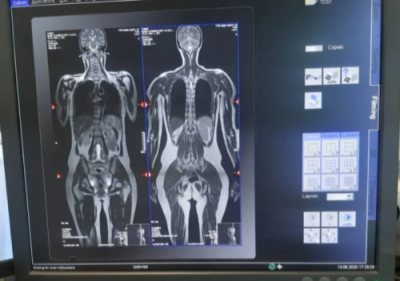
CT imaging is based on the varying densities of organs and tissues through which the X-rays pass. During the examination, an X-ray tube with a narrow beam of X-rays rotates around the patient. At the same time, many sensors record the slightest changes in density. Information from the sensors is processed by a powerful computer, after which it is sent to the monitor in the form of successive longitudinal and transverse sections (“tomography” literally means “image of a slice”). The thinner the section, the more accurate the diagnosis.
The University Clinical Hospital No. 2 has modern equipment - a high-class computed tomograph, which allows obtaining high-quality images with minimal radiation exposure.
Today, CT is one of the main diagnostic methods and is widely used both for making a diagnosis and for clarifying it. One of the main advantages of computed tomography is the speed of the study and the ability to scan several organs simultaneously. Modern tomographs can significantly reduce the duration of the examination and reduce patient exposure to a minimum, while obtaining hundreds of sections up to 0.5 mm thick. Thanks to high-precision three-dimensional projections, which make it possible to examine the position, shape, size and structure of various organs and structures of the body, the results obtained from CT examination are the key to success in making an accurate diagnosis, selecting the most effective treatment and planning surgery if necessary.
A significant advantage of this method is the ability to examine patients with pacemakers, artificial heart valves, metal clips in the body, etc., that is, those for whom MRI is contraindicated.
CT scanning is used to examine almost all parts of the body and organs: chest, abdomen, pelvis, limbs, liver, pancreas, intestines, kidneys and adrenal glands, bladder, lungs, heart, as well as blood vessels, bones and spine.
The range of diseases for which this research is necessary is extremely large. In each individual case, the need and feasibility of this study is assessed by your attending physician.
Advantages
- CT diagnostics provide more complete and accurate information about hard formations, bone density, and damage to lymph nodes in the body;
- MRI diagnostics will provide more information about the condition in the soft tissues of the body and the presence of an inflammatory process.
Please note that before making a final decision on which diagnostic method to use, be sure to consult with your doctor. There are situations when it is necessary to conduct both studies.
Second, conducting research on devices with low power
It is imperative to specify the number of slices for the CT machine. “To conduct modern research, the number of CT slices must be at least 64, the power of the MRI machine must be at least 1.5 Tesla. If the clinic staff rolls their eyes or refuses to name the parameters of the device, it is better to look for another place for diagnostics. Unfortunately, situations are still common when CT is performed on devices with a number of slices of 16.32. Using software and firmware, it is possible to obtain a number of slices close to 64, but the image quality will be quite low. The maximum quality is provided by a 128-slice device. It is mainly used for diagnostics with contrast and provides the best sharpness, but if the study is standard, you can get by with a 64-slice device,” advises Serebryansky.
MRI also has its own rules: “For MRI, one of the important parameters is power. It should be from 1.5 Tesla. Some institutions do not even disdain devices that are not intended for medical purposes. For example, they use an old, decommissioned device with a power of 0.35 Tesla, which was previously used in some zoo in Europe and miraculously then ended up in Russia. Special software allows you to increase the power, but it will still be insufficient to diagnose a person, no more than 1 Tesla.”
Indications for research
Magnetic resonance imaging indications:
- suspicion of a tumor in the internal organs (diagnosis can determine the type of tumor when it comes to benign and malignant formations);
- abnormal development of organs;
- presence of stones (abdominal organs);
- preparation for surgery (gathering diagnostic information).
Computed tomography is shown:
- in preparation for surgery;
- the patient is in a state of acute pain with a difficult diagnosis;
- with obstructive jaundice and sudden changes in weight;
- presence of acute injuries;
- suspicion of formations in internal organs.
Watch the video, after which you will have a better understanding of what CT and MRI diagnostics are.
How the research works
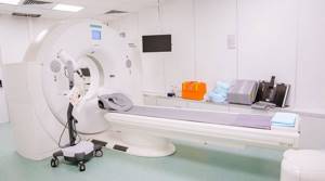
We ask you to come to the radiology department (9th floor) 10-15 minutes before the scheduled start time of the study and, having contacted the office, inform the staff of your arrival. Given the possibility of emergency research, there is a possibility of waiting for the procedure for some time. Experienced doctors and x-ray technicians will do everything to make you feel comfortable and safe throughout the entire examination.
Computed tomography is an absolutely painless procedure that takes no more than 30 minutes (including changing clothes and, if necessary, intravenous contrast). The scanning itself lasts several minutes. If you need a CT scan with contrast, you will first be invited to the procedure room, and a nurse will install an intravenous catheter for you to administer a contrast agent during the examination.
Before the examination, you will be asked to remove all jewelry and metal objects that fall into the scanning area; the x-ray technician who will carry out the procedure will explain to you the rules of conduct during the examination and will help you sit comfortably on the tomograph table.
If the study is carried out using a contrast agent, it can be introduced into the patient’s body in various ways depending on the purpose of the study: intravenously - in CT scans of the chest, abdomen and pelvis.
- orally - for some abdominal examinations, the contrast agent must be drunk
- through a special catheter into the bladder or intestines
During the procedure, the patient lies on a special table connected to a CT scanner, which is a large ring-shaped machine. The scanner rotates around the area of the patient's body being examined, taking layer-by-layer images of the corresponding organ. A faint hum or clicking sound may be heard.
Try not to move during the procedure, as moving your body may reduce the quality of the image.
Given that a CT scanner emits X-rays, the machine is usually located in a special shielded (protected) room. The device is controlled automatically from the adjacent office, which houses the tomograph’s computer unit, monitors and equipment for monitoring the patient’s condition. The doctor performing the examination sees and hears you. You will be able to talk to him through a special intercom. The specialist may ask you to hold your breath for a few seconds to get a clearer picture.
Based on the images obtained, the doctor at the radiology department gives a medical opinion. In addition, your doctor or surgeon can comment on the results of the study.
Cons of diagnostics
Computed tomography - disadvantages:
- mainly provides information about the structure of organs, and not about their functional state;
- X-rays are present, although less than regular X-rays;
- there are contraindications for pregnant women and children;
- Frequent diagnostics of this type are not recommended.
Magnetic resonance imaging cons:
- contraindications for allergies to medications and medications;
- cannot be done if there are metal products in the patient’s body (implants, pacemakers, vascular clips);
- if surgery on blood vessels (metal mesh) has already been performed before;
- the presence of an intrauterine device;
- in acute forms of exacerbation of the disease (kidney diseases, sickle cell anemia - the use of contrast is contraindicated).
- Available: doctor’s appointment from 1500 rubles
- Convenient: open daily from 8:00 to 21:00
- Quickly: we will carry out all the diagnostics at the first appointment
- Complete: has all the necessary equipment
What sensations does the patient experience during the CT scan procedure?
The procedure itself is absolutely painless. Some discomfort may be caused by the hard surface of the table, the inability to move, and the temperature in the CT room (the room may be cool).
Some patients feel nervous while inside the scanner. If contrast material enters a vein, you may experience a feeling of warmth, heat, or a metallic taste in your mouth. Sometimes patients experience nausea or headache. Be sure to tell your doctor or x-ray technician how you feel.
How does CT differ from MRI?
Each method is valuable, but has its own advantages.
Advantages and harms of CT:
- sufficient resolution;
- the ability to assess the relationship between the structures of the area under study;
- absence of areas closed to research;
- the radiation dose is lower than with fluorography;
- the ability to study in the presence of electrical and metal devices in the body;
- lower cost.
The harm of the method lies in the high risk of developing malignant neoplasms due to the harsh effects of X-ray radiation. Therefore, CT cannot be used repeatedly.
Advantages of MRI
The method complements information about problems detected by CT, ultrasound or x-ray. It is also characterized by a number of advantages:
- absence of dangerous x-ray radiation;
- the probability of error is close to zero;
- information is stored on a computer;
- three-dimensionality of the image;
- painlessness of the procedure;
- possibility of repeated use;
- safety for children and pregnant women (after 12 weeks);
- accuracy of diagnosing vertebral hernia;
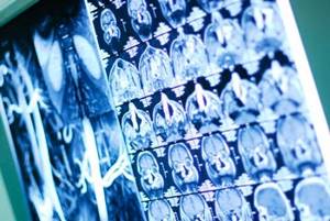
- the most informative method for pathologies of the nervous system;
- high research accuracy.
The cost of MRI depends on the quality of the images, and this depends on the power of the installation.
What is examined on an MRI
The main task of using magnetic resonance is to examine soft tissue internal organs:
- brain and spinal cord;
- articular areas;
- abdominal and retroperitoneal region;
- pelvis;
- soft tissues of the neck and limbs.
The images clearly display any degree of inflammation, tumors, ischemia, degenerative changes, aseptic and septic necrosis, hematomas and other pathologies developing in loose structures.
Fourth - the presence of unnecessary manipulations
Usually only a CT or MRI scan is sufficient to determine the problem. Unnecessary manipulations are required only in rare cases and only on the recommendation of a doctor. “Seeing the problem that a patient comes with, a good specialist can suggest expanding the area of research. This should not be taken with skepticism. Let’s say a patient with breast cancer comes in for a chest CT scan. In this case, examining the chest alone will not be enough; it is necessary to include both the breast area and the area down to the lower jaw. It is also necessary to understand where cancer usually metastasizes, and, accordingly, look at these areas to find out what other organs and systems are affected,” says Oleg Serebryansky.