August 11, 2016

Some of the myths about mammograms can have a serious impact on your long-term health. Find out what's really behind this vital research.
Think it's no big deal if you miss your annual mammogram this year? Or are you concerned that too much radiation is used during mammograms?
Annual mammograms for women 40 years of age and older help detect breast cancer at an early stage, which allows for less aggressive treatment in the future and ensures a higher survival rate.
Myth #2: Mammograms expose me to an unsafe dose of radiation.
Fact: “Mammograms do use radiation, but in very small amounts and according to medical protocols,” says Dr. Cakmakci. Because mammography is a diagnostic tool, the process is strictly regulated by established standards and is monitored by external regulatory organizations. Mammograms are safe as long as you get them at appropriate, regulatory-certified facilities. There is a constant background radiation in the world, to which we are exposed every day. The radiation dose from a mammogram is about two months of background radiation for the average woman.
Is it harmful to have a mammogram?
Many women are afraid whether mammography is harmful, because this study uses x-rays. Of particular concern is the presence of existing tumors - would mammography be dangerous in this case? The answer is more than clear: the dose of X-ray radiation received during the examination does not cause any concern at all. It does not exceed acceptable limits and does not have sufficient carcinogenic activity. As for the presence of neoplasms, mammography is used, among other things, to monitor their activity and growth. Before prescribing a study, the attending physician will definitely take into account all risk factors and refer you to the study that will be informative in a particular case.
For those who are terribly concerned about exposure to X-rays, there is another option - you can undergo a mammogram using a magnetic resonance imaging scanner. This test is called MR mammography or MRI of the mammary glands. MRI (magnetic resonance imaging) does not use X-rays; the method is based on the principle of interaction of magnetic fields. This procedure is absolutely safe for the patient and has virtually no contraindications (including age-related ones).
MRI of the mammary glands is recommended to be performed with the introduction of a contrast agent, which allows you to examine suspicious areas and detect a malignant formation even at the earliest stages.
Myth #3: 3-D mammography is the same as traditional mammography.
Fact: Three-dimensional mammography, or tomosynthesis, is the most advanced screening method and diagnostic tool for the early diagnosis of breast cancer. Compared to standard 2-D mammography, 3-D mammography displays more images of the breast and thin sections of breast tissue. “3-D mammography gives us greater clarity and the ability to tell the difference between normal tissue and cancerous tissue,” says Dr. Cakmakci. “With 3-D mammography, data is shown at 40 percent magnification, allowing detection of early stages of cancer and reducing false diagnoses (overdiagnosis) or unnecessary repeat screenings by 40 percent.”
Is mammography harmful for women under 35?
Typically, age 35 years is a relative contraindication to mammography. If complaints arise, the patient is usually recommended alternative research methods. But if there is a clear suspicion of oncology, mammography may be prescribed for clarifying diagnostics. For the most part, mammograms are not done at a young age, not because they are dangerous, but because of the high density of breast tissue. And this can hinder objective research. Therefore, to the question: is mammography harmful for young women, we can answer that it is rather insufficiently informative than dangerous.
Mammography to protect women's breast health
The likelihood of breast cancer is closely related to age: from 1 in 72 for women 40-50 years old to 1 in 29 for women over 70. At the same time, the mortality rate of women aged 35-64 years from breast cancer in Russia reaches 50% of cases. Each of these women could have lived another 20 years if the pathology had been identified earlier. There are no methods to identify the first tumor cells, but there is a test aimed at identifying not yet palpable breast tumors - mammography.
Early detection of breast cancer increases the chances of successful treatment, saving the lives of up to 97% of women.
Mammography is a method for early detection of breast cancer. The main goal of this study is to identify a malignant tumor as early as possible, stop the disease and, if possible, save the breast. To do this, you need to undergo regular examinations.
When to do it
The optimal time is days 5-11 of the monthly cycle. At this time, the mammary glands experience minimal hormonal influence. In the second phase of the cycle, the glands begin to swell. As menstruation approaches, the amount of fluid increases, the breasts swell and become denser. During this period, the number of false positive results increases, which entails unnecessary additional research and puts psychological pressure. During menstruation, mammography cannot be done; the pain of the procedure increases, and the information content decreases.
How to prepare
Mammography is based on X-rays. It implies some rules for preparing for research:
- A week before the procedure, it is advisable to give up coffee, as it helps to thicken the breast tissue, which makes it difficult to decipher the images.
- Before the procedure, you should not use deodorants or other cosmetics, as they can create suspicious shadows on the pictures, which will require unnecessary additional examinations. For the same reason, all moles and other formations on the skin should be covered.
- Take your previous research results with you.
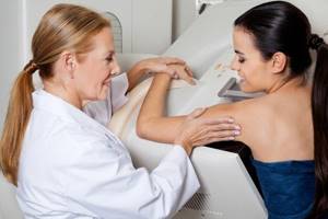
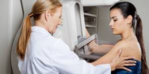
How is mammography performed?
One breast is placed on the mammography platform. The chest is compressed and an x-ray beam is passed through it. Then the mammograph is tilted, the woman changes position and another picture is taken - in the lateral direction. The manipulations are repeated for the second mammary gland. As a result, the doctor receives 2 images of each breast, in direct and oblique projections.
What will show
After studying the images, the doctor will indicate the breast density (the lower it is, the more informative the study) and assign a category from 0 to 5 according to the standardized Bi-RADS system.
- 0: the picture is not clear, additional examinations are necessary - ultrasound or repeated images with magnification.
- 1: normal, nothing suspicious.
- 2: benign lesions were detected, for example, fibroadenoma, cysts, calcifications...
- 3: a most likely benign formation was discovered; a repeat examination after 6 months is recommended for confirmation.
- 4: A suspicious malignancy has been identified and a referral for a biopsy will be given for confirmation.
- 5: with a high probability cancer is detected, a referral will be given to an oncology facility, where the necessary additional examinations will be carried out.
At the Toksovskaya MB you can get a mammogram for free as part of a medical examination or for a fee.
The main weapon against breast cancer is your vigilance! Take care of yourself!
Based on materials from the website MammographyAll.ru
Preparing for the examination
There is no specific preparation for mammography. The study is carried out on days 6-12 of the monthly cycle, during menopause and for menstrual dysfunction - on any day. During your consultation, be sure to tell your doctor:
- About all alarming symptoms.
- Past illnesses, surgeries.
- Having relatives diagnosed with breast cancer.
The doctor needs such information for the most correct interpretation of the nature of the detected violations. On the day of the procedure, it is not recommended to apply cosmetic products to the skin of the chest and armpits, since the components included in their structure may be erroneously visualized in the photographs, negatively affecting the reliability of the results.
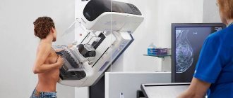
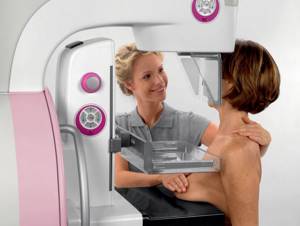
How to do a mammogram
During the examination, the patient's breast is placed between the mammography plates and compressed. In this way, the breast tissue is evenly distributed over the surface of the device, and the picture produces the clearest possible image. If the patient is concerned about the condition of one breast, doctors will still examine both, because for a more accurate diagnosis it is necessary to compare the mammary glands in both images to complete the picture. The procedure itself takes about 15 minutes, during which time the mammologist can examine both breasts.
Which diagnostics to choose
Technology does not stand still, and today medical centers use various types of mammographs. For example, in the middle of the last century, film diagnostics were widely used, today digital mammography is popular. Its results are more accurate, and if necessary, the doctor can change the contrast of the images and obtain a more detailed analysis. Also, during digital diagnostics, the resulting images are recorded in the computer’s memory, which subsequently makes it possible to send them by e-mail to any specialist and patient. There is also mammography with tomosynthesis, which takes a three-dimensional scan of the patient's breast. The result is a whole series of photographs in which the slightest tissue compaction is visible.
This technology is rightfully considered a breakthrough in the study of female breast pathologies.
Indications for the study
Mammography is performed for diagnostic and preventive purposes, for the early detection of breast diseases. Women over 35 years of age are recommended to undergo a similar study every two years; after 50 years of age, an annual examination is required. Direct indications for mammography are the following symptoms:
- Asymmetry, deformation, change in breast shape.
- Lumps in the mammary gland.
- Changing the shape of the nipple (retraction).
- Breast tenderness (unrelated to menstruation).
- The appearance of any discharge from the nipples (outside of lactation and pregnancy).
- Breast injuries.
- Enlarged axillary lymph nodes.
- Changes in the skin of the breast - the appearance of peeling, redness.
The innovative equipment used at GMS Hospital for mammography is equipped with a program for automatically identifying the densest areas of the mammary gland and selecting optimal exposure parameters, which significantly increases the information content of the study, taking diagnostic capabilities to a new level.
What is mammography?
What is mammography? This is a procedure that takes place using special x-ray equipment - a mammograph. As a final result, the patient receives an image of the breast, which shows the vessels and milk ducts. If signs of cancer are detected, the screening device will detect and record this. The device identifies the size and location of tumors, if any, regardless of their nature - benign or malignant. For example, a doctor can use a device to see the presence of cysts in the mammary gland, their number and size. Or find fibroadenoma - a benign formation caused by the rapid growth of connective tissue. Another common problem in women is the deposition of calcium salts in the mammary gland, hence the name - calcifications. These accumulations cannot be pulped, but they can be easily detected using mammography.
Many women are interested in which day of the cycle to do a mammogram. The best time is from the 5th to the 12th day of menstruation, since it is at this time that the mammary gland is the softest. When its structure is rigid and dense, as in the premenstrual period, the examination may be inaccurate, and the process itself may be painful for the patient. To avoid having to undergo the study again, it is better to first correctly calculate the day of your cycle. No other preparatory measures are required from the woman.