| Ultrasound during pregnancy in Moscow clinics. Ultrasound at home. | Reception is strictly by appointment, make an appointment by phone: +7 | Prices for services | Reviews about the clinic |
Determining the sex of the child using ultrasound is difficult during multiple pregnancies. A singleton pregnancy allows you to determine the sex of the future heir by obvious signs - the presence or absence of a penis.
Determining the sex of the fetus can also be difficult in this case if it is carried out in the early stages and using outdated ultrasound machines.
Make an appointment with a gynecologist by phone or by filling out the online form
| Select a clinic | Discharge in pregnant women | Gynecologist | HCG tests |
Answers to frequently asked questions on ultrasound:
- Is ultrasound harmful?
- Where can I get an ultrasound done?
- How to properly prepare for an ultrasound?
- What organs are examined by ultrasound?
- What diseases are ultrasound used to diagnose?
- Is it possible to do an ultrasound at home?
- What symptoms require an ultrasound?
- How often should an ultrasound be done?
- Do they do ultrasounds for children in your clinic?
- Can I get the transcript and ultrasound images in hand?
- What organs are examined during an abdominal ultrasound?
- What organs are examined during a pelvic ultrasound?
- Ultrasound in Moscow
- Paid ultrasound
- What other diagnostic methods are used in your clinic?
- How to sign up for an ultrasound?
When are signs of a baby’s gender determined by ultrasound?

In a girl, the ultrasound sensor detects the presence of the labia, and in a boy, the scrotum and penis. Examination of any part of the fetus depends on its position, the amount of amniotic fluid, as well as the thickness of the pregnant woman’s abdominal wall. The quality of the equipment and the experience of the ultrasound specialist are also important.
How accurately can you determine what gender the baby will be in the first trimester of pregnancy? You need to know that the genitals begin to form approximately from the sixth week of pregnancy, when a small bulge is formed - the genital tubercle. Until the ninth week, the genitals of a boy and a girl look the same. There are no noticeable differences, since the genital tubercle and genital folds are externally surrounded by labioscrotal tubercles.
Main signs and timing for determining gender
Starting from the 11th week, boys:
- the genital tubercle forms the penis, but its formation is not yet completed;
- the scrotum is formed from the labioscrotal tubercles;
- At this time, the testicles are located in the abdomen, and they descend into the scrotum.

At 11 weeks, the ultrasound doctor can only guess the gender of the baby, but the error is 50%. External sexual characteristics can be accurately determined 5-6 weeks after the formation of the genital organs.
No matter how much you would like it, at the first ultrasound, which is performed at 12-13 weeks, it is not possible to determine the sex of the child. It is possible to determine what gender the child will be starting from the 15th week, but even at this time it is not always possible to unmistakably discern any signs of gender and here’s why.
- Swelling of the labia majora in a girl, which occurs in the early stages, is mistaken for the male genital organ.
- Umbilical cord loops or fetal fingers may be mistaken for the penis.
- Very often, the tightly compressed legs of the fetus hide the male genitals, and the boy is mistaken for a girl.
However, experienced ultrasound specialists can more accurately determine the sex of the child at 14 weeks by measuring the angle formed between the genital tubercle and the back of the fetus. In boys, the genital tubercle forms an angle of 300 or more, in girls this angle is less than 300.
Why do errors occur?
- Ultrasound is a method that allows you to determine signs of gender if the genitals are available for examination.
- A baby swimming in the waters often changes his position, turns sideways, turns his back, he can close his legs. Such mobility can cause inaccuracies in determining the sex of the child.
- Features of the shape and size of the genital organs may be misinterpreted. The labia are often mistaken for the penis before 21 weeks.
- The presence of two or more babies in the uterus often makes it impossible to determine their gender. The tightness in the uterine cavity, mobility and frequent contact with body parts of their “neighbors” do not allow babies to settle in a position convenient for the diagnostician.
- Errors or difficulties in determining the sex of a child in later stages are due to the large size of the fetus and the rounded shape of the limbs, which may obscure the genitals from view.
Useful information on the topic:
- Calling a gynecologist to your home
- Medical abortion
- Consultation with a gynecologist
- Surgical abortion
- HCG tests
- Screening during pregnancy
- Intrauterine device
- Diagnosis of sexually transmitted diseases
- Fetal ultrasound
- Pelvic ultrasound
- False pregnancy
- Transvaginal ultrasound
- Abortion pills
- Discharge in women
How long does it take to determine the sex of a child?
Do you want to know who you are having, a boy or a girl? It is better to go to a specialist no earlier than your pregnancy is approaching 18-19 weeks. There is an opinion that it is easier for boys to identify during this period, but to “follow” a girl no earlier than 20-25 weeks.
The optimal period for determining the sex of the unborn child is the 20-24th week of pregnancy. At this time, gender differences are clearly distinguishable, the fetus is mobile, so there is a possibility that during the ultrasound it will take the desired position and its gender will be clearly visible.
The most important and interesting news about infertility treatment and IVF is now in our Telegram channel @probirka_forum Join us!
Features of determining the sex of the fetus
The structural features of the walls of the abdominal cavity, the thickness of the uterus, the quality of the placenta, polyhydramnios or oligohydramnios, and the location of the umbilical cord at the time of the study affect the reliability of ultrasound results.
If a family expecting a child has a hereditary disease transmitted through the male or female line, then determining the sex of the baby plays a decisive role in the question of whether to keep the child.
In these rare and sad cases, you can resort to examining the fibers of the placenta or amniotic fluid. This complex operation is performed by penetrating the uterine cavity and fetal membranes.
A woman who is in a tender and exciting period of life can choose a private clinic through the “Your Doctor” Help Desk and sign up for an ultrasound.
Useful information on the topic of gynecologist:
- How to get tested for gynecological diseases?
- What diseases does a gynecologist treat?
- What tests can be done by a gynecologist?
- How to prepare for an appointment with a gynecologist?
- Where to go with a gynecological problem?
- What symptoms should you consult a gynecologist for?
- How does an appointment with a gynecologist go?
- Female doctor
- Diagnosis of sexually transmitted infections in women
- Diagnosis of gynecological diseases
- Treatment of female diseases
- Pediatric gynecologist
- How is a gynecologist examined?
- Paid gynecologist
This article is posted for educational purposes only and does not constitute scientific material or professional medical advice.
Author:
Kasabov Alexander Vladimirovich Urologist, Candidate of Medical Sciences, Ultrasound Doctor
Back to section
How is an ultrasound examination performed?
Testicular ultrasound is a painless procedure. Pain in a man can only occur with acute orchiepididymitis. In this case, it is better to wait a few days and do a diagnostic procedure after the inflammatory process has passed. In other cases, no unpleasant sensations occur during ultrasound scanning. The patient is usually in a supine position. The doctor applies the gel to the area of study and to the sensor of the device for smoother gliding and greater information content. The gel is water-based and hypoallergenic, so there is no need to worry about an allergic reaction of the body. The sensor is installed on the organs, and with smooth movements in various directions, the diagnostician scans the area, receiving information in the form of an image on the monitor. The ultrasound procedure lasts 10-15 minutes. The examination time may increase if a tumor is suspected, since in this case additional examination of adjacent organs is carried out. The results are delivered to the patient 10-15 minutes after the examination.
Doctors' opinion
Doctors rightly consider attempts to guess the sex of a child by external signs to be ordinary fortune-telling. There is no evidence that the gender of the unborn child has such a significant impact on the appearance of the mother. Not a single study has found the presence of this dependence, and not a single scientist has been able to find a rationale for the signs.
Since there are only two options for the gender of the child, the effectiveness of this method is 50% - either they guess or they don’t. But the human psyche is designed in such a way that it more often remembers successful options, so it seems that there are more coincidences between what was predicted by signs and what was actually received than misses.
Features of scanning
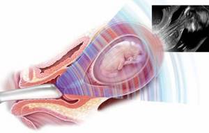
Transvaginal ultrasound during pregnancy
From about 12 weeks, the growing uterus begins to leave the pelvic area, rising above it. Therefore, at week 16, an ultrasound is performed through the abdominal wall. The doctor uses a special sensor that needs to be applied to the stomach. The diagnosis does not require any preparation . If it is very difficult to examine the baby, the specialist can replace the transabdominal sensor with a transvaginal one. In this case, the study involves inserting the device into the vagina. Women with threatened miscarriage or cervical insufficiency should not be subjected to such manipulations.
What happens to the fetus?
At 16 weeks of pregnancy, the unborn baby reaches the size of a ripe grapefruit. It already has distinguishable limbs, the legs become longer than the arms. The embryo's ears gradually move from the neck to their proper place. Changes also occur in the eyes; they take on a natural shape and position. The baby's eyelids are still closed, but he can clearly distinguish between light and dark, and his pupils move quickly. If you direct a bright beam at the belly, the baby will try to cover his face with his arms. The baby continues to move every minute: he kicks his legs, clenches his fists, rolls over. The mother may not feel this yet, but some women report the first movements at this time.
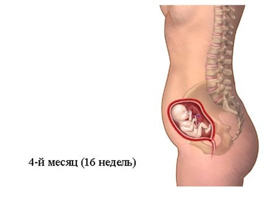
Internal organs continue to work at full capacity. Some of them change their function. The tiny heart beats almost twice as fast as mom’s: it pumps up to 28 liters of blood per day. The intestines and stomach work by digesting the amniotic fluid, releasing the necessary elements and nutrients. Urine is released hourly, but the baby is in no hurry to empty its bowels. Normally, the first feces will be released only after birth. The skin of the baby is still very thin and transparent; it has a reddish tint due to the fact that it is penetrated by small vessels.
Findings and next steps
After the 16th week ultrasound is done, the specialist will provide you with a protocol. It describes the results obtained during the study and measurements of the fetus. Normal values are also often indicated nearby. You shouldn’t compare them yourself, because you won’t be able to understand anything. A discrepancy in one or more values can be both normal and evidence of deviation. The ultrasound specialist's conclusion also cannot be considered a diagnosis .
The final verdict is made by the gynecologist, taking into account all the individual characteristics of the pregnant woman. Be sure to contact your obstetrician, who will help decipher the protocol.
Why is ultrasound necessary?
At week 16, an ultrasound scan is mandatory for those women who, for some reason, missed the first screening. The study allows us to identify embryo pathologies that are incompatible with life or require correction. Diagnostics is necessary to establish the exact duration of pregnancy and the expected date of birth.
Ultrasound allows you to find out the following:
- possible detachment of the ovum and hematoma formation;
- tone and hypertonicity of the uterine walls, distortion of the shape of the fertilized egg;
- the place where the placenta is attached, whether it is presenting;
- number of embryos and their gender;
- presence or absence of the corpus luteum;
- condition of the cervix, possible premature opening.
If the doctor recommends doing research at this time, then it is really necessary. Refusal to undergo an examination can have disastrous consequences.
Norms of values for a given period
As you understand, not all expectant mothers have an ultrasound scan at 16 weeks that shows the proper values. Some representatives of the fairer sex have deviations in one direction or another. Is this always a sign of serious pathology? Let's consider which values are considered satisfactory.
- The biparietal size of the baby's head is in the range from 3 to 3.7 centimeters.
- The fronto-occipital zone is 4.1-4.9 centimeters.
- The circumference of the head is 11.2-13.2 cm, and the tummy is 8.8-11.6 cm.
- The length of the femur is at least 1.7, but not more than 2.3 centimeters.
- The size of the shoulder and lower leg is approximately the same, it averages 1.5-2.1 centimeters.
- The forearm is about 1.5 centimeters (1.2 to 1.8).
- The baby's body weight can vary from 90 to 110 grams, and his height is already 10-12 centimeters.
- The amount of amniotic fluid is no more than 200, but not less than 70 milliliters.
- The placenta can have a size from 13 to 23 centimeters and can be located at any point of the reproductive organ.
An ultrasound performed at 16 weeks of pregnancy may show that the placenta is fully formed and the umbilical cord has three vessels. It is thanks to them that the baby receives the necessary nutrients, nutrients and vitamins.
Ultrasonography
If you are 16 weeks pregnant, you will most likely not be scheduled for a scan. Such a recommendation is given in connection with special circumstances. Diagnostics is indicated for women who are at risk of miscarriage. This condition manifests itself as pain in the body and lower back, and bloody discharge. Diagnostics are carried out to clarify the gestational age, determine the sex, and identify any pathologies in the child.

Transabdominal ultrasound
Many women worry that ultrasound can harm the fetus by irradiating it. But this is a strong misconception. During diagnosis, data is supplied to the monitor using ultrasonic waves repelled from the tissues of the uterus and embryo. Thus, no harmful radiation exposure occurs.
Diagnostics can be performed throughout pregnancy as many times as necessary. It is much more dangerous to miss the right moment and lose the opportunity to change or correct something.
The shape of the abdomen and the gender of the child
It is believed that by about the seventh month of pregnancy, the pregnant woman’s belly has already grown sufficiently and has taken its final shape, according to which, according to folk superstitions, one can determine which ribbon to prepare for discharge from the maternity hospital - pink or blue. Of course, you should not take the results of such “fortune telling” seriously, since there is no medical justification for it and there is not a single confirmation that the effectiveness of this determination is higher than 50%. But if the ultrasound has not yet given an answer as to who is inside, then it is difficult to resist such innocent entertainment.
Video: folk signs
First, it’s worth deciding on the terminology. So:
- high belly - the center of the circle formed by the protruding part of the abdomen is located high, closer to the ribs;
- low belly - it is shifted towards the hips;
- narrow belly - protrudes forward, the woman retains her waist;
- wide belly - spreads out to the sides, weakly protruding forward.
Signs of waiting for a girl
Folk signs say that a wide belly indicates that a girl is pregnant. With this type of pregnant belly, a woman looks fuller and shapeless than with a narrow belly, and this is considered an additional “plus” in favor of a girl - according to popular belief, “girls take away the beauty from their mothers.” The list of signs of “loss of beauty” includes:
- deterioration of skin condition;
- fragility and hair loss;
- the appearance of edema;
- a blurry figure with loss of waist.
Also, signs of pregnancy with a girl include a low belly. In addition, its location is not always symmetrical and the abdomen can be shifted to one side - protrusion to the left is considered a sign that a female child will be born.
Deviations from the norm: what is the danger?
At this time, some of the treated data may not fit into normal values. In this situation, doctors suspect congenital anomalies of the embryo. But these can only be confirmed with the help of additional research.

On the left is a normal fetus, on the right is a fetus with signs of Down syndrome: increased thickness of the nuchal space (TN) and absent septum
At 16 weeks of pregnancy, an ultrasound can determine the excess thickness of the space behind the neck: the collar zone. This is an indirect sign of Down syndrome. The same is indicated by the presence of a nasal bone. The absence of the neural tube or its improper closure indicates a dangerous anomaly. Scanning during this period can already determine the absence of the brain and cranial vault . This reports a rare disease: anencephaly. The absence of limbs, certain organs, established congenital anomalies - everything is an indirect indication for termination of pregnancy. The last decision always remains with the woman, think carefully several times.
If we talk about less serious pathological processes, we can mention detachment or placenta previa, its premature aging. These ailments can be easily corrected with the help of modern drugs and techniques. Timely intervention and therapy can help avoid more serious disorders in the baby’s development. This is another reason why you should definitely have a scheduled ultrasound at 16 weeks.
The baby's place, which covers the pharynx, can still rise quite high, because there are two whole trimesters ahead. If the size of the baby is smaller than the norm, then the baby may still be able to catch up. But it is worth taking this fact into account and periodically conducting unscheduled studies to exclude intrauterine growth retardation.
Indications
Specialists send patients for an ultrasound test of the testicles if there is:
- suspicion of a neoplastic process, signs of inflammation;
- absence of one or both testicles;
- enlargement of regional lymph nodes;
- for infertility;
- to monitor treatment and perform biopsies;
- when the testicles increase or decrease and their contours change;
- if the patient complains of painful manifestations in the testicular area;
- if there is a suspicion of torsion of the spermatic cord and with injuries in the scrotum area.