Although doctors do not have a consensus on the functions of the paranasal sinuses, if problems arise with them, any person experiences discomfort. In most cases, a standard medical examination using a special instrument allows you to find out the cause and prescribe treatment. In some cases, the doctor may order an x-ray of the paranasal sinuses.
– a simple and affordable procedure.
What is an x-ray of the paranasal sinuses?
X-ray of the paranasal sinuses is one of the types of X-ray examination that allows you to clarify the condition of the organ and identify the causes of unpleasant symptoms. X-rays of the paranasal sinuses are taken in two projections: chin and nasomental, which allows visualization of all structures of the paranasal sinuses. For already diagnosed tumors in the sinus cavity and paranasal sinus, this research method is not suitable, since the structure and size of the tumors are poorly captured by x-rays. Therefore, a study is ordered to confirm the occurrence of pathology.
X-ray of the maxillary sinuses
X-ray diagnostics are prescribed to confirm the assumption of sinusitis in patients. The procedure allows, in addition to determining the presence of a bilateral or unilateral inflammatory process, to additionally diagnose the presence of pathological “growths” of the mucous membrane such as polyposis, parietal inflammation (localized near the walls of the bones, has uneven boundaries), exudative darkening (demonstrates the level of filling of the sinuses with mucus).
It is important to understand that x-rays of the paranasal sinuses will be ineffective if there is a purulent accumulation in the sinuses - only CT can cope with this task.
Indications for the purpose of the study
Mainly, radiography of the paranasal sinuses is prescribed to confirm or exclude sinusitis, sinusitis and other inflammatory processes in the nasal cavity, as well as if cysts or neoplasms are suspected, bleeding, the cause of which has not been identified. X-rays of the paranasal sinuses are taken throughout treatment to monitor the disease, before and after nasal surgery. This is a mandatory procedure for injuries to the nose and face, as well as foreign objects entering the nasal cavity. Typically, x-rays of the paranasal sinuses reveal problems hidden from the doctor’s traditional equipment, so its importance cannot be overestimated.
What can diagnostics show?
X-ray of the paranasal sinuses reveals defects and anomalies in the structure of the bones and cartilage of the face, as well as the nature of their damage, if any. The x-ray clearly shows fluids and their quantity, which most often turn out to be purulent inflammation of the paranasal sinus, which invariably accompanies many diseases of the nose. An X-ray examination of the nose reveals tumors and cysts, which can be distinguished by their outlines. This is an indispensable diagnostic method when foreign objects enter the nasal cavity - to identify their location.
What does a sinus x-ray show?
An x-ray shows the human skull with bone structures, cavities and septa. The method is used at the preparatory stage in surgery.
What can be seen on a sinus x-ray:
- Foreign object in the nasal passages.
- Inflammatory process, thickening of the mucous membrane of the infected area.
- Consequences of injuries to the face and head.
- The presence of exudate (mucous, blood, purulent) in the paranasal and frontal cavities.
- "Airiness" of the sinuses.
- Neoplasms: polyps, tumors, cysts.
- Anomalies in the structure of the facial skeleton.
X-rays help to correct the diagnosis in case of fever or headache of unknown etiology.
Contraindications to the procedure
The procedure is contraindicated, like any other x-ray, for pregnant women and children under 15 years of age. However, if there are special indications, radiography of the paranasal sinuses can be prescribed for children, including preschool age: when the harm from it is less than from improper treatment. X-rays of the paranasal sinuses cannot be performed during nosebleeds. And the contrast agent is not administered to nursing women and patients with an allergy to iodine.
Is it possible to take x-rays of the sinuses during pregnancy?
For pregnant women, any x-ray is strictly contraindicated, as it can lead to fetal pathologies. X-ray radiation affects fetal development at the genetic level, disrupting DNA replication. A relatively small dose of radiation leads to malformations of the nervous system, and more serious consequences are possible. X-ray of the paranasal sinuses is a minimally dangerous type of examination during pregnancy, however, it is strictly not recommended to be carried out before the 16th week of pregnancy, if without obtaining an image the life of the expectant mother is not in danger. If an x-ray examination of the nose is nevertheless carried out, the “position” of the patient is taken into account and the radiation dose is reduced.
Diagnostic algorithm
There is no need to carry out any preparatory procedures before diagnosis. You just need to come to the X-ray room, provide the radiologist with a direction, remove all metal jewelry and clothing with metal inserts, put on a special apron - that’s all the preparation.
The manipulation algorithm directly depends on the indications:
- to examine the sinuses, pictures are taken from the occipitomental projection and the occipitofrontal projection;
- To study the presence of bone formations, photographs are taken in 3 planes: left, right and straight, sometimes diagnosticians make a fourth projection - nasomental.
The diagnostic radiologist provides precise instructions regarding the patient’s position. When taking the photo, you need to take a deep breath and hold your breath. The examination can last from one to five minutes. The results can be transmitted directly to the attending physician (indicated in the referral) or half an hour later to the patient himself. The attending doctor evaluates the images and makes a conclusion with further recommendations and treatment.
This type of examination is contraindicated to be carried out more often than once every six months!
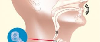
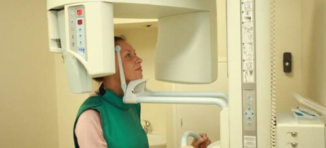
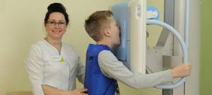
How are x-rays taken of the paranasal sinuses?
X-rays of the paranasal sinuses are carried out in specially equipped diagnostic rooms using an X-ray unit. Before the procedure, you will need to remove metal objects and outer clothing, and put on a protective apron with a lead coating. If it is necessary to take a picture in the nasomental projection, the patient presses his nose and chin to the device, opening his mouth. In the chin projection, the chin is pressed against the installation, and the nose is located 2 cm from it. Patient position for x-ray of the paranasal sinuses
corrected by the doctor. The patient is then asked to hold his breath and not move while a photo is taken.
X-ray with contrast
The contrast agent blocks x-rays and thus makes the image clearer. It is inserted into the paranasal sinus before the x-ray. The administration of contrast is an unpleasant procedure, so the doctor first administers anesthesia. However, recently, contrast X-rays of the paranasal sinuses have been used less frequently, as MRIs
And .
X-ray of the sinuses
X-ray of the sinuses is a fairly common procedure prescribed by otolaryngologists. With its help, you can identify most pathologies and diseases in this area.
The study is carried out to determine the condition of the bone walls and paranasal sections. The diagnostic method is based on the transillumination of tissues with short-length X-rays, which allows the specialist to clearly see the full picture of the pathological process in this area.
The X-ray method has been actively used since 1895, when it was discovered that fluids and bone tissue can retain gamma rays, which is why they have a different color in the picture.
Content
- Main indications
- What can be detected with x-rays?
- Contraindications to X-rays
- Preparation for the procedure
- Is it harmful?
- How is the procedure performed?
- Features of x-ray of the nasal sinuses for sinusitis
- Analysis of the information received
- What does a healthy x-ray of the nose look like?
- What to choose – CT scan or x-ray of the sinuses?
- Where can I do it?
Main indications
An X-ray of the nasal sinuses is prescribed at the slightest suspicion of foreign objects getting into the nose or the development of an inflammatory process.
Also, examination is mandatory if a person has suffered a traumatic brain injury, a fracture of the facial bones, after an accident, etc.
Thus, x-rays of the paranasal sinuses are performed in the following cases:
- Previous head injuries;
- Frequent nosebleeds;
- Uncomfortable sensations in the nose after suffering from acute respiratory viral infection;
- Headaches of different etymologies. They can occur when turning and tilting the head, localized in the eye area, bridge of the nose, etc.;
- Persistent nasal congestion;
- The simultaneous presence of photophobia, lacrimation and rhinorrhea;
- To confirm pharyngeal tonsil in children;
- Low-grade fever;
- If you suspect the presence of tumor formations.
An X-ray of the paranasal sinuses is also performed before an upcoming operation in this area. Diagnostics is also carried out as a control measure in the process of treating diseases.
What can be detected with x-rays?
A sinus x-ray is a very popular procedure, but what does it show? This is a question asked by many patients who are prescribed this type of examination.
The symptoms that we discussed above may indicate inflammation of the sinuses, so an x-ray examination can reveal accumulated fluid in them.
Thus, with the help of x-rays you can detect:
- Frontit
- Sinusitis
- Sphenoiditis
- Ethmoiditis
The X-ray procedure is carried out not only to detect these pathologies, but also to monitor the quality of further treatment.
X-rays of the sinuses are also performed to identify the following changes:
- Osteomyelitis (damage to the bone marrow and bones).
- Deviation of the nasal septum.
- Osteoporosis (a chronic disease characterized by increased bone fragility).
- Various neoplasms.
- A cyst may also be detected on an x-ray of the sinuses.
Contraindications to X-rays
X-ray of the paranasal sinuses is considered a relatively safe procedure. Although the radiation dose is minimal (about 0.02 millisieverts in a digital image), exposure to gamma rays can negatively affect the tissues of the human body.
When answering the question whether x-rays of the nasal sinuses are harmful, it is necessary to note a number of absolute restrictions on this procedure. First of all, the procedure is contraindicated for pregnant women in the first trimester. This period is extremely important for the normal development of the fetus, so any outside interference is unacceptable. But there is one caveat - if a woman’s health causes real concern, and alternative diagnostic methods are not possible, then an x-ray of the nose during pregnancy can be performed as prescribed by a doctor.
An X-ray of a child’s sinuses can only be done at a certain age – from 7 years. During this period, children are actively developing their skeletal system, so exposure to gamma rays can negatively affect growth. Again, if the situation is serious enough and there is a risk of complications, then the doctor allows an X-ray of the child’s nose.
Preparation for the procedure
Before x-raying the nose, minimal preparation for the examination is required, which boils down to the following points:
- Remove all metal objects that are near the area being examined (earrings, hairpins, piercings, etc.).
- Wear a protective lead apron, which is provided before the procedure.
There are no restrictions regarding eating and drinking.
Is it harmful?
The annual dose of X-ray radiation that is permissible for an adult is 150 millisieverts. In some cases, the frequency of examination is several times a year. Under this condition, the radiation dose will be insignificant, so there will be no negative effect on the body.
But another question is how often can a child’s nose be x-rayed and how harmful is it? Children are several times more susceptible to the effects of radiation than adult patients, so the advisability of the procedure is determined only by a competent specialist. Most doctors admit that after 7 years there are no restrictions on conducting the study.
As for frequency, the most optimal is no more than 2 times a year. It is advisable to carry out the procedure less frequently, or replace it with alternative diagnostic methods.
How is the procedure performed?
On average, an examination with modern equipment lasts for several seconds. The procedure looks like this:
- The patient enters a special room (puts on a protective vest).
- The head is securely fixed to avoid involuntary turns and tilts. X-rays of the paranasal sinuses are performed in different projections (mentoparietal, lateral or direct), so the head can be fixed in different positions.
- While remaining motionless, the patient stands in two positions (with his mouth open or closed).
- A photo is taken.
- Depending on the research method, the result will be ready in a few minutes.
Features of x-ray of the nasal sinuses for sinusitis
X-ray of the nose for sinusitis is a mandatory diagnostic procedure. In addition to identifying the inflammatory process, it is possible to determine its form:
- Parietal. The edges of the sinus are directed inward and are uneven. Inflammation is localized in the area of the bone walls.
- Essudative. The dark area is visible up to the upper transverse border of the sinus.
- Polypous. Protrusions of the mucous membrane are visible.
Analysis of the information received
The finished X-ray image allows you to see the following changes:
- Foreign objects (this is especially typical for children).
- Cracks and broken bones.
- Benign and malignant formations.
- Fluid in the sinuses is also clearly visible on x-ray.
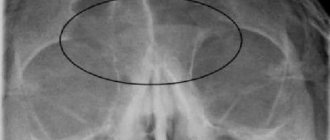
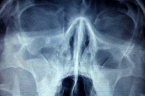
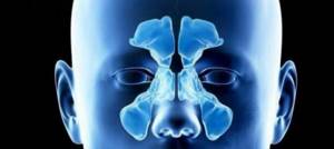
What does a healthy x-ray of the nose look like?
Darkening in the nasal sinuses on an x-ray always indicates the presence of a pathological process. The specialist’s task is to correctly interpret this information and make an accurate diagnosis.
An X-ray of the sinuses of a healthy person looks like this:
- The maxillary sinuses are presented in the form of triangular cavities on the sides.
- The nasal cavity is divided into symmetrical parts by the nasal septum.
- The nasal passages are represented by white stripes running along either side of the divided area.
- The cells of the ethmoid sinus should be clearly visible (it is located under the eye sockets).
- The sinuses should have smooth and clear edges.
What to choose – CT scan or x-ray of the sinuses?
Radiography is considered a fairly informative diagnostic method that is used everywhere. With its help, you can identify many pathological processes occurring in the area itself and nearby areas.
When answering the question of which is better - an X-ray or a CT scan of the sinuses, it should be noted that computed tomography is more informative, so it is prescribed if a traditional x-ray does not clarify the picture of the disease. Moreover, CT is better suited for diagnosing purulent, catarrhal and serous forms of sinusitis.
In general, x-rays are still considered one of the most popular and frequently prescribed procedures for diagnosing paranasal sinuses.
Where can I do it?
If you need to take an x-ray of your sinuses. Then you can do it in X-ray . Modern European equipment and qualified personnel allow us to obtain highly accurate results, significantly simplifying the diagnosis.
For your convenience, research results can be recorded on CD and DVD, or sent by e-mail. We constantly have discounts and promotions, so you have the opportunity to undergo examination on favorable terms.
You can make an appointment through the online registration form on the website, or by calling us at our contact phone number.
Sign up for a study by phone
(812) 332-52-54
Decoding the testimony
The radiologist deciphers what an x-ray of the paranasal sinuses shows. First of all, the condition and location of bone structures and cartilage is assessed. The liquid on the x-ray image appears as an intensely darkened horizontal boundary. Thickening of the mucous membrane indicates swelling in the paranasal sinus. You will receive a transcript of the research results in the form of a conclusion, but the final diagnosis is made by the doctor. Usually, when a pathology is identified that requires a more detailed study, another diagnostic method is prescribed.
How is a sinus x-ray performed?
The procedure is not painful and does not require preparation. Receive a referral at an appointment with an otolaryngologist, infectious disease specialist or surgeon.
How to do a sinus x-ray:
- The patient removes metal jewelry, glasses, and dentures.
- To protect against radiation, he wears a lead apron or vest with a collar.
- The laboratory assistant indicates how to position yourself correctly relative to the apparatus. Depending on the required projections, the position is changed at the command of a specialist.
- While taking the photo, you should not move and hold your breath.
- After development and decoding, the film with the description is given to the patient.
The conclusion is not considered a final diagnosis, but provides clarifying information for the treating specialist.
X-ray of the sinuses for children
Pediatricians try to protect preschool children from the harm caused by X-ray radiation to the fragile skeletal system. The procedure is allowed to be performed on patients over 7 years of age.
Before this period, a clear justification for the importance of intervention is necessary - for example, severe facial trauma or severe sinusitis with a risk of inflammation of the meninges.
It is difficult for a young child to sit or stand still during an x-ray. To distract him, they use toys, sedatives, and in emergency cases, anesthesia. At an older age, you can captivate your child with a game in which you should freeze for a short time.
Sign up for the study
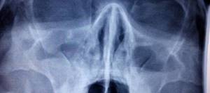
Darkening of the sinuses on x-ray
The method is based on the different permeability of hard and soft tissues by electromagnetic waves. Bones block radiation and appear white in the picture. Specialists are interested in blackouts, which determine the nature of the disorder.
Darkening of the sinuses on an x-ray indicates fluid accumulation. This is a sign of inflammation with the release of mucous or purulent secretion. Sometimes, the picture shows thickening of the walls of the mucous membrane lining the sinuses.
New growths stand out as clear dark spots with shadows. Polyps look like peas on a “pedicle”, and cysts have a cavity filled with fluid inside.