Ultrasound of soft tissues is performed to obtain information about morphological changes in these structures, especially when formations such as lumps or tumors appear. Ultrasound can determine whether the mass is cystic (fluid-filled) or solid. Ultrasound imaging technology allows detailed examination of soft tissue abnormalities around joints, tendons and muscles.
Who is recommended to have this ultrasound scan?
Indications for the procedure are:
- Presence of lumps in soft tissues
- Enlarged lymph nodes
- Infections
- Hematomas
- Volumetric formations
- Inflammation
- Cysts
- Abscesses
- Cancerous tumors
- Hernias
- Lipoma
- Gangliomas
- Ultrasound is also used to guide the administration of medications to specific areas: tendons, bursa, cysts, ganglia and neuromas. Ultrasound can also be used to aspirate tissue material from pathological formations
Indications for ultrasound of soft tissues of various parts of the body
- Belly. Ultrasound is prescribed for acute pain, suspected appendicitis, hernia or tumor, suspected injuries to internal organs, intestinal disorders, ingestion of a foreign body, unreasonable appearance of bruises on the skin, etc.
- Limbs. Ultrasound examination of the soft tissues of the arms, legs or thighs is carried out if there are seals of unknown nature in them, after injuries, if pain occurs at rest or with movement, if there is suspected inflammation of the joints or an abscess (purulent inflammation), etc.
- Groin area. Ultrasound is prescribed for enlarged lymph nodes as a result of infection or inflammation, suspected tumor or inguinal hernia, abscess of the iliopsoas muscle, hemorrhages, etc.
- Face, neck and maxillofacial area. An ultrasound examination is performed when signs of inflammation appear, cysts or other neoplasms on the face are suspected, or after plastic surgery. Ultrasound of the soft tissues of the neck is prescribed when the lymph nodes are enlarged, there is a need to assess the condition of the arteries, etc.
How is soft tissue ultrasound performed?
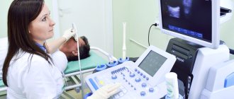
Ultrasound scanning of soft tissue is a fairly simple procedure. A small amount of gel will be applied to the area to be examined. The sensor will then move across the skin and this will create images that will appear on the ultrasound monitor. These images are stored in a computer for further interpretation. This painless scan usually takes 10 to 15 minutes.
After the procedure is completed, the patient may receive a brief explanation of the results. A full description of the scan results is sent to the attending physician.
Advantages of performing ultrasound in the network of multidisciplinary clinics "Alfa Health Center"
- Affordable prices. Multidisciplinary clinics of the Alfa Health Center network are open in 12 cities of Russia. In any of our branches you can perform an ultrasound of the soft tissues of the neck, face, abdomen and other parts of the body at an affordable price.
- High information content of the study. Our clinics use modern high-precision ultrasound equipment. It allows you to examine soft tissues in detail on ultrasound. High accuracy of research results is an important condition for quickly making a correct diagnosis.
- Attentive attitude of doctors. Our specialists do everything possible to ensure that your visit to the clinic leaves only a pleasant impression. Doctors conduct appointments strictly by appointment. You come directly to the appointed time and do not wait in line.
- Absolute safety of the procedure. Ultrasound, unlike many other methods of examining soft tissues, has no contraindications and is suitable for all patients without exception. The examination is successfully used even in pregnant women, newborn children, people with chronic diseases, installed pacemakers or metal implants.
- Quick results. Ultrasound of soft tissues takes no more than half an hour, and it takes only 15 minutes to decipher the data. You receive diagnostic results and a doctor’s conclusion immediately after the procedure. The examination data is entered into the medical record.
results
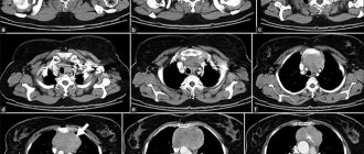
Ultrasound results are determined both in real time (observed during the examination) and as final static images. After the examination, this information will be studied by an ultrasound specialist and a written description of the study results will be generated, and the protocol will be transferred to the attending physician. If fluid is aspirated during the test, the sample will be sent to a laboratory for analysis and the results will be sent to your doctor. Copies of the study protocol, if necessary, can be given to the patient.
Ultrasound examination of soft tissues
If any pathological process is suspected or in order to control the dynamics of treatment or surgical intervention, the attending physician may prescribe an ultrasound of soft tissues.
What is soft tissue ultrasound?
The ultrasound research method is based on the ability of the tissues and organs of our body, depending on their density and structure, to reflect ultrasound waves differently. Thanks to this ability, tumors, neoplasms, and various deviations from the norm are displayed on the screen of the ultrasound machine. Ultrasound of soft tissues
is an accessible and effective method for studying the pathological processes that occur in them. This method allows you to examine even pregnant and lactating women, as it is absolutely safe. In addition, it has no contraindications, with the exception of violation of the integrity of the skin in the area of study (wounds, abrasions, rashes). When the skin is restored, you can sign up for the procedure.
What reveals
Diagnostics reveals the presence of:
- ordinary and dermoid cysts;
- abscesses: examination not only detects the process, but also allows you to determine the stage of the process;
- connective tissue pathologies;
- benign and malignant neoplasms (lipomas, fibromas, lymphangina, atheromas, etc.);
- hernias - umbilical, inguinal, in the back;
- lymphostasis - local edema caused by impaired lymph outflow;
- rheumatoid diseases - degenerative, inflammatory rheumatic, metabolic, periarticular;
- damage to thin bones (hands, feet);
- inflammation in the periarticular tissues;
- myositis - inflammatory muscle disease;
- hematoma.
Especially often during ultrasound diagnostics the following is detected:
- lipomas - neoplasms from benign adipose tissue (uniform structure, poor echogenicity);
- hygromas - dense formations filled with fluid, often forming in the joints;
- hematomas - cavities with liquid or coagulated blood resulting from injuries;
- chondromas are benign tumors in cartilage tissue;
- hemangiomas;
- atheromas;
- tendon injuries;
- connective tissue diseases (scleroderma, lupus erythematosus, recurrent panniculitis, relapsing polychondritis, polymyalgia rheumatica, Sharp's syndrome, Sjögren's syndrome, idiopathic dermatomyositis, diffuse fasciitis, etc.
Ultrasound diagnostics of soft tissues reveals hundreds of pathological processes and allows you to track them. Thus, most cysts - cavities with serous fluid - are benign, but some, under a combination of circumstances, can degenerate into metastatic cancerous tumors. The specialist decides whether the tumor requires immediate surgical intervention or whether other treatment methods can be used.
What pathologies can be detected?
When conducting an ultrasound examination of soft tissues, it is possible to identify pathologies such as lipoma (adipose tissue that forms a benign tumor that does not contain fluid and blood vessels), hematoma (due to bruises a cavity filled with blood appears), hygroma (a benign subcutaneous formation that is filled with serous fluid) , atheroma (suppuration of the sebaceous gland), tumors, cysts and abscesses.
Ultrasound can also be used to examine the contents of the hernial sac.
Interpretation of ultrasound results
The patient receives the results after the procedure. The ultrasound protocol of soft tissues describes their structure. If neoplasms are present, the location, size, structure, shape and how deep they go are described. Based on the results obtained, the attending physician makes a conclusion about the presence or absence of pathologies. Normally, an ultrasound scan should show nothing but the tissue itself. No inclusions, formations, accumulations of liquid, etc.
If a focus of inflammation or any formation is detected, but it is located deep in the soft tissues or the patient is overweight, an MRI of soft tissues
.
When it is necessary
It is almost impossible to list all the indications for this universal examination. It provides the most valuable information in the following cases:
- suspected violation of the integrity of soft tissues;
- suspicion of a hematoma deep in the tissue;
- diagnosis of inflammatory processes in muscles, ligaments and tendons;
- identification and clarification of the nature of neoplasms;
- suspicion of an abscess in soft tissues, determination of its stage;
- diagnosis of inguinal and umbilical hernias at the initial stage of development;
- diagnosis of changes in lymph nodes;
- clarification of the position of the foreign body.
There are practically no contraindications. Occasionally, ultrasound may be interfered with by inflammatory skin diseases at the site of the examination. It is important that the attending physician indicates in the referral the preliminary diagnosis and the place to be examined. You can also come to see a medical specialist if you notice alarming symptoms or for a routine check-up. Based on the results of the examination, the doctor will write out a referral for the necessary research.
FAQ
- Is it painful to have an ultrasound examination?
No, this is a non-invasive and painless diagnostic method.
- Will ultrasound be enough to make a diagnosis?
It all depends on the disease; for some diseases, only an ultrasound is sufficient, while others will require a more in-depth examination, such as magnetic resonance imaging or blood tests.
- How often can an ultrasound be done?
The ultrasound diagnostic method is absolutely harmless; if necessary, it can be done as many times as necessary.
- Is it possible to consult a specialist after an ultrasound?
Yes, you can get advice from a specialized specialist - a traumatologist, orthopedist, physiotherapist or sports doctor. You can also undergo treatment with us, and you can start on the same day you contact us.
Relevance of the problem
The urgency of the problem is due to the popularization of cosmetology, the increase in the number of contouring procedures, the use of gels based on hyaluronic acid in “forbidden” areas of the face, which, in turn, has led to a sharp increase in cases of complications of contouring, undesirable effects and overcorrection. The presence of a large number of low-quality products on the market contributes to the formation of foci of acute and chronic inflammation in the areas where gels are introduced, requiring both surgical and other methods of removing the gel [].
To correct the undesirable consequences of contouring with gels based on hyaluronic acid, it is proposed to use various drugs whose active ingredient is hyaluronidase. It is recommended to inject drugs with hyaluronidase activity into the defect area []. Moreover, the vast majority of drugs currently on the market, the active ingredient of which is hyaluronidase, do not have instructions for use in cosmetology. At the same time, there are no recommendations for each of the drugs regarding doses, frequency of administration, or volume of the drug administered. There are also no clear protocols for administering the drug: to what depth, in what way. Thus, specialists inject drugs with hyaluronidase uncontrollably, almost blindly, into the thickness of the facial tissues where the gel is supposed to be located, without taking into account the anatomical features of the patient and clearly determining the location of the filler, its shape, size, topography relative to other facial structures. With this administration of drugs, there is a high risk of damage to the vessels, nerves and other structures of the treated area located in the tissues, as well as the possibility of introducing hyaluronidase into the lumen of the vessel or into the nerve, bypassing the gel itself. In the intervention zone, an inflammatory reaction of the surrounding tissues is formed, accompanied by swelling and pain.
According to the results of our observations, the disadvantages of treating the undesirable consequences of contouring with gels based on hyaluronic acid by introducing hyaluronidase into the correction area are the lack of a pronounced clinical effect and a large number of side effects from the therapy. These include acute swelling in the injection area, long-lasting pastiness of surrounding tissues, the risk of allergic reactions, and pain during injection.
The lack of diagnostic standards, clear schemes, instructions and recommendations for the treatment of patients in cases of hypercorrection and edema served as the basis for the search for effective and safe methods for diagnosing and correcting the negative consequences of contouring with gels based on hyaluronic acid.
An alternative method is the technology we propose, which is similar to the effect on hypertrophic scars by phonophoresis with one percent hydrocortisone ointment, when the mechanical energy of an ultrasonic wave and the chemical composition of the drug administered into the lesion are simultaneously combined [, ].
Why are neck vessels examined?
The study can be planned or emergency. If you do not pay attention to the state of your health in time, you will have to find out what an ultrasound of the neck vessels shows as an emergency. The procedure may be prescribed for:
- pain in the neck and crunching when moving;
- osteochondrosis;
- the presence of bad habits, especially smoking;
- diabetes mellitus;
- increased ICP;
- neck and head injuries;
- strong pulsation in the neck;
- identifying a strong difference in pressure on both hands.
In each of these cases, what an ultrasound scan of the neck
in an adult shows will help identify the cause of the disease and treatment options. Thanks to this research method, any narrowing of blood vessels, their elasticity and tone, the presence of atherosclerotic plaques and blood clots, as well as the likelihood of rupture and blockage of a blood vessel will be identified. It will also be possible to obtain detailed information regarding the degree of narrowing of blood vessels by blood clots and plaques, learn about the presence of additional vessels and dilations, as well as their connections with veins and arteries. In each of these cases, adequate treatment is needed, which can only be prescribed if the diagnosis is highly informative.
It is worth knowing that such a study is practically uninformative if it is necessary to examine the vessels of the brain, since it is only suitable for soft tissues. In such a situation, MRI, CT or other diagnostic methods will be prescribed.
Method of performing ultrasound of the neck
1-2 days before diagnosis, drinking alcohol is not advisable. If an ultrasound scan of the neck includes diagnosis of the thyroid gland and blood vessels, you should stop taking medications that may affect local circulation. Therefore, you must first inform the doctor performing the ultrasound about all medications you are taking.
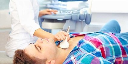
Diagnosis is carried out by an ultrasound specialist. The patient assumes a supine position. The doctor moves the device’s sensor over the skin, in the projection of the structures being studied. For better contact with the sensor, gel is pre-applied to the skin.
The neck ultrasound procedure is absolutely painless and has no contraindications, like other types of ultrasound examinations. In some patients, due to individual characteristics, touching the sensor to the projection of the larynx may cause a gag reflex. In these cases, we recommend going for an ultrasound scan on an empty stomach.
During an ultrasound, the doctor focuses on the following indicators:
- size and shape of anatomical formations
- tissue structure: homogeneous, heterogeneous
- echogenicity, the ability to reflect ultrasound
- presence of abnormal structures, space-occupying formations
- vessel diameter
- volumetric and linear blood flow velocity.
Depending on the indications, we perform a comprehensive ultrasound of the neck with diagnostics of the spine, soft tissues, thyroid gland, and lymph nodes. Or we diagnose these structures in isolation.
Spine
Here we evaluate the condition of the vertebrae and intervertebral discs. We diagnose cervical curvatures (scoliosis, increased lordosis), osteochondrosis and its complications - protrusions, disc herniations with vertebral subluxations. Of course, radiography, computed tomography and magnetic resonance imaging are indicated for diagnosing the spine. These methods are no less, and in some ways more informative, than ultrasound. But the advantage of ultrasound is its simplicity, low cost and lack of contraindications. That's why we perform neck ultrasounds even for children. And if changes are detected, we refer them to other, more complex and expensive studies.
Lymph nodes
According to ultrasound data, enlargement of lymph nodes, changes in their structure and shape are recognized. These disorders indicate inflammatory processes (lymphadenitis) or tumors. Moreover, lymph node tumors can be primary and secondary (metastatic). For a final diagnosis, we resort to targeted biopsy of nodes , which we also carry out under ultrasound control.
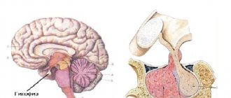
Thymus
Visualization of the thymus gland using ultrasound is possible in the first months of a child’s life. At an older age, ossification of the sternum is observed, while the lateral sections of the gland are covered with lung tissue, so a reliable determination of the size of the organ becomes impossible. A transverse scan of the thymus is obtained by placing the sensor transversely above the sternum at the junction of the manubrium and the body. To obtain longitudinal scans, the transducer is placed longitudinally along the parasternal lines on the right and left. The thymus is divided into two lobes. The most common pathology of the thymus is thymomegaly, which is not accompanied by structural changes in the gland tissue. Less commonly, a decrease in the size of the thymus is observed, indicating hypoplasia or accidental involution of the organ. Ultrasound does not reliably distinguish between hypoplasia and involution.
Thyroid
The relevance of ultrasound scanning of the thyroid gland is especially important due to the high prevalence of pathology of the thyroid system. The incidence of iodine deficiency conditions (endemic goiter) and autoimmune diseases of the thyroid gland (mainly autoimmune thyroiditis) has been especially high in recent years. During an ultrasound examination, the patient lies on a couch and a small cushion is placed under his shoulders. Scanning is carried out in three main planes - transversely, measuring the maximum thickness of the width of each lobe and isthmus, and oblique longitudinally (for both lobes), measuring their thickness and length. An ultrasound examination allows one to evaluate the echogenicity and echostructure of the thyroid tissue, the features of its contours, the relationship with nearby organs and the volumetric characteristics of the organ. Using Doppler techniques (pulse Doppler, color flow, ED), an objective assessment of the blood supply to the organ is carried out.
Ultrasound determines the location of the gland, the structure, size and echogenicity of its lobes, as well as the presence of abnormal inclusions. Based on available data, we diagnose:
- inflammatory processes ( thyroiditis )
- cavity formations (cysts)
- benign tumors, adenomas
- malignant tumors, cancer
- congenital anomalies.
Here, a puncture biopsy may also be required to definitively confirm or exclude a tumor process. The ultrasound picture of the thyroid gland would not be complete if we did not evaluate the local blood flow in the gland tissue. Standard ultrasound is not suitable here, and we resort to the triplex scanning technique with color duplex mapping (CDC). Ultrasound with color flow allows you to obtain a visual image in color of blood moving through the vessels.
Ultrasound of the salivary glands
Ultrasound examination may reveal sialadenitis. It is characterized by an increase in the size of the glands, uneven contours, reduced echogenicity due to edema, and moderate dilation of the ducts. When a lymph node is inflamed inside the gland, a hypoechoic lymph node is located in its tissues. On the affected side, signs of submandibular lymphadenitis may be detected. In chronic sialadenitis, a mosaic structure and uneven dilatation of the ducts are determined. Salivary stone disease on ultrasound is characterized by an increase in the size of the organ (mainly in thickness), uneven contours, “graininess” of the parenchyma, and an uneven increase in echogenicity. The calculus and dilatation of the ducts are visualized.
Ultrasound of the larynx
The study allows you to visualize the vestibular and vocal folds of the larynx, determine their mobility and symmetry. Using ultrasound, you can evaluate the lumen of the organ and the thickness of the cartilage covering the larynx. Echography can reveal space-occupying formations, adhesions, paresis or paralysis of the larynx, papillomatosis with papillomas larger than 2 mm.
Vessels
Here we can diagnose:
- inflammation of the vascular wall ( vasculitis ) in various diseases
- structural changes in the vascular wall (aneurysms, diverticula)
- atherosclerosis of the arteries with slowing of blood flow
- compression of the vertebral artery as a result of curvature or complicated osteochondrosis of the cervical spine.
Ultrasound of the neck vessels is also performed in triplex scanning mode.
Soft fabrics
Here our specialist can diagnose some structural changes in muscles and fascia. Reasons for these structural changes:
- congenital abnormalities of muscle structure
- curvature of the spine
- cervical osteochondrosis
- disturbances of innervation due to pathology of the central nervous system (CNS).
In addition, ultrasound of the neck allows you to identify inflammatory changes in muscles and fascia ( myositis , fasciitis ), as well as detect abscesses in the cellular spaces. All data is logged. A written conclusion along with a digital photograph is issued in person. For all questions related to diagnosis, the doctor gives oral consultations.
Indications for the study
The neck is bounded from above by the chin, the lower edge of the lower jaw, the mastoid region, and the lower edge of the sternum.
The lower border is the manubrium of the sternum, the upper edge of the clavicle, and the acromial process of the scapula. This entire space includes:
- cervical spine from the atlanto-occipital joint to the VII vertebra;
- skin and soft tissues: muscles, tendon sheaths (fascia), and the tissue spaces located between them;
- lymph nodes: cervical, mental, submental, submandibular, paratracheal, supraclavicular and subclavian;
- main vessels: external and internal jugular veins, common, external and internal carotid arteries, vertebral artery;
- thyroid gland and parathyroid glands.
Pathological changes in these structures are accompanied by a number of clinical manifestations. Among them:
- visible deformation, curvature of the cervical spine;
- diffuse or focal swelling of soft tissues, redness of the skin, increased local temperature;
- general weakness, headache , dizziness;
- sensation of tinnitus, loss of coordination of movements, unsteady gait;
- sleep disorders;
- depression, irritability, and other mental changes;
- painful sensations in the neck, radiating to the external auditory canal, to the shoulder girdle, to the arm, and to the lower jaw;
- painful or painless enlargement of lymph nodes;
- difficulty swallowing;
- visible enlargement of the thyroid gland , the appearance of focal compactions on its surface;
- changes in the level of thyroid hormones according to laboratory tests.
- increased organ size;
- signs of the inflammatory process;
- detection of pathological formations;
- excess body weight;
- weight loss;
- tachycardia.
We also conduct repeated ultrasound for already identified diseases of the cervical structures in order to assess the dynamics of these diseases and the effectiveness of treatment.