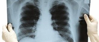
Chest X-ray
(radiography, fluorography) is a highly informative diagnostic method that uses X-rays - electromagnetic radiation with a wavelength of about 10-8 cm.
The method is based on the ability of radioactive X-rays, passing through human tissues and organs, to be partially or completely retained, leaving an image on the X-ray film. X-ray diagnostics
is widely used in clinical medicine and is also an important component of the minimum required testing in adults and children.
When is fluoroscopic examination indicated?
X-rays of the lungs and other organs of the chest cavity may be prescribed when a number of symptoms indicating developing diseases appear. As a rule, the study is prescribed to patients who complain of severe shortness of breath, pain in the chest or cough.
Accumulation of fluid or air in the pleural cavity
The accumulation of air is characterized by protrusion of the diseased side, the intercostal spaces are smoothed out, and when breathing, the diseased side “lags behind” the healthy side - asymmetry is observed. This condition can be combined with the accumulation of fluid in the pleural cavity. Often a consequence of injury. Performing an X-ray of the lungs in such a situation allows you to save the patient’s life.
Clarification of the localization of foreign bodies in the trachea or bronchi
This condition is dangerous due to the development of lightning asphyxia, as well as other severe complications. Therefore, it is necessary to find out how to get an X-ray of the lungs in such a situation, how often it can be done, and what will become known based on the results of the diagnosis. Identifying the position of a foreign body and its size allows you to plan its removal surgically or otherwise
Pleural adhesions or moorings
Adhesions are overgrown connecting cords, the location of which is usually between the serous membranes in the pleural cavity. Such adhesions can also be called pleurodiaphragmatic; they can be total, occupying all the pleural sections, or single, when the layers of the pleura are fused. A chest x-ray is necessary when the patient complains of chest pain, shortness of breath, and fever.
Determination of diaphragm pathologies
X-rays can detect all kinds of tumors, diaphragmatitis or hernias. Diaphragmatic dystopia, dyskinesia and dystonia, as well as damage, are common.
Examination of the mediastinal organs
Often, x-rays are prescribed in situations where it is necessary to determine the localization of a formation in the chest cavity. This is more appropriate for examining the anterior mediastinum. In the study, this cavity is divided into a retrosternal section (anterior) and a central (posterior) section. Chest X-ray allows you to examine the cavity and identify the presence of pathological processes.
Description of the study
Mammographic examination is a non-invasive method aimed at identifying various pathological processes and congenital or acquired anomalies in the structure or functioning of the mammary gland. How is a breast mammogram done? Most often, X-rays are used, but in some cases additional diagnostics are necessary, for example, with a tomograph or using an ultrasound sensor. However, the last two methods have more disadvantages than advantages. Yes, they do not expose a woman’s body to harmful radiation, but they are not as informative as breast x-rays. Mammography gives a very accurate result - the accuracy is about 90%.
to do a mammogram correctly
, diagnosticians of any medical institution know. However, before the procedure, pay attention to the experience of the employee, as well as the modernity of the equipment. The last factor becomes decisive - the reliability of the results, radiation exposure and the appropriateness of the prescribed treatment depend on it.
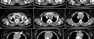
Diseases diagnosed by chest X-ray
It is necessary to find out what a chest x-ray shows if there is a suspicion of lung disease. A large number of diseases occur without any symptoms at all, so a routine examination and questioning of the patient does not provide the necessary amount of information. Using an X-ray, it will be possible to examine all the structures of the lungs and identify the cause of the pathology, after which the doctor will be able to prescribe treatment.
Pneumonia
It is an inflammatory disease characterized by significant disturbances in the breathing process. The disease requires quick diagnosis and timely treatment. If you do not know how often an adult can have an x-ray of the lungs in such a situation, it is worth finding out that no more than once a year.
Congestive heart failure
Failure is a complex failure in hemodynamics. Its cause is a dysfunction of the heart, which is provoked by overwork of the cardiomyocyte, as well as defects that are present from birth or acquired with age. A violation of this plan can be detected using a chest x-ray.
Pneumothorax
Such a deviation can be traumatic and spontaneous, as well as artificial. If the artificial type of disease is a consequence of treatment, then the traumatic and spontaneous type must be treated urgently. Plain radiography of the lungs allows you to diagnose the disease and assess its severity.
Pleural effusion
It is the accumulation of fluid in the pleural cavity. The cause of the effusion allows us to determine its type - exudate or transudate. As a rule, it is not an independent disease, but a complication. It can appear due to pneumonia, congestive heart failure, tuberculosis, pulmonary embolism, as well as HIV infection and oncology. X-ray of the lungs is performed in a vertical position in frontal and lateral projections.
Cardiomegaly
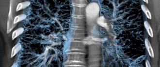
X-ray of the chest organs
may be prescribed due to an increase in heart size. The following indicators are normal:
- 10-11 centimeters in length;
- 8-11 centimeters wide;
- 6-8.5 centimeters in thickness.
The disease can otherwise be called “bull heart” and is characterized by an increase in the size and weight of the organ due to an increase in the width of the muscle wall, as well as expansion of the chambers. This phenomenon is not always a sign of illness. For example, in people who are professionally involved in sports, it is natural and understandable, indicating the general endurance of the body. However, this can also be a manifestation of diseases, so a chest x-ray will be needed to identify the cause.
Pneumoperitoneum
This pathology means filling the abdominal cavity with gas. Finding out what an x-ray of the lungs shows is necessary in order to identify the cause of the disease and prescribe treatment that is adequate to the patient’s current condition. The disease is extremely serious; untimely treatment can become life-threatening and lead to various complications.
Emphysema
It is characterized by a chronic course and a disorder of normal gas exchange in the lungs. Occurs due to overstretching of the alveoli and their excessive filling with air. Emphysema is caused by chronic bronchitis, but it can also be caused by other lung diseases or heredity. To understand what caused the disease, you need to take a chest x-ray.
Contraindications
There are also contraindications to chest x-rays. They do not interfere with deciphering the results of mammography, but are harmful to the patient. If a woman is contraindicated from undergoing an X-ray examination, then other diagnostic methods are used, which were already mentioned earlier.
Pregnancy
Here the risks are associated with the need for radiation exposure, which, although the dose is small, can affect the child. It is especially dangerous to take x-rays during the first trimester, when all the main systems of the fetus are formed. X-rays are done only when the potential danger to the mother's health outweighs the risks to the fetus.
Lactation period
For the same reason, nursing mothers should not agree to the procedure. Even preliminary milk expression will not be a solution here.
How is the examination carried out?
It is best to find out how often a chest x-ray procedure can be done, as well as how the examination is carried out, in advance. The patient must undress to the waist and remove all metal jewelry and accessories. The doctor is often asked to take the chain between his teeth so as not to remove it. After this, you can proceed with a chest x-ray and find out what the procedure shows.
The picture may need to be taken in two projections - lateral and frontal. Each one is done in just a few seconds, and the X-ray of the lungs itself, regardless of the diagnosis, will not cause any discomfort.
Preparation and methodology for conducting the survey
How to prepare for mammography of the mammary glands, and what is the preparation procedure? The examination is painless, but due to the individual characteristics of the body, it may cause some discomfort. In this case, it is possible to take painkillers before the study. Directly on the day of the examination, it is prohibited to use deodorant and other cosmetics that should be applied to the skin of the armpits. This is due to the fact that they may cause dark spots to appear on the film, which will prevent an accurate diagnosis.
When X-rays are contraindicated
Because the imaging process relies on radiation exposure, not every patient can undergo this procedure. Although the dose of X-ray radiation is small and does not cause damage when used correctly, such a study is carried out only when there are no contraindications.

During pregnancy, X-rays of the thoracic spine are not taken. Radiation exposure can cause irreparable damage to the development of the fetus. This procedure is avoided even in later stages, replacing it with alternative methods, for example, MRI of the thoracic spine.
To obtain clear images, the patient during the diagnostic process must not move for some time and follow the radiologist’s commands, for example, hold his breath. With severe mental disabilities and mental changes, a person cannot fulfill this requirement, so this may become a contraindication for the study.
A barium examination is considered a temporary contraindication. X-rays can be taken after such a procedure only after 4 hours.
In most cases, the examination is postponed to the next day. For people for whom this technique is contraindicated, other examination methods are recommended - MRI, CT, angiography, etc. In extreme cases, doctors are forced to examine the problem area only during surgery.
3.Is chest x-ray dangerous for human health?
Essentially, X-rays are radiation that, in high doses, is dangerous to the human body. But the amount of radioactive radiation that a person receives during an X-ray is negligible. Therefore, you should not worry about your health if your doctor has prescribed an x-ray for you. Remember that, if necessary, it is prescribed even to small children. To protect your body from unnecessary negative effects, adhere to the following rules:
- Rule No1 - it is better to do x-rays no more than 2 times a year.
Therefore, if you had an x-ray the day before, and another doctor prescribes this procedure for you again, be sure to tell him about it. In this case, it does not matter which part of the body was previously examined, because you were completely exposed to radiation. - Rule No2 - X-ray is a necessary measure, which is carried out only if necessary.
Therefore, you should not insist on taking an x-ray if the doctor believes that your diagnosis is confirmed by another method.
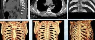
Where is the best place to take pictures of the spine?
The radiography procedure is considered the most accessible and easiest to use. Compared to other diagnostic methods, this can be done in any clinic. Similar devices are available in public clinics and hospitals, as well as large diagnostic centers. The process will be the same in both a private clinic and a public one.
The only differences are the price and waiting time for results. Thus, the state X-ray room will take pictures in a few minutes, but decoding may take several days. By law, this service in a government agency must be free.
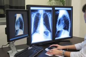
Private diagnostic centers provide examination results within an hour. In addition, the X-ray machine can be digital, and such a device transmits the image to a computer monitor. The doctor can examine the condition of the organs and spine immediately during the diagnostic process; this is very important when identifying dangerous pathological conditions.
The results of this technology are transferred, at the patient’s request, to an electronic medium - a flash card or DVD. The health worker can also send them by email, which is much more convenient.
Feedback from those surveyed shows that there is not much difference between having an X-ray done in a public clinic or in a private one. If comfort and level of service are important, you need a private one. The price for one photo will be from 10 to 40 dollars. Before your appointment, it is important to ask if transcription is included in the price. As a rule, in private centers you have to pay an additional 10-15 dollars for this.
However, modern equipment makes it possible to view the examined area with multiple magnification. The doctor can examine in detail even small cracks or tumors. Old-style equipment takes pictures only on a film matrix, which narrows diagnostic possibilities.