Magnetic resonance imaging
Magnetic resonance imaging is a highly informative non-invasive method for studying the human body. The advantages of the technology include:
- absence of ionizing radiation;
- the ability to take layer-by-layer images of tissues and organs;
- extensive diagnostic capabilities;
- no pain.
For maximum accuracy of results, proper preparation for MRI is necessary.
What is spinal tomography?
Magnetic resonance imaging of the spine is a high-tech method for diagnosing back pathologies. The operating principle of the tomograph is based on the effect of nuclear magnetic resonance. The scanning device itself consists of an installation that creates a powerful magnetic field and radio frequency signal detectors. When a patient gets inside the scanning part of the device, under the influence of a strong magnetic field and RF signals, the hydrogen atoms in the body's cells begin to resonate, that is, make oscillatory movements. The tomograph's computer translates these impulses into three-dimensional images, which the radiologist sees on the monitor screen.
What will an MRI of the spine show?
Deciphering the tomography of the spine will allow:
- Assess the condition and anatomy of vertebral structures.
- Identify congenital anomalies and pathologies.
- Diagnose degenerative diseases of the spine.
- Assess the level of compression of nerves and spinal cord.
- Determine the presence of neoplasms and infections.
- Detect inflammatory processes, damage to blood vessels and spinal membranes.
- Identify areas of injury, as well as changes that occurred after surgery or medical intervention.
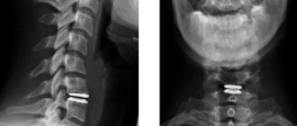
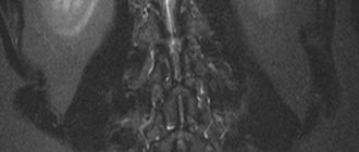
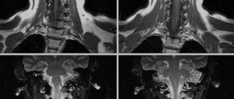
What does the spine look like on MRI images?
Indications
- Severe back pain;
- Trauma, bruise of the spine;
- Exclude malignant or benign neoplasm;
- Inflammatory process.
MRI should be taken regularly for people with diagnoses such as osteochondrosis, protrusions, hernias, and vertebral displacements. If the patient has ever had a birth or congenital injury in any part of the spine, this diagnosis will also be very useful to monitor the consequences of birth pathology.
Free diagnostic consultation
If in doubt, sign up for a free consultation. Or consult by phone
+7
How often can a spinal tomography be done?
Since magnetic resonance imaging does not involve radiation exposure to the body, it can be performed an unlimited number of times for any category of patients. This study can be combined on the same day with any type of diagnostics: ultrasound, radiography, computed tomography. The decision on the frequency of MRI of the spine is made by the attending physician, based on the diagnosis and course of treatment. For preventive purposes, it is recommended to have a spinal tomography once every two years for all people over 35 years of age.
Advantages and disadvantages
Magnetic resonance imaging of the spine has a number of advantages:
- high quality and informative images;
- display of back structures in a three-dimensional model;
- absence of pain and discomfort during scanning;
- no radiation exposure;
- the ability to detect neoplasms and pathologies at the development stage.
The disadvantages of this method include:
- higher cost of MRI compared to radiography and CT;
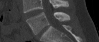
- poor visualization of bones (computer tomography is better suited to assess the composition of the bone tissue of the vertebrae);
- incompatibility with metal in the body and electronic implants.
- MRI
- Ultrasound
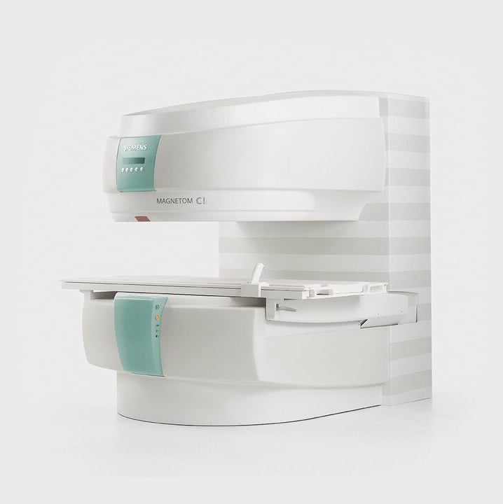
MRI tomograph:
Siemens Magnetom C
Type:
Open (expert class)
What's included in the price:
Diagnostics, interpretation of images, written report from a radiologist, recording of tomograms on CD + free consultation with a neurologist or orthopedist after an MRI of the spine or joint
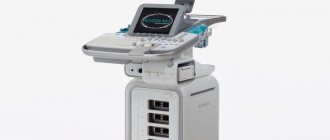
Ultrasound machine
HITACHI HI VISION Avius
Class:
Expert (installation year 2019)
What's included in the price:
Diagnostics, interpretation of images, written diagnostic report
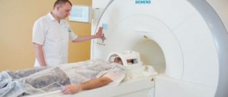
Tomography procedure
Magnetic resonance imaging of the spine does not require preparation. The patient simply needs to sign up for the examination in advance and arrive at the medical center 15 minutes before the start of the examination to complete all the documents. You can undergo diagnostics in your own clothes. For a visit to the MRI center, it is best to choose clothes that are comfortable for lying down for a long time and do not restrict movement. There should not be any metal elements on it, for example: zippers, rivets, buttons, rhinestones. Before the scan begins, the patient will be asked to remove all metal from the body and remove all electronic devices from their pockets.
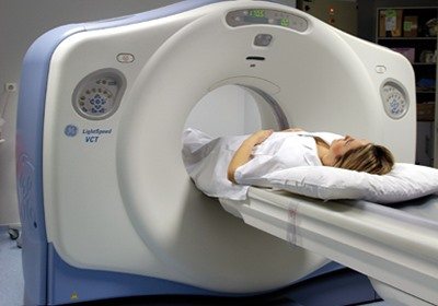
In the diagnostic room, the patient will be helped to take a horizontal position on the tomography table. A special MRI coil will be installed in the examination area. The table is then slid into the scanner. The operator will leave the room and launch the scanning program in the control room.
The patient will understand that the tomograph has started working by the noise of the operating machine. This will be the sounds of periodic tapping. If this noise annoys a person, they will be offered special noise-canceling headphones. During the procedure, two-way communication is maintained with the subject. He can report his discomfort at any time via the microphone inside the tomograph. The operator will immediately stop the scan and try to remove the cause of the discomfort.
The duration of examination of one part of the spine is on average 10-20 minutes. All this time, the person being diagnosed must remain motionless. Any movement can cause the final tomograms to have artifacts. This will reduce the information value of the images.
Questions about diagnostics
Dress code
You can enter the MRI room in any clothing that does not contain metal. When going to the clinic, it is best to wear loose, non-restrictive clothing without metal elements (zippers, rivets, hooks), in which you can lie comfortably. For women, we recommend bringing a T-shirt or not wearing a bra with metal wires or hooks.
Preparation
This tomography does not require any preparatory steps from the patient.
Is MRI harmful to health?
MRI is a completely harmless diagnostic method for the human body. This method of examination can be carried out at any age and for any disease an unlimited number of times, unless you have contraindications.
Contraindications
Some pacemakers and foreign objects in the body may pose serious limitations to tomography. In particular, cochlear implants, vascular clips, stents, heart valves and insulin pumps, pacemakers, neurostimulators, steel screws, staples, pins, plates, joint endoprostheses may be a contraindication to diagnosis. The patient must notify the radiologist about all implanted objects in the body. The diagnostician will be able, based on information about the composition and model of the implant, to assess the possibility of conducting diagnostics.
If you are having an MRI with contrast, be sure to report any allergies to medications or kidney problems. It is also necessary to inform the radiologist about a possible pregnancy.
Is it possible to do an MRI with braces and dental implants?
Dental implants and crowns are not a contraindication to magnetic resonance imaging. The magnetic field does not have any negative effect on them. Fixed brace systems can produce artifacts on tomograms during MRI of the head. If the light effect is too strong, the doctor will stop the study and offer the patient alternative diagnostic methods.
Is the device noisy?
Any MRI machine in working condition makes noises reminiscent of tapping. The open tomograph is one of the quietest installations. The noise from its operation is significantly lower compared to closed tomographs. If the sounds of the operating unit cause you anxiety, you will definitely be offered special noise-canceling headphones.
What should I do if I have claustrophobia?
An open tomograph is the optimal solution for patients suffering from panic attacks in a closed space. It is open on the sides on three sides and does not create a claustrophobic feeling.
Can I take sedatives before an MRI?
If you are a little nervous, before the tomography you can take mild sedatives, for example, valerian, motherwort infusion or afobazole. Taking sedatives does not have a negative impact on the quality of MRI.
Why is it important not to move during the test?
Any movement during the examination reduces the quality of the resulting images. Multiple motion artifacts may appear on the images, and the MRI results will be uninformative.
Can I do the research with an accompanying person?
Absolutely yes. You can invite any accompanying person from among your family and friends to the MRI room. It is important that your companion does not have metal implants or artificial pacemakers in his body.
Preparation and execution
Patients who have been prescribed an MRI are interested in how the procedure goes. To get accurate results, you need to properly prepare for the examination. No special preparation is needed; a person can continue to follow their usual regimen, not change their diet, and continue to take medications. Only about the last point you need to warn your doctor.
Doctors advise not to eat before an MRI, namely 3 hours before the examination. If you plan to remain in your clothing during the procedure, make sure that it is loose and does not have metal parts. In addition, you need to remove all metal jewelry and removable dentures from your body.
Attention. Before an MRI with contrast, it is recommended to have blood tests to make sure you are not allergic to the chemical compound used. This is important because some patients experience a rash, mild headache, and dizziness after contrast administration. However, adverse reactions occur rarely and disappear quickly.
If the patient has claustrophobia, he should warn the doctor about this. People with unstable mental health, as well as children, are asked to take medications with a sedative effect before being examined in a closed-type tomograph.
During an MRI, you need to lie still to ensure clear images.
Before the MRI scan, the diagnostician tells the patient how the procedure is done and how long it takes. The patient enters the office, takes off unnecessary items, puts on hospital clothes if necessary, and lies down on a pull-out couch. Depending on the area being examined, belts are fixed on it and rollers are placed. Then the diagnostician goes into the next room and turns on the tomograph. The table slides into the machine, which begins to make loud noises. To relax, the patient can wear earplugs or headphones with music. During the procedure, the patient can communicate with the doctor through a special microphone.
Important. During the examination, while the table is inside the device, the patient is prohibited from moving. This condition must be met to get clear images.
MRI with contrast differs from standard MRI only in that before the procedure the patient is given an infusion of a dye solution.
In most cases, the MRI scan goes without problems, and there are no negative reactions after its completion.
The video below will show how an MRI occurs.
MRI of the spine with contrast
If the examination is carried out according to the MRI protocol of the spine with contrast, before scanning the patient will be injected into a vein in the arm with a special contrast agent based on gadolinium salts. The main task of the dye is to maximize tissue contrast. Contrasting will allow doctors to identify tumor pathologies and judge the malignant potential of the tumor based on the type of drug accumulation. The duration of an MRI with contrast is about 40-60 minutes.
Contrast enhancement
To ensure that the pathology lesion has clear boundaries in the image, the doctor may prescribe an MRI with a contrast agent administered intravenously.
In modern clinics, the new generation drugs most often used are Omniscan and Magnevist. These are products with minimal toxicity, and when used, the risk of allergic reactions is almost zero.
The contrast penetrates the tissues through the vessels and visualizes them. Within 24 hours, no more than 5% of the administered dose of the drug remains in the patient’s body.
You can now find out information about contrast MRI in Moscow, find out addresses and prices on our website. Here you will find up-to-date information on where it is best to get diagnosed.
Interpretation of MRI of the spine
The result of spinal tomography is a series of tomographic images that are interpreted by a radiologist. He compares the anatomical features of the spine under study with the norm and notes all deviations in his conclusion. The decryption procedure takes on average 40-60 minutes. Upon completion, the patient receives the entire package of documents - MRI images recorded on electronic or film media, a written report from a radiologist and recommendations for further steps. The patient should contact his or her physician with the results of the tomography. His tasks include, based on the summary data of the medical history, initial examination and tomography results, making a final diagnosis and suggesting treatment options.
It is difficult for an ordinary person to independently understand and interpret the results that, after an MRI, will be given to him on a digital medium. Therefore, with the conclusion of the radiologist and the photographs, he should go for a consultation with the attending physician, who will make a final diagnosis based on the summary data of the examination, medical history and tomography data. In our clinic , after an MRI, you can have a free consultation with a neurologist or orthopedist . Doctor
- will answer all questions based on the results of the research and the conclusion received
- Helps explain tomography results without using complex radiological terminology
- will conduct an examination and, if necessary, offer treatment.
How to prepare for an MRI
There are a number of mandatory rules and recommendations, without which it is strictly prohibited to conduct an examination:
- The patient must remove items containing metal and leave personal items, including jewelry and electronic devices, outside the cell in a separate room;
- You should tell your doctor if you have electronic devices in your body that support your heart function or improve your hearing.
Make an appointment now!
Contraindications
Usually your primary care physician or neurologist will be able to tell you whether an MRI of your spine is necessary. This procedure is absolutely safe and painless. But you need to understand that, like any other diagnostic method, it has its contraindications. This type of examination cannot be carried out:
- In the presence of metal-containing implants - prostheses, pins, clips, shunts.
- If there are electronic devices and pacemakers in the body, for example: cochlear implants, insulin pumps, cardiac pacemakers.
- If the patient’s body contains large metal elements - fragments, bullets.
If you do not know the composition of metal structures in the body, you should undergo radiography before diagnosis. It will show whether there is a metal component in the inclusions. Contrast-enhanced spinal tomography should not be performed on pregnant women. Before undergoing a contrast study, you should consult with your doctor if you have asthma, kidney failure, or allergic reactions to any medications. Sometimes these conditions may be a contraindication to MRI of the spine with contrast. If you have any doubts about the possibility of conducting an MRI examination when making an appointment for diagnostics, you can always clarify all questions with your doctor. We will help you find the best diagnostic solution for your individual case.
Where to get an MRI and how much does it cost?
If you are interested in where you can get an MRI of the spine, then contact a public or private clinic.
The table below will indicate the cities and medical institutions where you can get an MRI of the spine:
| City | Medical facility | Procedure name | Price |
| Moscow | Laboratory "SklifLab" Invitro Center Dikul in the South-Eastern Administrative District Rehabilitation clinic in Khamovniki on Arbat | MRI of one part of the spine Examination of 1 part, scanning of three parts MRI of the entire spine Examination of all parts of the spine | From 4600 to 5400 rubles 4900 rubles 11800 rubles 10850 rubles 10500 rubles |
| Saint Petersburg | Center for Heart, Blood and Endocrinology named after. Almazov MRI Center | MRI diagnostics of 1st segment Study of 1st segment | 4500 rubles 3500 rubles |
| Tula | AlfaMed | Scanning one department | 2700 rubles |
| Rostov-on-Don | Diagnostic | MRI 1st department | 2800 rubles |
| Novosibirsk | Examination of each part of the spine | 5000 rubles each | |
| Ekaterinburg | Tomography LLC | MRI 1 segment | From 2600 to 2800 rubles |
In public hospitals, MRI of the spinal column is cheaper, but private clinics have more modern equipment, qualified staff and better office conditions.
Which tomograph is best for MRI of the spine?
| OPEN TYPE MRI | SEMI-OPEN MRI | CLOSED MRI |
The quality of magnetic resonance imaging of the spine depends on three factors:
- tomograph power;
- personnel qualifications;
- application of contrast.
In medical centers in St. Petersburg, you can undergo examination using open and closed type devices.
Open-type devices are low-field tomographs with an induction force of 0.2-0.5 Tesla. Diagnostics on them takes place in comfortable conditions for the patient, however, the scanning power of these machines is lower than that of closed-type devices. These devices will cope well with the diagnosis of degenerative changes in the spine - hernias, protrusions, spondylosis, osteochondrosis. If there is a suspicion of tumor lesions or demyelinating diseases, magnetic resonance imaging of the spine should be performed using closed tomographs. The closed-circuit device is a cylindrical tube into which the patient is placed for the duration of the study. It allows you to create a strong magnetic field, and the quality of the resulting images will be maximum. This level of detail will make it possible to identify space-occupying formations and foci of demyelination up to 1 mm in size. The final decision on the model and type of tomograph should be made by the attending physician. Based on the initial diagnosis, he will be able to indicate the minimum thermal power of the installation in his direction for MRI. If the patient independently decides to undergo diagnostics for preventive purposes, you can begin your diagnostic journey with an inexpensive open-type MRI or a high-field 1.5 Tesla unit. It is reasonable to resort to an expensive examination with 3 Tesla tomographs if you have serious medical conditions or are undergoing complex spinal surgery. Author: Belik Ekaterina Mikhailovna
Radiologist with 19 years of experience
Kinds
MRI allows you to notice pathological changes in the spinal column and surrounding soft tissues. This examination is used to identify infectious and oncological processes.
There are these types of MRI of the spine depending on the department:
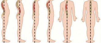
- MRI scanning of the cervical segment is prescribed to identify congenital malformations, infections, injuries, multiple sclerosis, severe scoliosis, and cancer.
- An examination of the thoracic region is prescribed for suspected tumors, diseases of the spinal cord, spinal column, or pinched spinal nerves.
- MRI of the lower back allows you to examine the “cauda equina” (lower spinal cord), spinal cord, epidural adipose tissue (the area between the hard shell of the spinal cord and the walls of the spinal canal), subarachnoid space (the cavity between the soft and arachnoid membrane of the spinal cord), cartilage pads between vertebrae.
Reference. An MRI scan of the head provides images of the brain as well as the cervical segment. This study is indicated for persistent headaches. MRI of the brain and spinal cord helps determine the origin of back pain that spreads to the legs, tumors, spinal cord conduction disorders, etc.
The study is carried out with or without contrast. To identify pathologies of blood vessels and tumors, MRI with contrast is prescribed. For this purpose, gadolinium-based solutions are used, which enhances the magnetic signal, which improves the quality of the images.
The following types of devices can be used for MRI scanning:
- Open type. They are used to examine patients who are overweight, tall, or afraid of confined spaces or children.
- Closed type. They look like a tunnel structure with a sliding couch. May provoke an attack of claustrophobia.
The decision about the type of MRI scan is made by the doctor together with the patient.
No ads 2
What determines the price of an MRI of the spine?
Prices for MRI of the spine in St. Petersburg largely depend on the need for contrast injection and the power of the tomograph. But these costs are fully justified: the earlier the disease is detected, the faster and more successful the treatment process will be. The cost of the study usually includes: preparing the patient, scanning, deciphering the results, issuing a written report and recording the tomograms on a CD.
| Service | Price according to Price | Discount Price at Night | Discount Price During the Day |
| from 23.00 to 8.00 | from 8.00 to 23.00 | ||
| MRI of the cervical spine | 3300 rub. | 2690 rub. | 2990 rub. |
| MRI of the thoracic spine | 3300 rub. | 2690 rub. | 2990 rub. |
| MRI of the lumbosacral spine | 3300 rub. | 2690 rub. | 2990 rub. |
| MRI of the craniovertebral junction | 3300 rub. | 2690 rub. | 2990 rub. |
| MRI of the sacroiliac joints | 4000 rub. | 3190 rub. | 3690 rub. |
| MRI of the coccyx (MRI of the sacrococcygeal region) | 3300 rub. | 2690 rub. | 2990 rub. |
| MRI of the cervicothoracic spine | 6600 rub. | 5380 rub. | 5980 rub. |
| MRI of three parts of the spine (MRI of the cervical, thoracic and lumbosacral regions) | 9900 rub. | 6900 rub. | 7900 rub. |
| MRI of the entire spine / MRI of the back | 9900 rub. | 6900 rub. | 7900 rub. |
| MRI of the central nervous system (MRI of the brain, MRI of the cervical, thoracic and lumbosacral regions) | 13200 rub. | 9590 rub. | 10890 rub. |
| Appointment with a neurologist | 1800 rub. | free after MRI | free after MRI |
| Comprehensive body diagnostics (MRI of the thoracic spine, MRI of the lumbar spine, ultrasound of the abdominal organs, ultrasound of the kidneys, ultrasound of the bladder, consultation with a neurologist, consultation with a therapist) | 11700 rub. | 7000 rub. |
What determines the cost of tomography?
![]()
Tomograph power
![]()
Applying Contrast
Personnel qualifications
![]()
Promotions and discounts