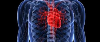
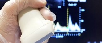
Doppler ultrasound of the vessels of the upper extremities is a diagnostic non-invasive technique aimed at recording the current in the blood vessels of the arms through the use of sound waves. The abbreviation used for the designation stands for Doppler ultrasound. The procedure is carried out using a scanner and makes it possible to correctly assess the condition of the vessels in real time.
The technique is based on the Doppler effect, which involves changing the frequency and length of emitted waves when the receiving party, i.e., blood cells, moves. They reflect the ultrasonic waves supplied by the sensor, in the process they are recorded and transmitted to the computer. The use of specialized software allows you to convert incoming signals into an image that can be studied by a specialist.
Thanks to it, the latter can accurately determine the condition of the walls of blood vessels and their pathological changes, identify plaques and occlusions, as well as strictures. According to the research results, it is possible to accurately determine the area in which there are violations and their extent. Having made the correct diagnosis, the specialist selects treatment tactics.
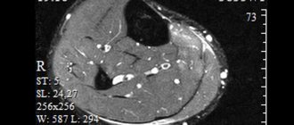
Duplex scanning of the vessels of the upper extremities - 4,000 rubles.
15-25 minutes
(procedure duration)
Indications
The list of diseases for which it would be useful to do this type of ultrasound is very extensive, although diseases of the veins and arteries of the upper extremities are much less common than vascular lesions of the coronary arteries, cerebral vessels and lower extremities. The main indications for the examination will be:
- Pain or tenderness in the shoulder or forearm and hardening of the veins.
- The appearance of swelling in the hands and soreness in the muscles.
- Convulsive movements (convulsions), numbness, decreased sensitivity of the fingers.
- Cold hands.
- Diabetes.
- Trophic ulcers.
- Increased cholesterol.
- Blood failure.
- Operations of the upper extremities.
What does diagnostics show?
The following pathologies are diagnosed using Doppler ultrasound:
- Thrombosis and embolism - blockage of blood vessels and circulatory disorders.
- Atherosclerotic changes in blood vessels, when cholesterol plaques form on their walls.
- Stenosis and occlusion are narrowing and blockage of a vessel.
- Phlebitis is inflammation of the venous wall.
- Thrombophlebitis - inflammation and blood clot formation.
- Aneurysm.
During Doppler sonography, it is only possible to estimate the speed of blood flow. There are also more informative methods - duplex and triplex scanning. With a duplex study, you can examine the condition of the veins and arteries, and with a triplex study, the vessels are tinted and the image is clearer.
Using ultrasound, you can study the following indicators:
- Assess the amount of blood in the veins and arteries.
- Identify blood flow disturbances and find out the exact location of the affected area.
- Assess how blood flows to tissues.
- Check the elasticity of the vascular wall.
Based on the results of the study, it is possible to make an accurate diagnosis and select effective treatment in the shortest possible time.
What will an ultrasound of the vessels of the upper extremities show?
The use of color Doppler ultrasound technology makes it possible to detect blood clots and dissection of vessel walls, and to determine the formation of atherosclerotic plaques. Stenosis . Carrying out an ultrasound scan of the arteries and veins of the upper extremities gives doctors the opportunity to diagnose narrowing of the lumen of blood vessels in the early stages. The presence of compactions and irregularities on the walls is a symptom of stenosis. But establishing only the fact of narrowing is not enough. The main thing is to determine the degree of criticality of the disease. Ultrasound screening data allows you to measure the level of the vessel lumen and stage the stenosis. This information is fundamental in deciding whether surgical intervention is necessary. A level of stenosis of 70% or higher is considered critical. Often, if a sufficiently strong narrowing in the upper extremities is detected, it is recommended to undergo an MRI of the main arteries of the head, especially if the patient suffers from headaches. Thrombosis . The presence of a blood clot in the lumen of a vein or artery is the main cause of blockage of blood vessels, which leads to impaired blood flow. Ultrasound examination techniques allow not only to find the exact location of the blood clot, but also to determine its structure and size and even determine the causes of blood clots. Problems with blood flow speed. Vascular ultrasound is one of the most effective methods for diagnosing blood flow speed. When scanning an area where blood flow is blocked, a sharp, high-pitched sound is produced and a sharp drop in the speed of blood flow is observed. Normal blood flow is characterized by a decrease in linear velocity in the direction from the proximal to the distal segments of the upper extremities and high peripheral vascular resistance. During an ultrasound scan, the doctor makes a comparative analysis of blood flow readings that were obtained at rest and under load. In the absence of pathologies, they should differ by at least 10-40%. If significant anomalies are detected, additional examinations may be ordered. Most likely, the patient will be offered to undergo an ultrasound of the lower extremities so that the diagnosis of the vascular system is complete.
| Service | price, rub. | Promotion Price |
| “Healthy Legs” program (duplex scanning of the veins of the lower extremities with assessment of valves and perforating veins and duplex scanning of the arteries of the lower extremities) | 3200 rub. | 3000 rub. |
| Ultrasound triplex scanning of N/C veins with assessment of valves and perforating veins | 2500 rub. | |
| Ultrasound triplex scanning of veins of the upper extremities | 2000 rub. | |
| Ultrasound triplex scanning of the arteries of the lower extremities with measurement of the brachiomalleolar index (BMI) | 2000 rub. | |
| Ultrasound triplex scanning of the arteries of the upper extremities | 2000 rub. | |
| Ultrasound duplex scanning of the arteries of the aorto-iliac segment | 1900 rub. | |
| Ultrasound duplex scanning of the abdominal aorta and visceral arteries | 1900 rub. | |
| Ultrasound duplex scanning of the inferior vena cava | 1700 rub. | |
| Ultrasound duplex scanning of the iliac vein | 1700 rub. | |
| Ultrasound duplex scanning of the renal arteries | 1700 rub. | |
| Intracranial duplex of cerebral vessels | 2500 rub. | |
| Ultrasound of neck veins | 2500 rub. | |
| Ultrasound of the brachiocerebral arteries (BCA) | 2500 rub. | |
| Heart ultrasound or echocardiography for adults | 2400 rub. | |
| Comprehensive neck diagnostics (MRI of the cervical spine, MRI of neck vessels, ultrasound examination of neck vessels, ultrasound of the thyroid gland and soft tissues of the neck, consultation with a neurologist) | 13200 rub. | 9500 rub. |
Advantages of performing ultrasound examination of hand vessels at the Doctor Nearby clinic
The quality of diagnostic results depends not only on the skill of the ultrasound specialist and the capabilities of the equipment, but also on how comfortable the patient feels during the process. At the Doctor Nearby clinic, all conditions have been created to ensure that patients feel as comfortable as possible. We guarantee high diagnostic accuracy and provision of all necessary data.
We do not have queues, since appointments are by appointment only. You can make an appointment at any time convenient for you by calling. Carry out diagnostics in a timely manner and trust them to the professionals at “The Doctor Is Nearby”!
Preparation for ultrasound of the upper extremities
Ultrasound of the arteries and veins of the upper extremities is a non-surgical method for examining the vascular system and does not require special training. It is not recommended to do it on an empty stomach; the patient needs to eat something light shortly before the procedure. The day before the examination, you should avoid strenuous physical activity. It's also important to get a good night's sleep. The day before the ultrasound procedure, it is advisable to avoid foods that can affect the condition of blood vessels, for example, energy drinks, caffeine (coffee, tea).
The role of elastography in the diagnosis of the thyroid gland?
Endocrinologists show less interest in elastography than in classical ultrasound, primarily due to the difficulty of obtaining reliable images. Multi-colored stains appearing on the screen are not always clear and understandable. To carry out elastography you need super modern and expensive equipment, which is rarely available.
The sensitivity and specificity of this test for detecting thyroid cancer is about 70-90%, the statistical accuracy parameter is estimated at 90%. Therefore, elastography cannot replace a thyroid biopsy.
The method is only one of the factors determining the endocrinologist's decision to prescribe a biopsy and surgical treatment. The combination of traditional ultrasound and elastography allows you to increase the sensitivity of cancer detection and narrow the group of changes classified as indeterminate. The prognostic value (probability of absence of disease) in this case is 99.8%.
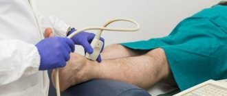
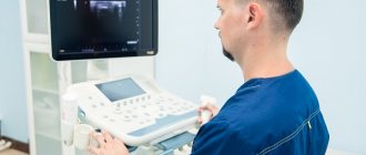
How is ultrasound of the vessels of the upper extremities performed?
The duration of an ultrasound scan of the veins and arteries of the upper extremities is from 20 to 30 minutes. The procedure is carried out in several stages:
- Consultation with a diagnostician and study of the medical history.
- Freeing the patient's hands from anything unnecessary (clothing, jewelry).
- The patient should assume a lying position.
- A special gel is applied to the area required for examination.
- The doctor attaches special sensors to the desired area and points and begins to study the image on the scanner screen, while at the same time listening carefully and monitoring the sounds of blood flow that the device records.
Different types of thyroid examination using ultrasound
There are several options for examining this organ. They all use ultrasound, but provide different data that is important in diagnosing organ pathologies.
- Classic examination.
Shows the structure of the thyroid gland. - Doppler
. Modern devices - Doppler sensors - allow you to obtain additional information about blood flow in the part of the organ of interest. - Elastography
. An examination that reveals mechanical properties and the presence of microcalcifications.
Possible contraindications
At the preliminary consultation, Dr. Khizriev S.M. explains possible contraindications for diagnostics. There are certain restrictions that make it difficult to carry out. These include thermal burn of the body at the point of contact with the sensor, mechanical damage to the skin.
It is not recommended to perform an ultrasound if the patient has increased motor mental arousal. It is also undesirable to perform a diagnostic procedure if you have an allergic rash on your hands.
Conducting an ultrasound also becomes impossible in acute emergency situations (asthma attack, bleeding, myocardial infarction). Diagnostics is also contraindicated in acute inflammatory processes and epileptic seizures.
Ultrasound of the thyroid gland with Doppler
Some ultrasound machines make it possible to determine the speed of blood flow in organs and tissues using a special Doppler sensor. In this case, the image is displayed in gray, and the areas of blood flow are colored. The intensity of the color depends on the direction and speed of the liquid.
In some devices, the color depends on the flow power. It is a physical quantity that depends on the flow volume relative to the surface and the test time. In this case, the endocrinologist receives information about an increase in blood flow - in the entire parenchyma of the thyroid gland, its fragments or in lesions. A generalized increase in blood flow is a symptom of inflammation in the thyroid gland.
Ultrasound with Doppler
Increased blood flow in the nodes is an eternal subject of debate. Theoretically, cancerous lesions should have increased blood flow in the central part of the lesion. But in fact, in most cases, lesions with clearly increased blood flow are not malignant.
Increased blood flow is usually associated with the rare medullary thyroid cancer, but the more common differentiated thyroid cancer usually does not show this pattern.
Today, the opinion that this method has limited value in the diagnosis of malignancy of nodes is becoming more and more justified.
Color or power Doppler ultrasound may be unreliable due to its physical limitations. The doctor can observe a similar picture in healthy areas and in real areas of increased flow due to errors - acoustic noise or motion artifacts. By increasing the signal strength, it is easy to confuse interference with lesions, and by decreasing the signal, there is a risk of missing a pathological focus.
The solution to the problem of correctly diagnosing thyroid pathologies using ultrasound is to choose an experienced specialist who can feel the difference between images, normal and abnormal, and who knows how to analyze the results.
ONLINE REGISTRATION at the DIANA clinic
You can sign up by calling the toll-free phone number 8-800-707-15-60 or filling out the contact form. In this case, we will contact you ourselves.
If you find an error, please select a piece of text and press Ctrl+Enter
Progress of the procedure
No special preparation is required for either duplex scanning or ultrasound scanning. It is not recommended to drink strong brewed tea or coffee before performing the procedure. They can cause vascular spasm.
Before the procedure, it is recommended to refrain from taking Nitroglycerin. This medicine helps dilate the arteries. But if taking a medication is necessary for health reasons, you should not refuse it.
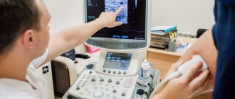
Special ultrasound signals that are reflected from blood vessels help to see possible abnormalities on the monitor. The sequence looks like this:
- You need to bare your hands and remove all jewelry from them.
- Diagnosis is performed in a lying or sitting position. To improve contact of the sensor with the skin, a transparent gel is applied to the area to be examined. Upon completion of the ultrasound, the product can be easily removed with a napkin.
- After installing the sensor on the area under study, the doctor continuously monitors the monitor. At the same time, the necessary marks are made to assess the condition.
It is considered absolutely safe for health. Its duration is 40 minutes. Interpretation of the ultrasound results is performed after completion of the procedure.