X-ray (x-ray) is a non-invasive medical test that helps doctors diagnose and treat various diseases. The images are obtained by exposing a specific part of the body to a small dose of ionizing radiation. This radiation is able to penetrate or be retained in the tissues of organs, thereby making it possible to obtain photographs or screen images. X-rays are the oldest and most commonly used form of medical imaging.
A chest x-ray is a very common test. This method is used to diagnose heart diseases separately or together with other chest organs. It is an X-ray examination that is among the first procedures that a patient will undergo if the doctor suspects a heart or lung disease. It is also used to test the effectiveness of treatment.
What abnormalities can be detected on an x-ray of the heart?
Everyone who suffers from heart disease has an x-ray of the chest taken in anterior and lateral projection. X-rays show the shape and size of the heart and the outline of the major blood vessels in the lungs and chest.
Content:
- What abnormalities can be detected on an x-ray of the heart?
- Indications for cardiac x-ray
- Contraindications for cardiac x-rays
- Types of research
- How to prepare for a chest x-ray
- What is the health threat from X-ray diagnostics?
- Pros and cons of cardiac radiography
The abnormal shape and size of the heart, calcium deposits in the blood vessels can be easily seen, and information about the condition of the lungs can be detected, such as whether the blood vessels are abnormal or whether there is fluid in or around them.
An X-ray will be able to detect an enlarged heart, which is often associated with heart failure or heart valve disease. The heart does not enlarge when heart failure occurs due to constrictive pericarditis. It causes scar tissue to form throughout the sac that envelops the heart (pericardium).
The appearance of blood vessels in the lungs is often more helpful in diagnosing heart problems than examining the heart itself. For example, dilation of the pulmonary arteries (the arteries that carry blood from the heart to the lungs) and narrowing of the arteries in the lung tissue suggests high blood pressure in the pulmonary arteries, which can lead to thickening of the muscle of the right ventricle (the lower chamber of the heart that pumps blood through the pulmonary arteries to the lungs ).

A chest x-ray may show changes or abnormalities in the lungs that are due to heart problems. For example, fluid in the lungs (pulmonary edema) may result from congestive heart failure.
Changes in the size and shape of the heart may indicate heart failure, fluid around the heart (pericardial effusion), or heart valve problems.
Because X-ray images show the outline of large vessels near the heart muscle—the aorta and pulmonary arteries and veins—doctors can identify aortic aneurysms, other blood vessel problems, or congenital heart defects.
A chest x-ray can detect the presence of calcium deposits in the heart or blood vessels. Its presence may indicate damage to the heart valves, coronary arteries, heart muscle, or pericardium. Calcium deposits in the lungs most often occur after a previous infection.
A chest x-ray is useful to monitor recovery after heart surgery. The radiologist may look at any lines or tubes placed during surgery to check for air leaks and fluid accumulation or air pockets. Pacemakers and defibrillators have special wires attached to the heart to monitor a person's normal heart rhythm.
When is fluorography enough?
Fluorography is performed annually - with the help of this preventive examination, tuberculosis, tumors (larger than with CT), fractures, and pathological changes in soft tissues are detected.
During fluorography, radiologists identify:
- Inflammatory processes in the lungs caused by fungi and bacteria (foci of viral pneumonia are much better visible on tomograms).
- Neoplasms (tumors from 1 cm) in the area of the lungs, mediastinum, heart, large blood vessels.
- Foreign objects in the respiratory tract.
To clarify the results of fluorography, the doctor may prescribe a computed tomography scan to the patient. A single or even double scan on a tomograph after fluorography is absolutely safe for a patient who has not had any other CT scans for a year.
Fluorography is mandatory for visiting the swimming pool and sports clubs, for future students and military personnel, workers in medical institutions, and catering establishments.
Indications for cardiac x-ray
A heart x-ray is performed for the following indications:
- planned treatment of patients suffering from coronary heart disease;
- suspicion of existing heart defects;
- asymptomatic, unstable angina;
- tracking conditions in the pulmonary circulation;
- asymptomatic hidden diseases of the cardiovascular system;
- to identify calcifications of the aortic valves, mitral valves, pericardial sac, myocardium after acute cardiac ischemia.

Cardiologists and surgeons refer their patients who complain of:
- having symptoms of angina pectoris - this is compressive pain in the chest, a feeling of lack of air, interruptions in heart function, rhythm disturbances;
- shortness of breath at rest and during physical activity, increased fatigue and severe weakness;
- tachycardia, arrhythmia - disturbances in the rhythm of heart contractions;
- swelling of the lower extremities and pronounced pallor of the skin;
- enlarged liver;
- persistent cough and fever.
X-ray of the heart makes it possible to suspect the following pathologies in a patient:
- exudative pericarditis - infectious inflammatory lesions of the pericardium (pericardium);
- abnormal increases in heart size called myocardial hypertrophy, which is found in coronary heart disease and hypertension;
- aneurysms of the walls after myocardial infarction, manifest themselves in the form of protrusions;
- dilated cardiomyopathy - damage to the heart muscles in the form of stretching of the heart chambers;
- a pronounced anatomical defect in the membranes of the heart muscle, most often these are various valve defects;
- calcium deposits on the walls of the coronary artery, the presence of seals, detection of atherosclerotic and thrombotic plaques;
- cloudiness, expansion of the root of the lungs, which means the possible presence of cardiac pathology.
Coronary diseases
Hypertension is the most common cause of heart enlargement.
This is explained by the fact that due to increased blood pressure, the organ is forced to pump large volumes of it and work in enhanced mode.
This causes the heart muscles to enlarge and the organ itself to expand.
Video:
If a person has ischemia, the heart muscle cells constantly do not receive enough nutrients, as a result of which they degenerate, and connective tissue appears in their place.
The latter, unlike muscle tissue, is not capable of contraction; as a result, the organ cavities become deformed and increase in size.
READ Is fluorography harmful during pregnancy?
What to do if an x-ray showed that the organ is enlarged, and the cause of this phenomenon is a disease of the cardiovascular system?
The answer to this question is simple and obvious - treat the root cause and return the organ to normal limits.
If a patient is diagnosed with hypertension, he is usually prescribed pharmaceuticals that lower blood pressure. The latter helps restore the normal size of the organ.
A patient with hypertension or coronary artery disease who has been diagnosed with an enlarged heart must take medications.
Video:
The fact is that despite the increased size of the organ, a large heart performs its most important function - pumping blood - much worse, which means that human organs and systems do not receive the nutrients they need - heart failure develops, and the whole body suffers.
That is, returning the organ to its normal size helps prevent heart failure, which in some cases can simply save a person’s life.
Contraindications for cardiac x-rays
Radiation examination is contraindicated for seriously ill patients, for example, with cancer. Doctors also prohibit carrying out this diagnosis if the patient has recently received a large dose of radiation. The total dose for the entire year should not exceed 5 millisieverts. The radiologist must indicate the radiation dose received at each patient diagnosis.
If the patient is under fourteen years of age, it is not recommended to undergo an X-ray examination of the heart. After all, the sensitivity of a young organism is several times greater and higher than that of an adult. Since the child’s organs are located close to each other, there is a high risk of irradiation of healthy internal organs.

Children are allowed to undergo X-ray examination in case of severe urination disorders, serious dental pathologies with the threat of accumulation of pus in the oral cavity, and with frequent and severe attacks of bronchial asthma. And it is strictly forbidden to do x-ray diagnostics if the Mantoux test is negative.
The doctor may order an x-ray of the chest organs, in particular the heart, in pregnant women quite rarely. Usually if there is already a suspicion of something very serious and necessary to determine the subsequent actions of the doctor. During the first trimester, a pregnant woman is strictly prohibited from undergoing X-ray diagnostics. After all, it is during this period that important foundations for the future health of the child are laid, and the formation of the main systems and organs of the fetus takes place.
If this study is necessary in subsequent months, it is carried out in this way: the abdominal area is covered with special protection from ionizing radiation, and the strength of the X-ray radiation itself is reduced.
It is interesting to consider the fact that in pregnant women, the size of the heart can usually increase and the position of the heart can shift, thereby arching the arch of the pulmonary artery. This is associated with increased blood flow to the fetus.
There are no contraindications to this type of examination during breastfeeding. After all, the rays do not affect breast milk and do not harm the baby.
What is fluorography and how does it differ from x-rays?
Fluorography and X-ray are similar methods for examining the chest organs; in both cases, X-ray irradiation is used. But with fluorography, the image is recorded on photographic film; it is small and not so informative. With X-rays, it is possible to obtain an image of the organ under study in almost life-size; it is more detailed, which affects the quality of diagnosis. In addition, with x-rays the radiation dose is slightly lower than with fluorography. Recently, chest X-ray has become a more common diagnostic method than chest fluorography.
Why do fluorography and chest x-ray?
Fluorography and chest X-ray are carried out for the following purposes:
- Detecting the causes of cough, shortness of breath, chest pain;
- Lung fluorography is aimed at diagnosing lung diseases such as pneumonia, tuberculosis (including early stage tuberculosis), sarcoidosis, lung cancer, COPD (chronic obstructive pulmonary disease), pneumothorax, cystic fibrosis, pulmonary edema;
- Heart examination, diagnosis of heart failure, atherosclerosis, aneurysm, aortic enlargement, heart valve diseases;
- Examination for traumatic injury to the chest (rib fractures, traumatic lung injury);
- Search for foreign objects in the stomach, esophagus, respiratory tract and lungs;
- Monitoring the correct placement of tubes, catheters in the respiratory tract, heart, vessels in the chest area;
Even in the absence of symptoms of disease, doctors recommend fluorography of the lungs or chest x-ray annually as part of a comprehensive therapeutic examination for early detection of the risk of developing many pathologies.
How to do fluorography of the lungs and chest x-ray?
Fluorography of the lungs and x-rays are painless procedures. As a rule, they do not require preliminary preparation. The dose of radiation received during fluorography is safe, but fluorography during pregnancy is contraindicated in most cases. You must inform your doctor about pregnancy.
Typically, fluorography and chest x-rays are performed in a standing position, but if necessary, fluorography or x-rays can be done in a lying or sitting position. You will need to hold still for some time so that the image does not turn out blurry. During fluorography of the lungs, the doctor may ask you to hold your breath for a few seconds. The doctor can take two pictures - from the back and the side, or only the frontal image. If you need to do fluorography quickly, the result of fluorography and x-ray can be ready within 15 minutes.
Fluorography of the lungs and x-rays may not show a number of diseases, for example, small cancer tumors, pulmonary embolism. In some cases, for a more accurate diagnosis of diseases, you may be recommended to undergo additional examinations - MRI, computed tomography, ultrasound, ECG, biopsy.
How to prepare for a chest x-ray
A chest x-ray does not require much preparation on the part of the patient. The person will need to remove any jewelry, glasses, piercings or other metal objects on the body before the procedure itself. Because this may make it difficult to make a correct diagnosis and obscure the image. Be sure to tell your doctor if you have a surgically implanted device, such as a heart valve or pacemaker. This determines which diagnostic method can be used in a particular case. Chest X-rays may be performed when the patient has metal implants. But other scans, such as MRIs, can be risky for people who have a metal device inside. Women with long hair are recommended to pin it up, since foreign bodies are prohibited from entering the diagnostic area. If hit, the quality and information content of the x-ray image drops significantly.

You will also need to undress to the waist before the x-ray and follow the instructions of the laboratory assistant. X-rays of the heart can be performed in three types of projections. During the procedure, the laboratory assistant will ask you to hold your breath for literally 10 seconds to get clear contours of the image.
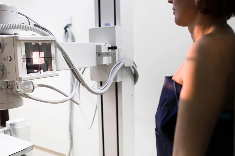
How does the research procedure work?
The X-ray image looks like a black and white photograph. Areas that are translucent and transmit X-rays are shown in black, and areas that cannot transmit ionizing radiation are highlighted in white. That is why the bones and heart muscle in the image are white.
The X-ray machine is located in a special large room with a movable X-ray camera attached to a large metal arm called an arm. The patient should stand next to the “plate”. This plate contains X-ray film or a special sensor that records the images on a computer. The laboratory assistant will tell you how to stand correctly - raise your arms and bend them at the elbows, and record in the front and side projections. Usually the manipulation is carried out in two projections, but for a more accurate and specific diagnosis it can be carried out in three or four - anterior, lateral left, oblique left and right, at an angle of forty-five degrees. It is the images in the oblique projection that help to view the walls of the aortic arch and the walls of the myocardium, which are not examined in the lateral view. For example, oblique right imaging makes it possible to examine all parts of the heart.
While the images are being taken, you will need to take a deep breath and hold your breath as requested by the technician, and to keep your body completely still. If you move, the pictures may turn out blurry. This procedure takes about ten minutes, after which you need to get dressed and go about your business.
This diagnostic method is one of the cheapest and most painless, non-invasive studies of the heart muscle. Only with an X-ray with contrast, the patient can taste the lime barium solution.
What is the health threat from X-ray diagnostics?
The effect of X-ray radiation on humans directly depends on the radiation dose. After all, exposure to large doses of radioactive ionizing radiation can have a bad effect on the general condition and health; it lingers in tissues, destroys deoxyribonucleic acid and, accordingly, provokes disruptions in the functioning of important organs and systems.
Because X-rays emit little radiation, people avoid and are afraid to do this type of research. But X-ray machines emit a very small dose of radiation and do not cause significant harm to human health. At the same time, the World Health Organization recommends not taking x-rays more than once a year, reducing the risks of radiation accumulation.