Reception is conducted by:
Nosova Svetlana Valerievna -
Ultrasound diagnostic doctor
Make an appointment
Ultrasound examination is a painless and informative diagnostic method. Using ultrasound, the condition of soft tissues, internal organs, blood vessels, joints and other structures is studied. The method is used to study paravertebral structures in children, even newborns.
An ultrasound of the child’s cervical spine can be performed at the Profimedica multidisciplinary clinic in St. Petersburg. The center has high-precision equipment for ultrasound diagnostics. The study is performed by sonologists with extensive experience working with children.
Indications for ultrasound of the cervical spine
An ultrasound scan of the spine provides information about the condition of the spinal cord and nearby soft tissues. The study is prescribed in the following situations:
- suspicion of a neck injury received during childbirth or at home;
- signs of abnormal development of the spinal cord;
- elucidation of the causes of congenital torticollis;
- suspicion of complications of injury;
- detection of spina bifida;
- pain in the head and neck area;
- impaired function of the upper limbs;
- numbness in the upper extremities;
- dizziness;
- disturbances of consciousness;
- instability of blood pressure;
- problems with memory and concentration;
- sleep disorders, etc.
This diagnostic procedure can be prescribed by a pediatrician, neurologist, or orthopedist after an objective examination of the child.
Indications
Indications for ultrasound examination of the neck may be:
- the presence of a palpable tumor formation, growth, swelling, or wen in the neck area;
- inflammation of the salivary glands;
- noise in ears;
- headaches and dizziness;
- enlarged thyroid gland;
- pain when turning the neck.
The decision about the need to do an ultrasound of the neck can be made by both the attending physician and the patient himself. This examination is safe for human health and does not require a referral from a medical specialist. However, in order to figure out which neck ultrasound is best to do, you should start your diagnostic journey with a visit to a neurologist or therapist. Based on the results of the initial examination, the doctor will be able to determine which form of additional examination should be chosen in your case.
5 most common causes of neck pain
- incorrect posture when working at the computer and with gadgets
- presence of drafts
- lack of gymnastics, which would strengthen muscle structures and improve nutrition of spinal tissues
- osteochondrosis
- herniated intervertebral discs.
What will an ultrasound of the cervical spine show in a child?
During an ultrasound of a child's neck, the condition of the spinal cord, intervertebral discs, main blood vessels, and surrounding soft tissues is assessed. As a result of the study, the following may be discovered:
- developmental anomalies;
- damage to spinal structures;
- changes in the width of the spinal canal;
- hernias;
- arterial pathology (stenosis, dilatation, abnormal location).
Conclusion Ultrasound is not a diagnosis. This is a protocol in which the sonologist describes the characteristics of the anatomical area. The attending physician identifies the pathology, taking into account medical history, complaints, laboratory results and other diagnostic procedures.
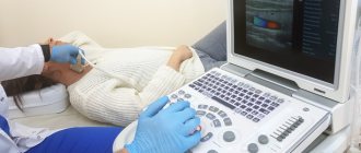
How is an ultrasound of the neck performed?
An ultrasound examination of the neck area takes no more than 15-20 minutes. It is performed in a supine position. The person is placed on a couch covered with a disposable sheet. A 10 cm thick pad is placed under the shoulder joint area, the head is turned in the opposite direction to the doctor so that there is maximum access to the neck area. The device's sensor and area of study are lubricated with a special gel for smoother glide and tight contact with the skin. This will allow the ultrasonic waves to pass through unhindered. During the examination, the diagnostician sequentially scans the areas, recording the data in the protocol, and also gives comments during and after the examination. Despite the sonologist’s explanations, the patient with the result obtained after the ultrasound is necessarily referred to the attending doctor. It is the attending physician who makes the final diagnosis and prescribes treatment.
Pathologies determined during the examination
What does an ultrasound of the soft tissues of the neck show:
- artery aneurysm;
- thrombosis;
- atherosclerotic plaques;
- degree of narrowing of the vascular lumen;
- tone, condition of vascular structures;
- focal or diffuse pathologies of the thyroid gland, neoplasms of any etiology;
- hematomas;
- cysts, abscesses, hemorrhages;
- lymphadenopathy;
- hygroma cysts;
- condition of the lymph nodes, presence of metastases, inflammatory processes.
Using an ultrasound examination, the doctor will determine the localization of the pathological focus or tumor and will be able to assess the structure and degree of correspondence with the surrounding healthy tissues. Also, during an ultrasound, you can preliminary assess the nature of the tumor.
If the tumor is malignant, its shape is irregular, its structure is heterogeneous, the blood flow is increased and pathological. However, at an early stage, it will be difficult to determine the degree of malignancy of the tumor using ultrasound. Therefore, to make a diagnosis, the doctor will give a referral for additional examination.
Indications for performing NSG and ultrasound examination:
- intrauterine hypotrophy;
- intrauterine infections;
- previously identified brain defects;
- pathology in hypoxic, ischemic, toxic and metabolic disorders of the brain;
- birth injuries of the brain and spinal cord;
- seizures and other symptoms of neurological diseases;
- multiple malformations of organs and systems;
- genetic diseases.
To assess the blood circulation of the brain in children after the “closure” of the fontanel, modern ultrasound techniques are used - transcranial Dopplerography. In some cases, this technique makes it possible to clarify the cause of headaches in children (angiospasm, increased intracranial pressure). Transcranial Dopplerography allows you to examine large vessels of the head and neck, see the lumen of the vessel, the degree of patency of veins and arteries. This method is absolutely painless and has no contraindications, which makes this procedure possible for young children.
We invite you to do a study at our medical center.
How is it carried out?
It is possible to perform an ultrasound of the child’s brain only until the age when the fontanelles close. The examination is carried out through the anterior, lateral fontanelles or foramen magnum at the base of the child’s neck.
During an ultrasound, the diagnostician moves a special sensor over the baby's head. The resulting pulses are sent to the device and displayed on the monitor in the form of images. This is a comfortable procedure that can be done in the clinic, even while the child is sleeping.
You can sign up for ultrasound diagnostics by calling the reception at (495) 951 87 63 or
In our center, infants undergo comprehensive diagnostics (NSH, ultrasound of the hip joints, EEG/video-EEG monitoring) and consultations with a neurologist, orthopedist, geneticist, and ophthalmologist.
Ultrasound diagnostics
Ultrasound examination (ultrasound) is one of the procedures necessary when examining internal organs of both adults and children. Almost all specialized specialists refer ultrasound, because with its help it is possible to diagnose diseases at the initial stages.
In our clinic, you can perform an ultrasound even on a newborn child - the examination is absolutely painless and harmless. For your convenience, we work both weekdays and weekends.
Ultrasound in children
A modern diagnostic method that allows a detailed assessment of the condition of internal organs and surface tissues, their structure, size and boundaries, identifying pathological changes in them and determining their exact location and extent.
Ultrasound is based on the ability of ultrasonic waves to be reflected when passing boundaries between different media. The advantages of the technique include its non-invasiveness, high information content and safety (no radio exposure). Ultrasound is a mandatory procedure performed as part of routine screening for all children aged 1-1.5 months.
NSG in children
Ultrasound examination of brain structures performed through the child’s fontanel. It is carried out in all children, without exception, in the 3rd month of life.
Indications for neurosonography in children:
- identification and exclusion of congenital abnormalities of brain development;
- prematurity;
- perinatal brain damage, birth trauma;
- signs of hypoxia and ischemia in a child;
- increased excitability or inhibition;
- convulsions.
There is no need to prepare for NSG in children.
[collapse]
Ultrasound of the hip joints in children
A diagnostic procedure that is required to be performed in the first year of a child’s life. It is carried out as part of the diagnosis of hip dysplasia and determination of its form (dislocation, subluxation). A routine ultrasound is performed at the age of 1-3 months, and if pathology is detected, it is repeated after 1-1.5 months.
The procedure is mandatory for children with breech presentation, birth injuries, unfavorable family history, impaired hip abduction, asymmetry of the folds of the buttocks, or shortened limbs. No preparation is required for the test.
[collapse]
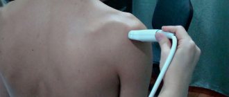
Ultrasound of joints and soft tissues in children
A modern method for studying joints and para-articular soft tissues (cartilage, musculo-ligamentous apparatus).
In addition to the mandatory examination of the hip joints, it is performed in children in the following cases:
- injuries;
- swelling, pain in the joint area;
- local hyperthermia;
- joint deformation, limited mobility;
- crunching sound when moving;
- identification of palpable pathological elements in the paraarticular region;
- autoimmune and endocrine disorders.
The advantages of this diagnostic method are its harmlessness (no radio exposure), high information content, and non-invasiveness. No special preparation is needed to perform it.
[collapse]
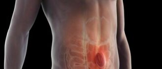
Ultrasound of the abdominal cavity and retroperitoneal space in children
A highly informative diagnostic procedure that allows a detailed assessment of the condition of the liver, gall bladder, pancreas, spleen, kidneys, adrenal glands and bladder. Ultrasound allows you to determine the shape and size of internal organs, assess the echogenicity of tissues and the homogeneity of their structure, identify pathological changes and determine their exact location and extent.
Ultrasound of the abdominal cavity in children is performed in the following cases:
- routine examination at the age of 1-1.5 months;
- nausea, belching, flatulence, bitterness in the mouth;
- diarrhea, constipation, unstable stool;
- pain, bloating;
- yellowness of mucous membranes and skin;
- frequent unmotivated rise in body temperature;
- swelling;
- changes in blood and urine tests;
- complicated childbirth, perinatal abdominal trauma.
The procedure is carried out 6-8 hours after the last meal.
[collapse]
Ultrasound of the esophagus and stomach in children
A modern method for diagnosing diseases of the esophagus and stomach in children, allowing to identify GERD, gastritis, gastric ulcer and other pathologies.
Indications for use in children:
- frequent regurgitation in infants, vomiting;
- heaviness and pain in the right hypochondrium;
- bad breath;
- sour belching, heartburn;
- weight deficiency;
- persistent diarrhea;
- frequent respiratory illnesses, unmotivated cough.
Mandatory preparation is required: strict fasting 6-8 hours before the procedure. During the study, the child must drink 200 ml of still water. It is prohibited to conduct examinations after FGS.
The most important advantages of the technique are its non-invasiveness, accessibility, absence of contraindications and ease of implementation.
[collapse]
Ultrasound of the thymus (thymus gland) in children
The study is aimed at identifying the pathology of the thymus gland, located behind the sternum. The thymus is the main organ of immunogenesis in children.
The procedure can be performed as part of preventive screening at the age of up to 1 year and at older ages according to the following indications:
- frequent colds with severe complications;
- tendency to allergies;
- prolonged unmotivated increase in body temperature;
- swollen lymph nodes;
- persistent cough, heart problems;
- superior vena cava syndrome.
Ultrasound of the thymus gland in children is performed to exclude thymomegaly, thymic neoplasia, its hypo- and aplasia, and congenital developmental anomalies.
[collapse]
Ultrasound of the thyroid gland in children
A highly informative study that allows you to assess in detail the size, shape and structure of the thyroid gland in children, determine the density and uniformity of its tissues, identify nodes, neoplasms and determine their size, boundaries, examine regional lymph nodes near the gland.
Indications for conducting research in children are:
- increase in size of the organ (visualized and palpable);
- the presence of palpable nodes and compactions in the tissues of the gland;
- discomfort when swallowing;
- increased sweating, nervousness, irritability of the child;
- sleep disturbances, shortness of breath, unstable stools;
- a sharp increase or decrease in body weight over a short period of time;
- living in iodine-deficient areas;
- family history;
- arrhythmia confirmed by ECG results.
No special preparation is needed before the procedure.
[collapse]
Ultrasound of the scrotum in children
A highly accurate diagnostic method that allows detailed examination of the testicles, epididymis and spermatic cords with their membranes. During the procedure, the shape, size of the testicles and appendages, their location are assessed, the structure of the tissues, their echogenicity and homogeneity are examined, neoplasms and developmental anomalies, functional disorders are identified.
Indications for ultrasound of the scrotum in children:
- testicular injuries;
- swelling, increase in size, pain, redness, local hyperthermia in the scrotum area;
- the appearance of palpable seals in the testicles;
- decrease in testicle size;
- delayed sexual development.
Using ultrasound, children are diagnosed with:
- inguinoscrotal hernia;
- varicose veins of the spermatic cord;
- cryptorchidism;
- inflammatory and neoplastic pathologies.
No special preparation is needed before the study.
[collapse]
Ultrasound of the pelvic organs in children
A method for diagnosing reproductive pathology in girls, allowing a detailed assessment of the condition of the cavity and cervix, fallopian tubes, ovaries, and bladder.
The study is carried out:
- for menstrual dysfunction;
- late or too early sexual development;
- for pain in the lower abdomen.
Ultrasound diagnostics makes it possible to identify congenital anomalies of the reproductive organs and bladder, their inflammatory, degenerative and tumor lesions.
Ultrasound of the pelvic organs in children is performed through the anterior abdominal wall. Satisfactory visualization requires that the bladder be full and the rectum previously emptied using enemas. For menstruating girls, the procedure is performed on the 5-10th day of the menstrual cycle.
[collapse]
Ultrasound of the cervical spine in children
Highly informative study of the atlas-axial joint and surrounding soft tissue structures.
Indications for its implementation in children are:
- complicated childbirth;
- obstetric care for large fetuses or fetal malnutrition, stimulation of labor;
- perinatal encephalopathy;
- diagnosis of osteochondropathy;
- torticollis;
- hyperexcitability syndrome in a child;
- cardiac arrhythmia confirmed by ECG results.
No special preparation is needed for the procedure.
[collapse]
Interpretation of ultrasound results of soft tissues of the neck
The doctor can evaluate the first results of the study directly during the procedure. All data is recorded in an ultrasound protocol, which must be shown to the specialist who gave the referral for diagnosis.
The results that meet the standards are:
- homogeneous structure;
- smooth contours;
- absence of any neoplasms or swelling;
- absence of inflammatory processes in soft tissues and lymph nodes;
- absence of foreign bodies in the larynx;
- size of the thyroid gland – no more than 2 cm, width – 2 – 2.5 cm;
- the length of the carotid artery on the right is 7 – 12 cm, on the left – 10 – 15;
- systole to diastole ratio 25 – 35%;
- the shape of the carotid artery is straight or slightly tortuous;
- pulsation in the vertebral artery is continuous.