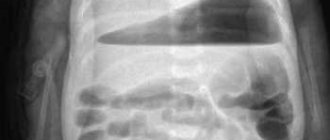
A barium pass is an x-ray of the small intestine. The study helps to assess the general condition of the organ, its motor and evacuation function. All data is recorded in photographs.

The small intestine has many bends and narrowings, which the barium passage helps to examine. The study is mainly carried out for tumors in the organ, obstruction or narrowing of the intestine.
Information! On this page read about chest x-rays
Indications
Passage of barium through the small intestine is used in several situations. First of all, it is prescribed in case of causeless diarrhea. If a person leads a healthy lifestyle and eats right, but has constant diarrhea, then this study is necessary to identify the cause of the disorder. In addition, such a symptom may indicate the development of a more serious pathological process.
The procedure is also necessary when blood impurities are found in the stool. However, in this condition, the study must be carried out with extreme caution, since incorrectly performed actions can provoke even more severe bleeding.
An indication for the passage is pain in the abdominal area. An X-ray with a contrast agent is performed if the pain syndrome is accompanied by other symptoms. However, it must be remembered that if the pain is pronounced, then this type of examination cannot be prescribed.
A specialist can refer you for a barium passage even if you are rapidly losing weight. This is explained by the fact that unreasonable weight loss along with proper nutrition may indicate the appearance of cancer.
The procedure is also used for congenital anomalies. Thanks to x-rays, it is possible to identify the abnormal structure of the intestines, as well as determine how it affects the digestive process.
In what cases is the procedure prescribed?
An X-ray of the intestines with barium is performed to clarify the diagnosis and evaluate the therapeutic measures taken for the following conditions and diseases:
- long-term defecation disorders (diarrhea, constipation);
- suspicion of intestinal obstruction;
- fistulas, diverticula;
- if it is impossible to carry out diagnostics using instrumental methods in the presence of disorders in the intestines (for example, due to anal fissures, hemorrhoids and other conditions);
- suspicion of the development of tumor processes;
- diaphragmatic hernia;
- sudden changes in body weight downward (possibly malabsorption of nutritional compounds);
- chronic colitis;
- presence of a foreign body;
- the presence of impurities (mucous, blood) in the stool, changes in their color and odor.
An X-ray may also be prescribed after the operation to ensure the patency of the organ, as well as to promptly detect adhesions and scars.
Contraindications
Despite the fact that this technique is effective and informative, there are certain limitations to its implementation. Experts identify a number of conditions and situations when examination cannot be carried out.
Pregnancy
Doctors do not recommend the procedure during pregnancy. This is explained by the fact that when the intestines are full, there is pressure on the uterine body. In addition, barium can negatively affect fetal development.
X-ray radiation also poses a danger to children. An examination can be carried out only when there is an emergency need, and the use of other diagnostic techniques is not possible for some reason.
Biopsy
This study involves taking a small piece of biological tissue for further study. If barium is present in this area of the mucosa, irritation or an inflammatory process may begin.
Severe pain
During the procedure, the patient must remain motionless. Severe pain usually disrupts this process. In addition, when exposed to a contrast agent, the pain can only intensify until loss of consciousness.
Intestinal perforation
If this pathology is suspected, the method is also not used, or the contrast is replaced with a water-soluble one. In case of perforation during the use of barium, the risk of developing peritonitis increases several times.
Possible complications of irrigography
If irrigography is performed by an experienced specialist, then no consequences for the patient are usually noted. The patient recovers literally within a few hours after the examination.
However, if a person undergoing diagnosis has gastrointestinal pathologies, in particular, thinning of the walls, colon rupture may occur. The situation requires urgent surgical intervention. This happens extremely rarely and this is not a reason to refuse an examination if the results of other studies are available.
Procedures such as MRI, ultrasound, CT and irrigography do not replace, but only complement each other and help to create an accurate picture of the disease.
Attention. The article is not a guide to action; be sure to consult your doctor.
You can learn more about preparations for the event from the video:
One of the most common methods for diagnosing the condition of the digestive tract is an X-ray of the intestines. Modern X-ray equipment makes it possible to quickly and painlessly identify the presence of many pathologies, carry out differential diagnostics in controversial situations, and evaluate the quality of the therapy provided. Barium is often used to take x-rays of the intestines. This contrast agent helps to obtain a clearer picture of the presence/absence of changes in the organ.
Preparation
Despite the fact that the examination is a painless procedure, during the procedure the patient may experience discomfort. To keep their manifestation to a minimum, it is necessary to follow some preparatory rules.
About four days before the appointed date, it is recommended to follow a slag-free diet. It is necessary to exclude fatty, fried, sweet foods, carbonated drinks, fresh vegetables and fruits from the diet, as they can cause increased gas formation.
Why can our articles be trusted?
We make health information clear, accessible and relevant.
- All articles are checked by practicing doctors.
- We take scientific literature and the latest research as a basis.
- We publish detailed articles that answer all questions.
The day before the examination, you are only allowed to have breakfast. After this, the patient cleanses the intestines using Fortrans. This medication is the only one that allows cleansing procedures to be carried out simultaneously for the large and small intestines.
The drug in the amount of one packet must be diluted in a liter of water and drunk within 60 minutes. Throughout the day you need to take 4 of these sachets.
After using this remedy, painless diarrhea appears. Colon cleansing ends at the moment when clean water flows out of the anus instead of feces.
It is prohibited to eat barium on the day of the passage.
Is it harmful to have an X-ray examination of the intestines?
You can often hear the opinion that an X-ray of the intestines is a very dangerous procedure for health. However, if all safety measures are followed, then the likelihood of developing any negative consequences of radiation is negligible. The rays themselves do not “get stuck” in the body, and their intensity is strictly controlled by a specialist and does not exceed acceptable standards.
In this case, the doctor selects the level of radiation in accordance with the individual characteristics of the patient. In addition, the device directs the rays exactly to the place that needs to be studied. The surrounding structures are either not exposed to radiation at all, or it is minimal.
Another concern relates to the effects of using barium on bowel x-rays. This substance rarely leads to allergies, but in most cases it causes difficulties with bowel movements. To be fair, it should be noted that not all patients follow the diet after undergoing diagnosis. Lack of enough water and poor diet in the first days after an x-ray naturally lead to constipation. To avoid this, you need to maintain a smooth transition to your usual food and monitor your water regime.
Intestinal X-ray is, in general, a safe, informative, inexpensive procedure. If you carefully read the reviews of patients, most of them note that the procedure is successful and completely painless only if you follow the doctor’s instructions. It must be remembered that no article from an Internet source can take into account the individual characteristics of the body. Therefore, every moment before and after the x-ray (and during it) must be agreed upon with the attending physician.
An experienced Russian doctor will tell you how to properly prepare for an x-ray procedure in a short video.
How to do it
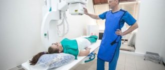
The patient removes all metal jewelry, clothing and underwear, and takes a position chosen by the doctor.
Depending on what kind of research will be carried out, this may be a horizontal or vertical position. In the first case, the examination of the intestines will be more qualitative and accurate, and the second allows you to speed up the process of barium dissolution.
In some situations, the specialist recommends taking the Trendelenburg position. The patient's pelvis should be located above the body at an angle of 45 degrees. This position allows you to more accurately examine the intestines, stomach and diaphragm.
There is a gradual passage and filling of all parts of the organ with barium. Thanks to the massage movements of the abdomen, the contrast agent is evenly distributed throughout the gastrointestinal tract.
Causes of thickening of the intestinal walls
Diseases of the gastrointestinal tract occupy one of the first places among other diseases.
When it is necessary to examine the large intestine, the drug is injected directly into the anatomical structure being examined.
In a comprehensive study, the solution is taken orally by the patient before the procedure.
The mixture is also prepared before starting the study. Its composition contains elements such as air, barium sulfate and aluminum gel. Additionally, sorbitol, sodium citrate or an antispasmodic drug may be added. A single dose is about 400-650 grams.
The resulting solution must be drunk in several doses. First, take a small sip. This is necessary so that the specialist can assess the quality of the mixture, as well as at what speed it moves through the esophagus.
Next, the patient drinks the remaining amount. After this, the entire gastrointestinal tract is filled with barium for some time.
The duration of radiography takes no more than forty minutes. During this time, it is possible to capture 6-10 images. The last picture is taken after the solution is removed from the human body.
What should you take with you to the examination?
Contrast radiography is a rather complex procedure that requires extensive preparation. It is not enough to just cleanse the intestines. It is advisable to take into account other points so as not to get into trouble during irrigography.
When going for irrigography, you need to take with you:
- Direction.
- Passport, medical insurance and results of past examinations, if any.
- A diaper or sheet (optional).
- Women's robe and slippers.
- Wet wipes or toilet paper.
- Spare underwear and clothing below the waist (preferably), since barium leaves white stains on things.
- Light snack.
After receiving the results, it is good to eat something nutritious, since enemas are very draining on the body.
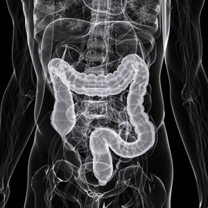
Decoding the result
The interpretation of the data obtained is carried out exclusively by a specialist. The examinee will receive the final conclusion and the photographs obtained during the examination. The doctor, as a rule, evaluates the entire process over time.
So, if barium is presented in the form of flakes in the image, this may indicate a violation of absorption by the small intestine. Also in the pictures you will be able to see diverticula, multiple neoplasms, and intestinal elongation.
The passage of barium through the intestines is the most informative technique used for a more accurate examination of the organ. Despite the high accuracy of the study, this method has certain contraindications.
Features of the technique
Barium passage is not carried out for preventive purposes. Diagnostics is used only for obvious disorders of the intestines, and if it is not possible to examine the organ by other methods. Barium passage is a highly informative technique, without which it is sometimes difficult to make a diagnosis.
The advantages of the study include less pain compared to colonoscopy (and this diagnosis applies only to the large intestine). With the help of a barium passage, the entire gastrointestinal tract, including the small intestine, is assessed. The method is inexpensive, but takes a long time.
When is a plain radiography of the abdominal organs with contrast done?
Survey radiography of the abdominal organs is done for the following diseases:
- pancreatitis - inflammation of the pancreas;
- cholecystitis – inflammatory changes in the gallbladder;
- abscess - a purulent cavity;
- urolithiasis, nephrolithiasis – stones in the kidneys and urinary tract;
- intussusception - twisting of the intestine around an axis;
- blockage of the lumen by a tumor;
- diverticulitis;
- traumatic injuries;
- stomach pain.
For these diseases, a survey x-ray of the abdominal organs is first performed. This requires preliminary preparation. It involves a cleansing enema 2 hours before the procedure.
After a survey image is taken and in the absence of X-ray signs of perforation of the intestinal wall, organs are contrasted with oral barium.
Attention! During a contrast study of the abdomen and large intestine (irrigoscopy), the contrast is administered through the rectum.
Important!
The reliability of the results depends on the correct preparation for the procedure. To ensure a comprehensive examination, the digestive tract is cleared of large volumes of feces, strictly following the prescribed diet.
You should follow a slag-free diet for two days before the test. Fatty and fried foods, coffee, fresh vegetables and fruits, as well as foods that promote gas formation (rye bread, legumes, baked goods, pearl barley and oatmeal porridge, milk, etc.) are excluded from the diet. Instead of fresh bread, you can eat Lenten cookies and crackers made from wheat buns.
It is recommended to drink plenty of fluids (up to 2 liters per day, in the absence of contraindications). Drinks such as fermented milk drinks, dried fruit compote, and weak green or herbal tea are allowed.
The day before the test, breakfast and lunch should be light. The last meal is the day before at 19:00, after which you are allowed to drink weak tea and water.
Radiography of the barium passage must be carried out! on an empty stomach, in the morning before the procedure it is forbidden to have breakfast.
To conduct a study, the doctor must first study existing information about your disease.
We ask you to bring your medical history or outpatient card with you to the study.
If there is no data on the course of the disease in the medical history or outpatient card, this information can be provided in any form - in extracts, research forms, copies of medical documents. The more the doctor knows about the patient's situation, the more effective the diagnosis will be.
You must also bring with you dry and wet wipes (for hygiene after bowel movements) and a clean sheet. You can ask for a sheet in advance from the nurses in the department where you are being treated.