Computed tomography is one of the proven and reliable methods for studying various pathologies of internal organs located in the abdominal cavity. This type of diagnosis allows you to quickly obtain three-dimensional images of the area under study, which allow you to quickly identify pathological formations of even small size. Thanks to this, the doctor can detect benign and malignant tumors, damage to the lymph nodes, changes in blood vessels, as well as other anomalies in the development of internal organs at the earliest stage of their appearance.
Features of computed tomography:
- high research accuracy. Using tomography, it is possible to accurately identify the organic causes of the disease, since this diagnostic method makes it possible to distinguish between tissues that differ slightly from each other in density;
- minimum time for processing results. This study does not require much time for preparation, so tomography is very often performed in emergency cases when there is a real threat to the patient’s life;
- safety for human health. Modern tomographs fully comply with safety requirements, guaranteeing the absence of any side effects after the examination procedure.
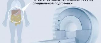
Diet rules before computed tomography of the abdominal cavity
- The day before the test, the patient must exclude gas-forming foods from the diet. These include primarily: beans, peas, cabbage, fermented milk products, fruits, with the exception of citrus fruits, baked goods, naturally fermented drinks (beer, kvass, champagne), carbonated water and juices. You can safely include water-based porridge, eggs, fish, chicken, and steamed meat in your diet.
- The evening before the procedure, a light dinner is allowed (until 21:00), and then only drink. After dinner, to relieve excess gas formation, you need to take enterosorbents - activated carbon, espumizan, enterosgel. The next day you need to come for the study strictly on an empty stomach. The minimum interval between the procedure and eating should be at least 6 hours.
Dietary ration
A diet prescribed by your doctor will help you prepare for testing. This rule reduces the likelihood of flatulence and improves stool output. The exact result of the test depends on the wave-like contraction of the walls of hollow organs of a tubular nature - the larger they are, the more opportunities for data distortion. Diet before diagnosis is considered a mandatory rule to obtain reliable information, and therefore must be strictly followed by the patient.
What can you eat before the procedure?
Doctors recommend minimizing the consumption of foods that can cause flatulence and increase intestinal activity: fresh vegetables and fruits, legumes, nuts, baked goods. Additionally, you will need to reduce food intake that affects stool retention: black tea drinks, dairy products, pasta, sweets. The dietary ration is calculated for at least three days.
Approximate diet of patients:
- Dishes from boiled beef, poultry, rabbit.
- Steamed cutlets.
- Casseroles or puddings.
- Cottage cheese with a minimum fat content.
- Boiled vegetables.
- Mashed potatoes.
- Fish products.
- Galette cookies.
Testing must be done on an empty stomach. It is recommended to stop eating 5-6 hours before the start of the session. For a study scheduled for the morning, dinner should be fairly light. In the future, food is not accepted until the diagnosis is completed. If checking your condition in the afternoon, you can have a snack six hours before the start.
Bowel preparation on the day of diagnosis
On the day of diagnosis, you will need to properly prepare your intestines. To do this, you should empty it naturally. If the patient suffers from constipation, a mild laxative can be taken. An enema should only be used as a last resort. Any mechanical intervention on the rectum leads to more severe intestinal spasms, which only complicates the diagnosis. On the day of the study, 30-40 minutes before the start of the procedure, you should take two No-Spa tablets. It will relax the smooth muscles of the intestines.
Features of diagnostics
The stages of performing a CT scan of the stomach and small intestine are not complicated, but must be followed in a clear sequence.
Best materials of the month
- Coronaviruses: SARS-CoV-2 (COVID-19)
- Antibiotics for the prevention and treatment of COVID-19: how effective are they?
- The most common "office" diseases
- Does vodka kill coronavirus?
- How to stay alive on our roads?
You need to arrive for the procedure 10-15 minutes before it starts; before doing this, you must remove all metal jewelry and a belt. Upon entering the office, the patient exposes the desired area of the body and lies down on a special table with his stomach down, sideways or on his back. Your arms need to be thrown back behind your head (if necessary, the specialist can secure the body with belts to avoid unnecessary body movements).
A tube will be inserted into the anus to a depth of 5 centimeters, through which the intestines will be filled with air. An electric pump may be used to fill the small intestine with carbon dioxide. Filling the intestines with air is necessary for its straightening and smoothing out folds and folds.
The table with the patient moves into the scanner. At this time, the patient should not breathe (hold his breath for 10-15 seconds). When the patient holds his breath, pictures are taken. Next, the table moves out from under the scanner and the patient turns over. Then the table slides under the scanner again and a similar manipulation is performed. When the patient lies on his side, the tomograph examines the stomach and determines its condition, size and other parameters.
The tube is removed only at the end of the procedure. The duration of the diagnosis is from 20 to 30 minutes. Using CT, the thickness of the walls of the small intestine is measured, and pathologies found in the stomach are assessed.
How to prepare for a CT scan of the abdomen, kidneys and bladder
If you plan to have a CT scan of the abdomen, retroperitoneum, and bladder, you need to make sure that the bladder is moderately full. To do this, an hour before the procedure, you can go to the toilet, and then drink two glasses of water and not urinate anymore. There is no need to overfill the bladder, since it will be quite uncomfortable to lie motionless for 20–30 minutes in the tomograph.
How is the procedure done?
The patient removes all metal objects from clothing, body jewelry, and a mobile phone in advance. Then you need to lie down on a special pull-out couch that slides into the tomograph tunnel. To ensure that the patient's body remains motionless throughout the entire diagnosis, it is secured with belts and a bolster.
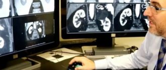
The information received from the scanner is transmitted to the screen and processed using a special program, and the doctor receives a high-definition three-dimensional image. Several photographs are taken per revolution of the device.
The Medunion clinic in Krasnoyarsk has installed the latest tomograph, built in 2021, for performing MSCT of the abdominal cavity and retroperitoneal space. This is one of the most convenient and versatile tomographs. It is equipped with a large number of applications, which expands the list of examined organs and research options.
What to take for examination
It is best to take with you to the examination all medical documentation that relates to the root causes of the tomography. This may be a doctor’s referral, extracts from medical records, examination results, results of previous tomographs or ultrasound. The radiologist will be able to take this information into account in his report and conduct a comparative assessment of the dynamics of the development of the pathology.
It is best to dress at the diagnostic center in clothes that do not restrict movement, in which it is comfortable to lie in a horizontal position. It is advisable that there are no metal elements on it.
| List of studies | Price | Stock |
| CT scan of the urinary system (kidneys, adrenal glands, bladder, urinary tract) | from 2500 rub. | At night |
| CT scan of the abdominal cavity (part of the esophagus, stomach, liver, gallbladder, pancreas, spleen, small intestine) | from 2500 rub. | At night |
| CT scan of the retroperitoneum (kidneys and adrenal glands, ureters, soft tissues and lymph nodes) | from 2500 rub. | At night |
| CT virtual colonoscopy (large intestine) | from 4100 rub. | At night |
Indications and contraindications for CT scan of the small intestine and stomach
A patient should be prescribed a CT scan for a number of diseases and ailments: tumors, polyps, bleeding, severe abdominal pain, the appearance of blood clots in the stool, sudden weight loss, flatulence, and inflammatory processes. Also, an indication for examination using a tomograph is the inaccuracy of analyzes made by other methods, for example, conventional X-ray examination or magnetic resonance imaging.
Contraindications for CT scanning of the stomach and small intestine:
- severe stage of diabetes mellitus;
- allergic reactions to medications that contain iodine and barium;
- pregnancy and lactation period (you can breastfeed your baby at least one day after the test);
- renal and liver failure;
- heart failure and coronary heart disease;
- tumor in the bone marrow;
- immune system dysfunction;
- excessive fatigue, tiredness and lack of sleep;
- partial hearing loss or complete deafness.
If a patient’s blood pressure has increased or decreased before the procedure, or has complaints about their health, this should be reported to the doctor immediately.
Preparing for a CT scan of the abdomen with contrast
Computed tomography of the abdominal cavity with contrast requires the use of a special iodine-based contrast agent. Contrasting adds some additional diagnostic limitations and training requirements. Because contrast is eliminated from the body through the kidneys, if a patient has kidney failure, they should have a creatinine blood test done in advance in preparation for an abdominal CT scan with contrast. The data from this test will assess the readiness of the kidneys to cope with the additional load. Some medical centers in St. Petersburg require a creatinine test for all patients over 65 years of age before contrast tomography.
If you have a history of thyroid disease, you should consult an endocrinologist as preparation for a contrast tomography.
Contraindications to MRI of the gastrointestinal tract
Absolute:
- 1st trimester of pregnancy;
- wearing a pacemaker, middle ear implants;
- foreign metal objects (shards, bullets, shavings);
- metal clips/clamps on cerebral vessels.
Relative:
- allergy to iodine, breastfeeding, severe renal or liver failure (with contrast administration);
- inability to remain still during the study (children, mental illness, claustrophobia, panic attacks);
- decompensated cardiovascular pathology;
- joint prostheses; insulin pump; vena cava filters; neurostimulators.
Preparing for a CT scan of a child's abdomen
If you are planning to have your child have a CT scan of the abdomen, the preparation requirements are no different from the recommendations for adults. However, a small child additionally needs to be psychologically adjusted to the examination. Children may be afraid to be in an examination room where there is noisy equipment. Therefore, the child will need to be explained in a playful way how the CT machine works and what will happen to it during the scan. The tomography procedure itself can be played out like a flight in a spaceship or like a ride on an amusement ride.
What does a CT scan of the intestines show?
What does a CT scan of the intestine show? The only difference between this method and colonoscopy is that it is less accurate, since with a CT scan it is impossible to see the color of the mucous membrane or take a biopsy from pathological lesions. However, with the help of three-dimensional scanning, you can view the organ from all sides, which is the great advantage of CT. The technique allows you to study the relief of the intestinal mucosa, the thickness and structure of its walls. If diffuse thickening is detected, it is easy to diagnose lymphoma, leiomyoma and leiomyosarcoma. It is also possible to examine the small intestine to determine its patency.
Contraindications
Today, CT scan of the abdominal cavity is considered one of the safest examinations.
The functional diagnostics clinic of the Yusupov Hospital has the latest generation equipment, the software of which provides for a reduction in radiation dose by 50%. An absolute contraindication to CT scanning is the presence of pregnancy in a woman. Relative contraindications include:
- acute kidney disease;
- age up to 14 years;
- lactation;
- decompensated diabetes mellitus;
- individual intolerance to the components of the radiopaque substance;
- severe diseases of the heart, liver, blood.
Indications
Indications for CT scanning of the abdominal cavity are varied. Among them are the following:
- preoperative preparation for major abdominal surgery;
- assessing the patient's operability (surgical treatment);
- development of acute conditions and pain syndrome of unknown origin;
- abdominal trauma;
- development of obstructive jaundice and sudden changes in weight;
- emergence of suspicions of the presence of a space-occupying formation in the abdominal cavity;
- chronic dysfunction of the digestive and urinary system;
- control of treatment dynamics.
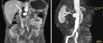
CT scan with contrast of the abdominal cavity determines:
- Tumors (malignant and benign);
- Vascular abnormalities and atherosclerosis;
- Abscesses;
- Echinococcosis;
- Hemorrhages and injuries;
- Conglomerates (stones) in the gallbladder and kidneys;
- Lesions of the lymph nodes;
- Extensive processes of pathological changes (volume of formation, its border and degree of prevalence);
- Liver pathologies (hepatosis, cirrhosis, hepatitis);
- Congenital developmental anomalies;
- The structure of parenchymal organs (organs consisting of continuous tissue), its painful changes;
- Cysts;
- Blood diseases;
- Normal or pathological patency of hollow and tubular organs;
- Foreign bodies.