Home » Diagnostics » Colon X-ray
X-ray of the intestine is a method of diagnosing an internal organ using an X-ray machine. The study is the most informative and does not cause complications. To carry out X-ray diagnostics, modern equipment is used, which allows you to obtain a clear image of the internal organs and make the correct diagnosis.
What does a colon x-ray show?
Radiation diagnostics makes it possible to conduct a full assessment of the functionality of the intestine and identify the nature of interaction with other organs located in the peritoneum.
X-ray of the intestines is an informative diagnostic procedure that:
- will show the diameter, shape of the lumen and location of the small and large intestine;
- will establish stretchability and elasticity level;
- will evaluate the characteristics of the motor functions of the internal organ and the area of inflammation;
- will determine the presence of developmental anomalies, polyps, tumors, ulcerated areas and diverticula.
Which technique is preferable?
What's better
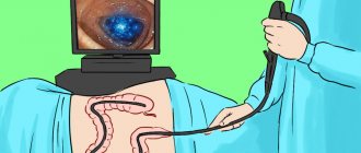
: X-ray examination of the intestines or colonoscopy - the doctor decides depending on the individual characteristics of the patient. Irrigoscopy with barium is often preferable, as it does not create discomfort for the patient.
But often it is impossible to do without endoscopic examination. This technique is the “gold standard” for diagnosing colon cancer; it allows detection of tumors at the earliest stages of development. X-ray methods do not provide such accuracy; their resolution is much lower.
Methods of X-ray examination of the intestines
Diagnostic testing is divided into two types:
- Radiography is the taking of photographs of a specific area of the body and printing of the images onto film. In this case, it is possible to make a video recording.
- X-ray. Processing of the internal organ in real time using a special screen. Using the equipment, you can take several pictures to display on the monitor.
With barium
An X-ray of the intestines using a contrast agent is called irrigoscopy.
Contrast is used to:
- obtaining an accurate outline of images;
- checking the patency of the rectum;
- determining the nature of intestinal functioning.
Today, intestinal X-rays with barium are performed 87% more often than without it.
Colon examination
Barium is mixed with warm water (3 liters) and slowly introduced into the intestine using a special device.
Pictures are taken in different projections:
- on the stomach;
- on the side;
- on the back.
After the procedure, the doctor recommends taking laxatives to speed up the removal of barium from the body.
X-rays of the colon can cause complications. That is why it is recommended to closely monitor your health in order to take therapeutic measures in time.
Small intestine examination
The patient takes a special solution orally and after 4-7 hours comes to the clinic for diagnosis. If the contrast agent is unevenly distributed, the specialist performs a light massage of the abdominal cavity, which helps correct the situation.
The duration of the procedure is 30-60 minutes. The conclusion is made based on how the contrast is distributed in the intestinal area: the presence of lumens, obstruction.
Indications for the procedure
A referral for a barium X-ray of the intestines is issued by an oncologist or gastroenterologist; in some cases, a general practitioner can refer you for the procedure. An examination is prescribed in cases where the doctor suspects a disease that poses a threat. Therefore, a referral for an X-ray of the intestines with barium should be taken with particular seriousness.
There is a whole range of signs of dangerous diseases of the digestive tract, in which a specialist prescribes an examination of the intestine with a barium-based contrast agent using radiography.
Serious weight loss for no significant reason
Severe weight loss without lifestyle or diet changes clearly indicates pathology. The patient often experiences lethargy and apathy. The source of the symptom may be a disruption of the intestines. To find out the cause, your doctor may order a barium X-ray of your small or large intestine.
The occurrence of loose stools for no reason
Loose stools for no apparent reason indicate a disruption in the digestive process. With long-term problems, it can cause intestinal damage. Therefore, the symptom requires prompt diagnosis and proper treatment.
Change in stool color
Loose, black stools are also called “tarry” stools. The discharge resembles a black paste with a pungent, unpleasant odor. The symptom indicates bleeding in the initial part of the duodenum or in the small intestine. If the bleeding is heavy, the patient may show signs of blood loss.
These conditions are extremely dangerous. Therefore, at the first appearance of tarry stools, it is better to immediately contact a specialist and undergo an intestinal examination with barium as prescribed; X-rays will show the source of bleeding and help the doctor make a diagnosis.
The appearance of severe pain in the abdominal area
Pain in the abdominal area can be caused by intestinal deformation or dysfunction. Often pain in the abdominal area indicates an ulcer. To determine the source of pain, various procedures are prescribed to examine the intestines, including the use of barium during x-rays.
Serious pathologies (emerging tumor processes)
Various neoplasms can interfere with normal intestinal motility, leading to pain, impaired defecation, loss of appetite, etc. To localize the neoplasm and establish its nature, the patient is prescribed an intestinal X-ray; the use of barium in an X-ray helps to obtain a contrast and informative image.
Preparing for an X-ray
To obtain more accurate results from an X-ray of the intestine, it is necessary to prepare for the procedure, following the recommendations of a specialist. Preparation includes following a special diet.
It is mandatory to carry out bowel cleansing activities before the procedure.
Diet
It is recommended to refrain from spicy and fatty foods. You should start preparing 2 days before the procedure.
It is recommended to avoid foods that cause flatulence:
- black bread;
- whole milk;
- legumes;
- fruits and vegetables.
16-18 hours before the procedure, it is recommended to exclude solid foods, carbonated drinks and coffee from the menu. It is permissible to eat only broths, drink tea and water.
Purgation
The day before the procedure, you should take laxatives prescribed by your doctor, which will help clear the intestines of the accumulation of feces. 2-3 hours before the intestinal x-ray, it is imperative to do a cleansing enema.
Who reviews the research results and where can they be obtained?
The images are analyzed by a radiologist: a doctor who specializes in performing x-ray examinations and interpreting their results. After examining the images, the radiologist draws up and signs a report, which is sent to the attending physician. In some cases, the report can be collected from the radiology department itself. The results of the study should be discussed with your doctor.
A follow-up x-ray examination is often required, the exact reason for which will be explained to the patient by the attending physician. In some cases, additional examination is carried out when doubtful results are obtained that require clarification during repeated images or the use of special imaging techniques. Dynamic observation allows timely identification of any pathological abnormalities that arise over time. In some situations, repeated examination allows us to talk about the effectiveness of treatment or stabilization of tissue condition over time.
Up
How does the procedure work and how long does it last?
An X-ray of the intestines is done in a room equipped with all the necessary equipment and protective equipment.
Patients are under the strict supervision of a specialist who follows a certain sequence of actions during the examination:
- The subject takes off his clothes with metal fasteners and jewelry and lies down on the couch.
- Using special belts, the specialist fixes the limbs to immobilize them.
- The table is set in a vertical position and the initial image is taken.
- The patient is injected with a contrast agent, after which photographs are taken in various projections.
- The study is carried out until the barium fills the entire area of the small intestine.
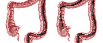
When examining the large intestine, double contrast can be used: with barium and air, which is pumped inside using the Bobrov apparatus. As barium and air are distributed, the specialist takes pictures in different projections.
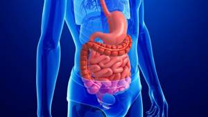
How does the human large intestine work, why is it examined?
The main stages of digestion associated with the primary processing and digestion of incoming food occur in the stomach and small intestine. However, after the food bolus passes into the large intestine, the process of its processing does not end - it is the large intestine that is responsible for the further entry of remaining beneficial substances into the blood from processed food.
Content:
- How does the human large intestine work, why is it examined?
- What diseases can affect the large intestine?
- X-ray of the large intestine: irrigoscopy and irrigography
- Indications and contraindications for irrigoscopy
- How to properly prepare for a colon x-ray
- Irrigoscopy for adults and children: technique
- Possible risks and consequences of the procedure
Anatomically, the department is represented by the cecum, colon, sigmoid and rectum. The colon, in turn, has three subsections - ascending, transverse and descending. The rectum has a wider part - the so-called ampulla of the rectum, and a tapering section located closer to the anus - the anus. Visually, the large intestine looks like a loop, the shape of which is similar to a square with an open contour. On average, its diameter reaches 6-6.5 centimeters, and its length is about two meters.
The large intestine contains a wide range of different beneficial bacteria. Its special flora contributes to the further processing of the food bolus and its transformation into feces. What is useful that remains in the contents of the food bolus at the stage of its presence in the thick section is processed and converted by bacteria into vitamins, sugars and amino acids. They are then absorbed by the mucous walls of the intestine, from where the beneficial substances enter the blood.
Leftover food that can no longer be digested accumulates in the large intestine in order to leave the body during bowel movements.
The large intestine, as part of a well-functioning mechanism, performs its functions in conjunction with the rest of the digestive tract, so in the body of a healthy person, food passes a continuous path from the mouth to the rectum, through which the body leaves what is left of this food after processing.
What happens if pathological changes appear in the large intestine? The patient experiences characteristic symptoms, deterioration in health, weight loss, abdominal pain and other ailments. In such cases, it is necessary to prescribe diagnostic procedures, in particular, x-rays of the large intestine.
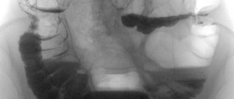
Interpretation of results
The data obtained is deciphered by a specialized specialist - a radiologist. It takes no more than half an hour to obtain the results of the study. Normally, the pictures should have a speckled image.
In the presence of flakes we are talking about the development of:
- lymphosarcoma;
- lymphogranulomatosis;
- malabsorption syndrome.
Polyps
An X-ray examination of the intestines can reveal polyps. The formations are localized on the mucous membrane and do not cause much harm. Despite this, they must be identified and removed, which eliminates the possibility of developing malignant degeneration.
Signs of intestinal obstruction on x-ray
Intestinal obstruction has peculiar symptoms: pain in the abdomen and vomiting. In this case, intestinal motility is also impaired. The x-ray shows small intestinal obstruction in the form of a stop in the movement of contrast agent or air to other parts.
Intestinal dyskinesia
The disorder may be asymptomatic and detected during an x-ray for the first time. Intestinal dyskinesia is accompanied by insufficiency, that is, functional disorders.
X-ray signs of hypomotor dyskinesia include weakened peristalsis and intestinal tone. This is fraught with a slowdown in metabolic processes and contributes to weight gain. The development of intestinal obstruction should be highlighted as complications.
Contraindications
Methods for examining the intestines using barium, including x-rays, have a number of contraindications. One part of them is dictated by the impact of ionizing radiation, the other – by the contrast agent.
Pregnancy
Ionizing radiation and barium can have a negative effect on the development of the fetus. Therefore, during the period of bearing a child, it is impossible to conduct an examination of the intestines using barium, especially using X-rays.
Intestinal perforation
Perforation (through damage) of the intestine makes it impossible to use barium, as it can enter the abdominal cavity. If there is a suspicion of such damage to the large or small intestine, contrast is not used during the examination.
The patient is in a serious or even unconscious state
When the patient is in serious condition, especially with acute inflammatory processes, the contrast can be harmful to his health. Therefore, examination of the intestines using barium using radiography in a pre-fainting state or in acute inflammatory processes is not carried out.
Ulcerative colitis
With ulcerative colitis, inflamed areas appear on the intestinal mucosa, which makes the use of barium in X-rays impossible. In this case, conventional radiography is prescribed.
Tachycardia
If barium is used to examine the intestines, various complications are possible that will lead to stress on the heart, so the use of contrast when X-raying a patient with tachycardia is not recommended.
Benefits and risks of lower gastrointestinal radiography
Advantages:
- X-ray examination of the lower gastrointestinal tract is a minimally invasive procedure with rare complications.
- X-rays of the lower gastrointestinal tract often avoid more invasive procedures such as colonoscopy.
- Allergic reactions accompany the study extremely rarely, since barium is not absorbed into the blood.
- After completion of the examination, no radiation remains in the patient’s body.
- When used for diagnostic purposes, X-rays do not cause any side effects.
Risks:
- With excessive exposure to X-ray radiation on the body, there is always an extremely small risk of developing malignant tumors. However, the benefits of accurate diagnosis significantly outweigh this risk.
- The effective dose of radiation is different for all patients.
- In rare cases, barium can leak through undetected holes in the intestinal wall, causing inflammation in the surrounding tissue.
- Even more rarely, barium can cause gastrointestinal obstruction.
- With rectal administration of iodine-containing contrast material, allergic reactions are possible, but extremely rare.
- A woman should always tell her doctor or radiologist about the possibility of pregnancy.
A few words about reducing the effects of radiation on the body
During an x-ray examination, the doctor takes special measures to minimize radiation exposure to the body while trying to obtain the best quality image. Experts from international radiological safety councils regularly review radiology standards and produce new technical recommendations for radiologists.
State-of-the-art X-ray machines allow you to control the dose of X-ray radiation and provide filtration, which minimizes beam scattering. In this case, the patient’s organs and systems that are not examined receive a minimal dose of radiation.
Up